Endocrine System
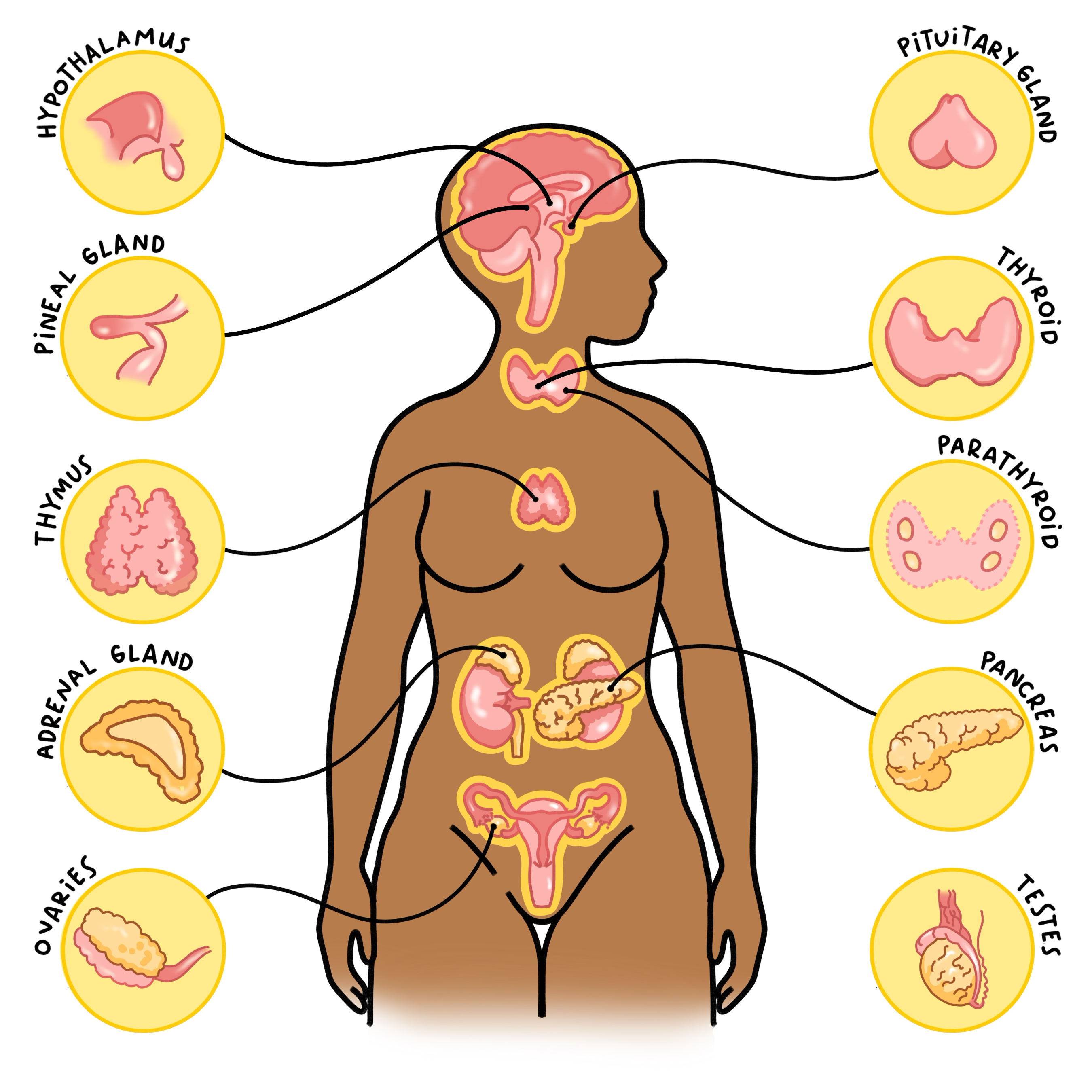
Both the nervous system and endocrine system are involved in controlling important body functions and maintaining homeostasis. While the nervous system uses electrical impulses and chemicals called neurotransmitters to send messages, the endocrine system uses chemicals called hormones that carry messages through the blood to the body’s organs and tissues. The endocrine system (Figure 1) is a complex network of glands/organs which include the pituitary gland, thyroid gland, parathyroid gland, adrenal gland, pancreas, and gonads (ovary and testis). For the purpose of simplicity, this chapter will discuss the cellular composition of each endocrine organ as well as the hormones produced by each, but not hormone functions.
Figure 1: Endocrine glands of the body

Pituitary Gland
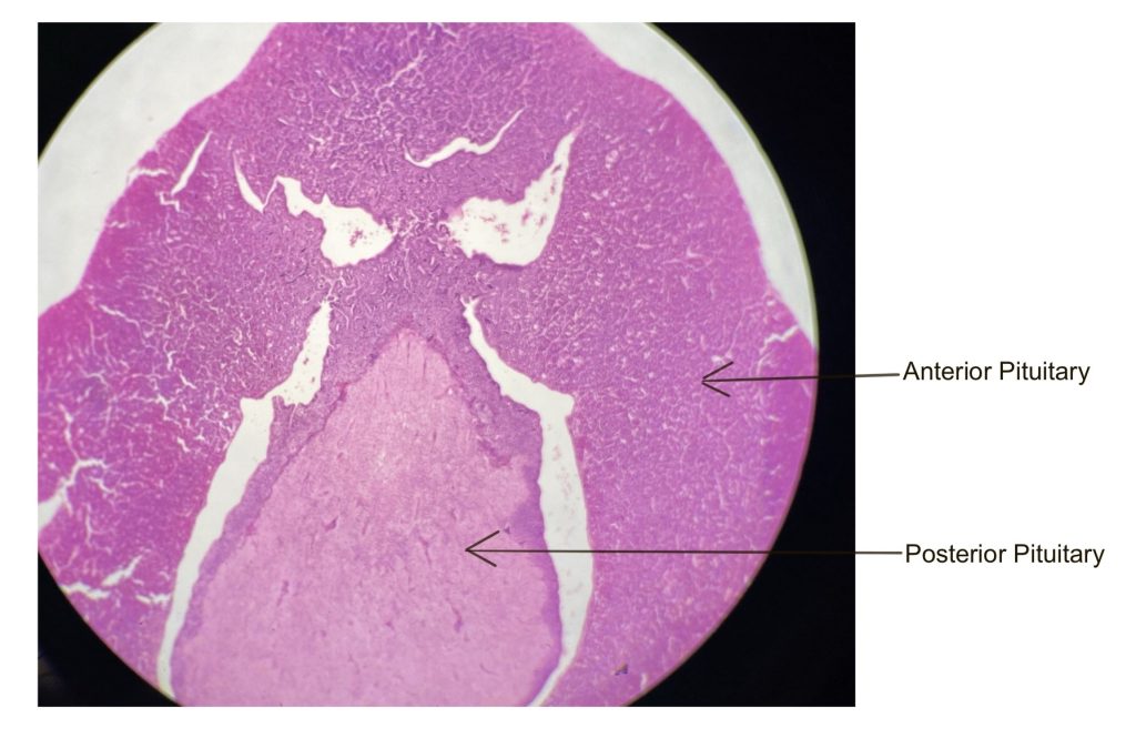
The pituitary gland is divided into the anterior pituitary (adenohypophysis) and the posterior pituitary (neurohypophysis). As seen in Figure 2 and 3, the anterior pituitary consists of glandular tissue, while the posterior pituitary consists of mostly nervous tissue.
Figure 2: Anterior and posterior pituitary gland under scanning power (version A)

The anterior pituitary contains two major cell types that secrete hormones, acidophils and basophils, distinguished by how they stain with Hematoxylin and Eosin (H&E) staining (Figure 4, Table 1). The acidophils stain pink/red and produce the growth hormone (GH) and prolactin (PRL). The basophils stain blue/purple and produce tropic hormones: follicle stimulating hormone (FSH), luteinizing hormone (LH), adrenocorticotropic hormone (ACTH), and thyroid stimulating hormone (TSH). A tropic hormone is a hormone that regulates hormone secretion by another endocrine gland. A third cell type may be visible, the chromophobes. These cells do not stain well (have clear cytoplasm) and may play a local role in regulation of pituitary hormones.
The nervous tissue of the posterior pituitary is composed of many axons with their axon terminals (Herring bodies) and surrounding glial cells (Figure 3, Table 2). These axons are part of neurons that originate in the hypothalamus. The glial cells are called pituicytes, they play a key role in the maintenance of blood vessels. The hormones, oxytocin and antidiuretic hormone (ADH, also known as vasopressin), are synthesized in cell bodies of neurons in the hypothalamus but stored and released at axon terminals in the posterior pituitary.
Figure 3: Pituitary gland with and without illustration overlay (version B)
Figure 4: Anterior pituitary with and without illustration overlay (version A)
Table 1: Cells of the anterior pituitary and their hormones
| Cell Type | Description | Hormones Produced/Released |
|---|---|---|
| Acidophils | Cells that stain pink/red and contain prominent round nuclei. These cells tend to appear larger than the surrounding cells. | Growth hormone (GH) and prolactin (PRL) |
| Basophils | Cells that stain blue/purple and contain prominent round nuclei. | Follicle stimulating hormone (FSH), luteinizing hormone (LH), adrenocorticotropic hormone (ACTH), and thyroid stimulating hormone (TSH) |
| Chromophobes | Cells that stain poorly and contain prominent round nuclei. | Little to no hormone content |
HINT: To remember the hormones that are produced and released by the basophils of the anterior pituitary, remember B-FLAT (first letter of hormones produced).
Table 2: Cells of the posterior pituitary and their hormones
| Cell Type | Description | Hormones Produced/Released |
|---|---|---|
| Hypothalamic neurons | Axons appear as wispy strands. Cell bodies of these neurosecretory neurons are located in the hypothalamus, and axons project down into the posterior pituitary where the hormones are stored at the axon terminals until released. | Oxytocin and antidiuretic hormone (ADH) |
| Pituicytes | Nuclei of these cells typically stain dark purple. | Little to no hormone content |
Thyroid and parathyroid gland
The thyroid gland and parathyroid glands are both located in the neck just below the larynx. The parathyroid glands are approximately four (up to eight) tiny glands located on the posterior surface of the thyroid gland.
The thyroid gland is made up of many thyroid follicles, which carry out the synthesis of thyroid hormones (T3 and T4). The follicles consist of a single layer of cuboidal cells called follicular cells. The lumen (space within the follicle) is filled with a substance called colloid. Between the thyroid follicles are slightly larger cells called parafollicular cells. These cells are important for the synthesis and release of calcitonin (Figure 5, Table 3).
Figure 5: Thyroid gland with and without illustration overlay
Table 3: Cells of the thyroid gland and their hormones
| Cell Type | Description | Hormones Produced/Released |
|---|---|---|
| Follicular cells | Part of the thyroid follicle, a single layer of cuboidal cells surrounding the colloid filled lumen. | Thyroid hormones (T3 and T4) |
| Parafollicular cells | Rounded cells located between the thyroid follicles. | Calcitonin |
The parathyroid gland consists mostly of chief cells. These densely-packed rounded cells are responsible for the synthesis and secretion of parathyroid hormone. While the cells in this micrograph are stained very darkly, this is not always the case (Figure 6, Table 4). Additionally, you will often see larger cells that do not stain well. These are oxyphil cells, whose exact function is currently unknown.
Figure 6: Parathyroid gland with and without illustration overlay
Table 4: Cells of the parathyroid gland and their hormones
| Cell Type | Description | Hormones Produced |
|---|---|---|
| Chief cells | Most abundant cell type. Rounded cells that have a prominent nucleus with little cytoplasm. | Thyroid hormones (T3 and T4) |
| Oxyphil cells | Large rounded cells that stain lightly. | Little to no hormone content |
adrenal gland
The adrenal glands (or suprarenal glands) sit on top of the kidneys. The adrenal gland consists of two main regions, the adrenal cortex and adrenal medulla. These glands are surrounded by a layer of connective tissue that acts as a protective capsule.
The adrenal cortex, the outer region of the adrenal gland, is glandular and divided into three layers or zonas (singular, zona) (Figure 7, Table 5). The superficial layer, the zona glomerulosa produces the mineralocorticoid hormones, such as aldosterone. The cells of zona glomerulosa tend to be small and arranged in small clusters. The next layer, the zona fasciculata produces the glucocorticoid hormones, such as cortisol. The zona fasciculata is composed of long mostly straight cords of large, light-staining cells. The deepest layer of the adrenal cortex, the zona reticularis produces androgens (sex hormone precursors).
The adrenal medulla is the innermost region of the adrenal gland and is a mixture of glandular and nervous tissue (Figure 7, Table 5). The secretory cells of the adrenal medulla (chromaffin cells) synthesize and release the catecholamine hormones, epinephrine and norepinephrine, into the blood in response to the activation of the sympathetic nervous system.
Figure 7: Adrenal gland with and without illustration overlay
Table 5: Regions of the adrenal gland and their hormones
| Region | Description | Hormones Produced |
|---|---|---|
| Cortex – zona glomerulosa | Thin outer most layer, small cells arranged in ovoid clusters/arches. | Mineralocorticoids (e.g., aldosterone) |
| Cortex – zona fasciculata | Middle cortex layer, larger light-staining cells arranged in long straight cords. | Glucocorticoids (e.g., cortisol) |
| Cortex – zona reticularis | Inner cortex layer, small dark-staining cells. | Androgens (e.g., dehydroepiandrosterone (DHEA)) |
| Medulla | Inner most region of the adrenal gland, contains clusters of large spherical (chromaffin) cells and preganglionic axons. | Catecholamines (e.g., epinephrine and norepinephrine) |
Pancreas
The pancreas is a gland posterior to the stomach that has both exocrine and endocrine functions. The exocrine tissue contain acinar cells that produce digestive enzymes and duct cells that produce bicarbonate to be secreted into the small intestine. The endocrine tissue of the pancreas, called the islets of Langerhans (pancreatic islets), appear as clusters of lightly-stained cells. The islets of Langerhans contain cells important in blood glucose regulation; alpha cells produce glucagon, while beta cells produce insulin (Figure 8, Table 6). It is difficult to differentiate the cells of the islets with the staining technique (H&E) used to produce these slides.
Figure 8: Pancreas with and without illustration overlay
Table 6: Cells of the pancreas and their hormones
| Cell Type | Description | Hormones Produced |
|---|---|---|
| Acinar cells | Large cuboidal-shaped cells that stain both pink and purple | No hormones, secrete digestive enzymes and bicarbonate |
| Alpha cells – pancreatic islets | Rounded cells, stain lighter than acinar cells | Glucagon |
| Beta cells – pancreatic islets | Rounded cells, stain lighter than acinar cells | Insulin |
Gonads (ovary and testis)
The gonads, the primary reproductive organs, are the testes in males and ovaries in females. These organs are responsible for producing the germinal/sex cells (sperm and ovum/egg). However, they also secrete hormones and are therefore part of the endocrine system.
The ovary is comprised of many ovarian follicles, each containing an oocyte (immature egg) (Figure 9, Table 7). The granulosa cells of the follicles surround the oocyte. They assist in development of the oocyte and produce the steroid hormones estrogen and progesterone. Theca cells, which surround the granulosa cells, produce androgens (sex hormone precursors) important in the production of estrogen. You will be learning more about the different stages of follicle development in the female reproductive system chapter.
Figure 9: Ovary with and without illustration overlay
Table 7: Cells of the ovary and their hormones
| Cell Type | Description | Hormones Produced |
|---|---|---|
| Oocyte | Very large pink cell surrounded by granulosa cells | Immature female germ/sex cell |
| Granulosa cells | Cuboidal shaped cells | Estrogen and progesterone |
| Theca cells | Elongated cells on the outer portion of the ovarian follicle | Androgens and small amounts of progesterone |
The testes are the male gonads containing seminiferous tubules that produce the male germ/sex cells – spermatozoa/sperm (Figure 10, Table 8). You will be learning more about the cells of the seminiferous tubules later in the male reproductive system chapter. Between the seminiferous tubules are interstitial (Leydig) cells that produce and secrete the steroid hormone testosterone.
Figure 10: Testis with and without illustration overlay
Table 8: Cells of the testes and their hormones
| Cell Type | Description | Hormones Produced |
|---|---|---|
| Spermatozoa/sperm | Oval shaped cell with long flagella | Male germ/sex cell |
| Leydig/Interstitial cells | Cells located in the areas between the seminiferous tubules | Testosterone |
Chapter Illustrations By:
Aislin Sparrow, B.A.
Juan Manuel Ramiro Diaz, PhD
Georgios Kallifatidis, PhD
epithelial-derived cells that produce and secrete a product
Acidophilic cells stain pink or red with H&E
basophilic cells stain purple or blue with H&E staining
a hormone involved in the regulation of growth and metabolism
a hormone whose most well understood function is in promotion of breast milk production. The role of prolactin in men is poorly understood.
A trophic hormone responsible for promotion of gamete production (sperm in males and oocytes in females). Stimulates androgen production (testosterone in males and estrogen/progesterone in females)
A trophic hormone which stimulates androgen production (testosterone in males and estrogen/progesterone in females). In females, triggers ovulation.
A trophic hormone that stimulates the cells of the adrenal cortex to secrete their respective hormones
A trophic hormone that stimulates the thyroid gland to produce thyroglobulin precursors to T3 and T4 (thyroid hormone)
support cells in the central and peripheral nervous systems
A hormone primarily responsible for stimulation of uterine contractions. Also serves as a chemical messenger governing social interactions.
A hormone produced in response to high blood osmolality (solute concentration), which causes the kidney to produced more concentrated urine and thereby keeping more fluids in the body.
A hormone with numerous functions in the body, including growth and metabolism.
para- (near, beside, around) and -follicular (follicle) = cells beside or near the follicle
A hormone involved in calcium homeostasis
A hormone involved in calcium homeostasis.
superior (supra-) to the kidney (-renal)
a layer of dense connective tissue that surrounds an organ and serves a protective function
layers or zones of tissue in the adrenal cortex
A hormone involved in sodium and water regulation by the kidneys
A hormone involved in the body's stress response.
a class of neurotransmitter - epinephrine and norepinephrine are major examples of catecholamines. Because they are secreted and released into the blood stream by the adrenal medulla, epinephrine and norepinephrine are considered hormones or neuroendocrine molecules.
Part of the autonomic nervous system - that mobilizes the body during periods of stress or activity (fight or flight).
secretions are transported to the surface of an epithelial tissue or into a hollow organ
A hormone produced in response to low blood glucose levels, responsible for releasing stored glucose.
A hormone produced in response to high blood glucose levels, responsible for storing excess glucose.
female germ cell containing half the number of chromosomes (n) that somatic (body) cells contain (2n). Germ cells are haploid and somatic cells are diploid.
a type of lipid with a 4-ring chemical structure. Cholesterol is a classic example of a biologically relevant steroid
female sex hormone
female sex hormone
