1 Arthritis of the Hand
Arthritis of the hand is a common and often disabling condition. There are several distinct types of arthritis affecting the hand; of these, degenerative arthritis (osteoarthritis) is the most common. Also seen are inflammatory arthritis (including rheumatoid arthritis), crystalline arthritis (gout, psuedogout) and infectious arthritis. The features common to all forms of hand arthritis are pain, stiffness, and swelling of the joints, and, in turn, impairment of function.
Structure and function
Osteoarthritis (OA)
The cause of hand osteoarthritis, while multifactorial, is often attributed to simple wear and tear caused by repetitive use and aging. In point of fact, however, abnormalities of the cartilage surfaces, kinematics and soft tissue forces, as well as inflammation and hormonal influences can all cause or promote the disease. For example, ligamentous laxity may lead to abnormal joint loading. In turn, this causes accelerated wear of the cartilage surface. (The process is similar to what may be seen with an automobile tire that is not secured tightly to the vehicle: it will wobble and erode prematurely.)
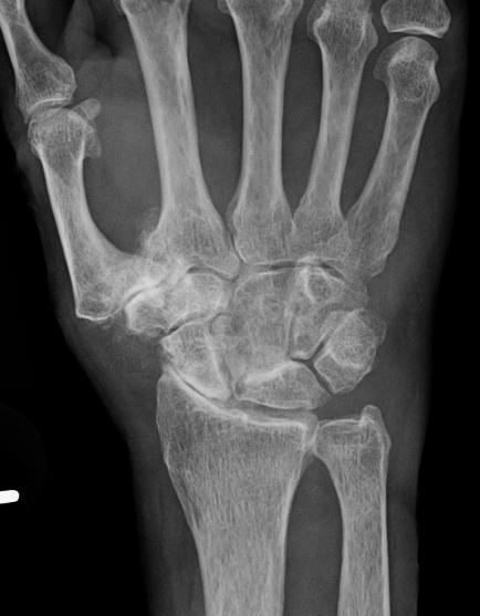
The classic radiographic hallmarks of osteoarthritis (subchondral sclerosis, joint space narrowing, synovial cysts, and osteophytes) are usually apparent in arthritis of the hand, however, the extent of these radiographic signs does not always correlate well with patients’ symptoms.
The characteristic features of a joint afflicted with osteoarthritis are hyaline cartilage degeneration; sclerosis of the subchondral bone; formation of osteophytes at the periphery of the joint; and cyst formation within the bone. In the hand, so-called mucinous cysts can form outside of the joint as well.
Although osteoarthritis is considered a disease of wear and tear, clearly genetic factors are involved. Twin studies have demonstrated a 59% concordance for hand osteoarthritis, and familial predispositions to osteoarthritis have long been recognized.
Interestingly, elevated body mass index is a risk factor for osteoarthritis of the hand. Given that the hand is not (typically) a weight-bearing structure, this risk suggests a possible systemic effect of an elevated BMI.
Rheumatoid Arthritis (RA)
The mechanism of joint destruction in rheumatoid arthritis is incompletely understood. The etiology appears to comprise both genetic and environmental components. Rheumatoid arthritis may generally (and incompletely) be described as an immune attack originating in the synovial cells against adjacent cartilage, tendon, bone, and soft tissues.
Rheumatoid arthritis begins in the synovial membrane with activation of the innate immune system, leading to loading of antigen presenting cells (APCs) with auto-antigens. APCs then migrate out of the joint and present these antigens to T-cells, which can then activate B cells or act directly on the synovium. There is likely no specific “rheumatoid arthritis antigen,” and the immune response seems to be directed at multiple targets, which vary across individuals. The wide variety of targets may explain the wide variety of treatment responses.
Patient presentation
The hallmark of hand arthritis, regardless of cause, is pain, stiffness, and swelling. To be sure, hand arthritis is not the only explanation for this presentation, and the examiner must be mindful of gout/pseudogout and infection (discussed below).
Osteoarthritis of the hand is typically seen in patients older than 50 years of age; the onset is often insidious. The classical symptoms of osteoarthritis are pain, tenderness, and stiffness worsened by activity and relieved by rest. Osteoarthritis most commonly involves the distal interphalangeal joints (DIP). The thumb carpometacarpal joint, (CMC) and proximal interphalangeal joints (PIP) are the next most commonly involved joints, but OA can be present in any joint of the hand.
Although osteoarthritis may occur in one joint in isolation, it is often symmetric and involves a “row” of joints.
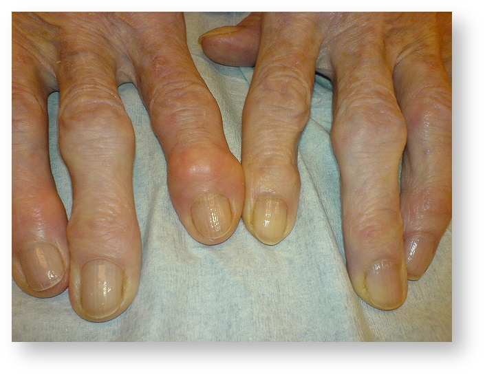
Osteoarthritis of the hand often produces deformity in its more advanced stages, though these are less dramatic than the deformities induced by rheumatoid arthritis. The most commonly seen osteoarthritis-related deformity is that produced by prominent exostoses (bone spurs) in the fingers. When these are present at the distal interphalangeal joint they are termed Heberden’s nodes; Bouchard’s nodes are similar exostoses seen at the proximal interphalangeal joint (as seen in Figure 1).

Hypertrophy of the soft tissues (coupled with osteophyte formation) can give a bulbous appearance to joints affected by osteoarthritis.
Basal thumb (CMC) arthritis may present with decreased pinch strength, and in advanced stages an adducted CMC joint with a compensatory MCP joint hyperextension deformity.
Additionally, mucous (mucinous) cysts, similar to ganglion cysts, may form in severe osteoarthritis, occasionally rupturing at the skin surface.
Rheumatoid arthritis in the hand most commonly involves the MCP and PIP joints. An insidious onset of pain, swelling and stiffness of multiple joints of the hand that is not related to activity are the distinctive features of the disease. Morning stiffness, particularly such stiffness lasting greater than one hour, is classic for rheumatoid arthritis. Affected joints will have an effusion (fluid within the joint capsule) and soft tissue swelling. Some additional features characteristic of this disease are inflammatory nodules, tenosynovitis, and tendon rupture.
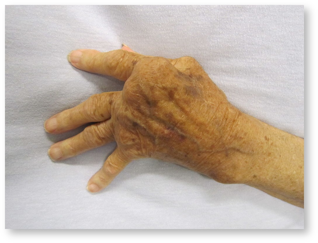
Chronic rheumatoid arthritis classically results in ulnar deviation of the MCP joints in addition to swan neck and boutonniere deformities of the interphalangeal joints (due to weakening of the ligaments and capsule, as well as intrinsic muscle imbalance).
Tendon ruptures, particularly of the extensor tendon to the thumb (EPL) or to the small finger (EDQ) are also common.
Clinically, three phases of rheumatoid arthritis are typically observed. The initial proliferative phase includes the onset of pain and effusion corresponding to synovial proliferation (described below). The second is the destructive phase in which the synovial pannus invades cartilage and soft tissue. Finally, there is the late or “burnt out” phase in which inflammation is replaced by fibrosis, scarring, and fixed deformities (Figure 2).

OBJECTIVE evidence
The radiographic findings of osteoarthritis include joint space narrowing, subchondral sclerosis and cysts, osteophyte formation as seen in Figure 3. Advanced disease may demonstrate deformity including subluxation and adjacent joint involvement.

There are no laboratory tests to confirm osteoarthritis; inflammatory markers are expected to be normal. Also, the synovial fluid should have a leukocyte count less than 2000 WBC/uL and without any crystals.
Any abnormal laboratory test results in the presence of presumed osteoarthritis should prompt the exploration for other diagnoses.
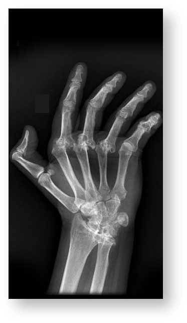
On plain x-ray, early rheumatoid arthritis will demonstrate soft tissue swelling. This will progress to joint space narrowing, subchondral cysts, peri-articular erosions and diffuse osteopenia. Erosions occur first in the “bare areas” of bone that are intra-articular but not covered in articular cartilage. In its advanced stages, the principle findings are gross deformity and auto-fusion of the joints.

MRI may additionally demonstrate effusion, synovitis, tenosynovitis, or tendon rupture.
Laboratory values in rheumatoid arthritis typically demonstrate a positive Rh factor and anti-cyclic citrullinated peptide (CCP) antibodies, though these can be negative in 20% of cases. The erythrocyte sedimentation rate (ESR, also known simply as the “sed rate”) and C-reactive protein (CRP) levels are typically elevated, though such elevation is a non-specific finding. Fluid analysis will typically demonstrate 3,500-50,000 WBC/uL.
Epidemiology
Primary osteoarthritis of the hand is common, with radiographic evidence of OA found in approximately 75% of females and 65% of males older than 55 years of age. Not all of these people have symptoms, however, symptomatic OA of the hand has a prevalence of about 25% among women and 13% among men (about 33% and 20%, respectively, of those with radiographic changes) Indeed, as you see, it is common for radiographic evidence of OA to not be symptomatic.
Radiographic OA is most commonly seen in the DIP joints (more than 50% of cases). The thumb CMC joint is the next most common (20%), followed by the PIP (8%).
RA is the most common inflammatory arthritis affecting 0.5-1% of the population worldwide. 70% of patients with RA report hand and wrist dysfunction. Women are 2-3 times more commonly affected. The age of onset is typically about 60.
Differential diagnosis
Hand pain and swelling can be caused by OA, RA, other inflammatory conditions (such as psoriatic arthritis and gout/pseudogout), infection and, rarely, a neoplastic process.
The location of the pain and its time course, the degree of inflammation, the number and symmetry of joints involved, the presence of systemic disease, and the associated signs of infection or trauma can aid in determining the diagnosis.
Carpal tunnel syndrome is a common cause of hand pain, but it is (by definition) limited to the median nerve distribution (the palmar side of the thumb, index and long fingers, primarily). In addition to pain, carpal tunnel syndrome can be detected by the presence of sensory changes and muscle atrophy (neither features of arthritis). Note that carpal tunnel syndrome can co-exist with arthritis and other diagnoses.
Pain at the base of the thumb may be from arthritis or tendinitis of the extensor pollicis brevis and abductor pollicis longus (de Quervain’s disease). Finkelstein’s test may help differentiate the two. In this test, the thumb is flexed and grasped by the patient’s four other fingers. Then, with the thumb stabilized, the examiner ulnar-deviates the hand, thereby stretching the extensor pollicis brevis and abductor pollicis longus tendons attached to the thumb. A positive test is characterized by reproduction of pain at the distal radius.
Hand infection may be a cause of acute swelling and pain. (The presence of a puncture wound, insect bite, or abrasion, of course increases the likelihood of this diagnosis.)
Gout and pseudogout are also causes of swelling and pain. In brief, gout and pseudogout are so-called “deposition diseases” in which crystals (monosodium urate and calcium pyrophosphate, respectively) are deposited in the joint and irritate it. Gout tends to affect the fingers, whereas pseudogout more commonly involves the wrist. The deposition of crystals can make the joint red, hot, tender and swollen – mimicking infection. A medical history, lab tests and x-rays can help differentiate these conditions from arthritis. The best means to make the diagnosis definitely is to examine fluid from the joint for uric acid or calcium pyrophosphate crystals.
| Feature | Osteoarthritis | Rheumatoid Arthritis |
| Patient demographics | Older; genders roughly equally affected | Younger; female |
| Time of maximal discomfort | End of day, pain with use | Morning stiffness, pain upon awakening |
| Joint(s) involved | interphalangeal joints; base of thumb | MCP joints |
| Tendon involvement | No | Yes |
| Visible deformity | Nodules at IP joint | Ulnar deviation of fingers; swan neck and boutonniere deformities of the interphalangeal joints |
| Radiographic features | osteophyte formation, sclerosis common Asymmetric joint space narrowing | osteopenia; symmetric joint space narrowing |
Red flags
Patients who present with acute swelling, warmth and erythema must be examined closely to rule out infection.
Patients with RA must be monitored for incipient tendon or ligament failure. Tendon rupture is an absolute indication for surgery, not only to restore lost tendon function, but to eliminate bone deformity or synovitis. This latter step will help prevent subsequent ruptures of neighboring tendons.
For patients with RA, systemic manifestations of disease must be considered as well. Children with juvenile arthritis must be monitored for uveitis (inflammation of the eye, including the iris, ciliary body and choroid).
Treatment options and outcomes
Non-operative Treatment
The mainstay of treatment for mild to moderate OA of the hand involves non-surgical measures aimed at reducing symptoms. These include hand therapy, NSAIDs, activity modifications, and local corticosteroid injections. Hand therapy includes splinting, strengthening exercises, and modalities such as hot soaks, and paraffin baths.
Several of the non-surgical strategies used in OA of the hand are employed in the treatment of RA of the hand as well. In addition, proper medical management of the disease, particularly using Disease Modifying Anti-Rheumatic Drugs (DMARDs), has significantly reduced the disease burden in patients with RA and reduced the need for surgical intervention in these patients.
The results of hand therapy in treating mild OA are good, with the majority of patients experiencing improvements in both pain and function. Those individuals requiring surgical intervention typically report high rates of improvement in pain and reduced deformity.
Operative Treatment
In more advanced disease that has not responded to initial treatment measures, surgery may help. Surgical options generally include tendon releases and transfers, cheilectomy (removal of excess bone), arthrodesis (joint fusion) and arthroplasty (joint replacement). While the specific details of the surgical management of hand arthritis is beyond the scope of this chapter, some common approaches and procedures warrant specific mention.
Synovectomy
Synovectomy is routinely performed in the surgical management of RA. Synovectomy alone, however, is rarely indicated (as there is 30-50% recurrence rate of synovitis after this procedure alone), but it is often applied in combination with tendon transfers and arthroplasty.
Arthrodesis
Surgical fusion of the joint (arthrodesis) is generally a very effective procedure in relieving pain. The resulting joint fusion provides a stable and pain free appendage at the expense of motion. Fusion is tolerated quite well in joints such as the DIPs and is particularly useful in joints that present with instability. Arthrodesis of an unstable index finger PIP joint can allow for significantly improved pinch strength and function. Arthrodesis of the MCP joint is poorly tolerated and is generally a last resort.
Arthrodesis of interphalangeal joints is extremely effective in improving pain and function.
Reconstructive Surgery
The thumb CMC joint is the most frequent site of reconstructive surgery in the upper extremity. Common approaches include volar ligament reconstruction, trapeziectomy with or without interpositional tendon graft, or arthroplasty.
The most common reconstructive procedure performed for RA of the hand is MCP joint replacement (arthroplasty), typically with a silicone implant. With the use of DMARDs, however, the need for this procedure has decreased dramatically in recent years.
The gold standard for treatment of advanced thumb CMC arthritis is trapeziectomy with or without interpositional graft and or ligament reconstruction. This method reliably relieves pain and improves function. Synthetic implants have a relatively high complication rate compared to excisional arthroplasty.
Arthroplasty
MCP arthroplasty in RA has demonstrated improved short-term functional and cosmetic outcomes in addition to reliable pain relief. Long-term studies have shown a high failure rate of the silicone implant, however, in such cases the implant can often be retained and retain adequate function. Recurrent ulnar drift is common, but usually less severe that the original deformity.
Risk factors and prevention
Risk factors for OA include advanced age, female gender, obesity, repetitive use, and family history. Prevention is based on avoiding risk factors such as repetitive activities. Often, such activities are associated with the patient’s work, and therefore, difficult to avoid.
Risk factors for RA include gender (female > male, as noted), family history, and smoking. Some studies have shown oral contraceptive use to be protective, as well as breastfeeding and childbirth. There is currently no preventive strategy, but early diagnosis and treatment can prevent many of the complications of the disease.
Miscellany
The use of chondroitin sulfate and glucosamine may provide benefit to some patients with osteoarthritis and carries a low risk profile if purchased from a reputable source. Studies have failed, however, to establish a proven benefit to these supplements.
Key terms
Osteoarthritis, rheumatoid arthritis, carpometacarpal (CMC) arthritis, Heberden’s nodes; Bouchard’s nodes
Skills
Recognize and describe deformities of the hands caused by arthritis. Recognize the signs and symptoms of infection. Examine the hand to determine if there is full and painless range of motion, normal strength and intact motor and sensory function.
