12 Flexor Tendon Injury
Injuries to the flexor tendons of the hand can be particularly challenging. Without finger flexion, patients will have difficulties with many tasks of daily living. The challenge to the surgeon is to re-attach the muscle to the bone in a way that glides smoothly within the pulley system of the hand. Flexor tendon injuries to the index and long finger tend to impede tasks that require fine motor skills; injuries to the flexor tendons of the ring or small fingers usually have a greater impact on grip strength.
Structure and function
Finger flexion occurs at three joints: the distal interphalangeal joint (DIP); the proximal interphalangeal joint (PIP); and the metacarpophalangeal joint (MCP).
Flexion at each of these joints is powered by a distinct muscle group:
- the flexor digitorum profundus flexes the distal interphalangeal joint;
- the flexor digitorum superficialis flexes the proximal interphalangeal joint; and
- the intrinsics (lumbricals and interossei) flex the metacarpophalangeal joints
The flexor digitorum profundus and the flexor digitorum superficialis attach directly to the distal and middle phalanx respectively (Figure 1). These tendons are held close to the bone by a system of annular (ring-like) and cruciate (cross-forming) ligaments or pulleys.
The lumbrical muscles do not attach to the proximal phalanx directly, but rather connect the tendons of flexor digitorum profundus (below, i.e. volar) to the extensor tendons (above, i.e. dorsal) and produce flexion by pulling on the extensor tendons as they pass over the proximal phalanx.
The flexor digitorum superficialis [FDS] and flexor digitorum profundus [FDP]) originate proximally in the forearm, at the medial epicondyle of the elbow. They, along with flexor pollicis longus (FPL) and the median nerve, travel through the carpal tunnel at the wrist and enter the palmar surface of the hand. From there, FDS and FDP send individual tendons to the index, long, ring, and small fingers.
Each pair of FDS and FDP tendons runs together through a tendon sheath into the fibro-osseous canal of each digit. This canal is formed by the metacarpals/phalanges, and by the pulley system and tendon sheath. The pulley system keeps the flexor tendons snug to the bone, preventing “bowstringing” during active flexion and converting a linear force into rotation and torque at the finger joints. The tendon sheath provides both lubrication as well as an important source of nutrition to the largely avascular tendons.
The superficialis lies, as its name implies, superficial to profundus (i.e. “deep”) throughout its course until it reaches the first annular pulley (“A1”) in the vicinity of the metacarpal head. At that point, the profundus emerges through a split in the superficialis (known as Camper’s chiasm); this allows the profundus to continue to its attachment on the volar surface of the distal phalanx where it flexes the distal interphalangeal (DIP) joint. The superficialis attaches onto the base of the middle phalanx, and thus flexes the proximal interphalangeal (PIP) joint.
Note that while the profundus distinctly flexes the distal interphalangeal (DIP) joint, it also indirectly flexes the proximal interphalangeal (PIP) joint as well. (This has important implications for physical examination, as the mere ability to flex the proximal interphalangeal (PIP) joint does not prove that the superficialis is intact; rather, such motion may reflect the indirect action of the profundus.)

Innervation to FDS and the radial-sided two FDP tendons (e.g., those to the index and long fingers) is provided by the median nerve, while innervation of the ulnar two FDP tendons (ring and small finger) is provided by the ulnar nerve.
Each digit also has a neurovascular bundle running along both its radial and ulnar borders. While these are not involved in finger flexion, per se, because of their proximity to the flexor tendons, they may also be injured when the flexor tendons are lacerated.
Injuries to the flexor tendons can be classified by the general location of the injury. Five zones have thus been identified. The first zone is distal to the FDS insertion. This is typically an avulsion of the profundus, known as a “Jersey finger,” as it may occur when a rugby player grabs the jersey of an opponent and pulls too hard. Zone II spans the PIP joint to the distal palmar crease near the metacarpal head. In Zone II the FDP and FDS travel in the same tendon sheath and are typically injured together. Zone III is within the palm itself and is often associated with neurovascular injury. Zone IV is the carpal tunnel itself and Zone V is any area proximal to the wrist.
Patient presentation
Patients will most frequently present with a laceration to the palmar aspect of the hand and an inability to flex one or more digits. It is important to ascertain the mechanism of injury, as it may impact treatment. For example, animal or human bites should prompt antibiotics, and any penetrating injury to the hand should prompt an inquiry of whether the patient’s tetanus is up to date.
Identification of which tendons are injured requires an accurate and complete physical exam, documenting the presence or absence of FDS and FDP function in each digit.
To evaluate the superficialis (Figure 2), begin by having the patient extend all digits. Then, immobilize all other fingers (besides the one being tested) in this position of full extension. (This holds the profundus out to length via the “quadrigia phenomenon” (see Miscellany, below) and thus prevents the profundus from indirectly flexing the proximal interphalangeal (PIP) joint.) Instruct the patient to attempt to flex the finger. If the FDS is intact, the patient will flex at the PIP joint.

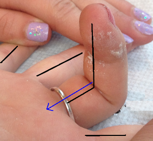
To evaluate the profundus, have the patient extend the digit. Grasp the finger to immobilize the PIP and MCP joints. Instruct the patient to attempt to flex the finger. If the profundus is intact, the patient will flex at the DIP joint.
Additionally, ensure the integrity of the digital neurovascular bundles (that course near the flexor tendons and may be injured concomitantly) by testing two-point discrimination. Doppler exam of the digital arteries may also be helpful.
objective Evidence
While flexor tendon injuries cannot be diagnosed via x-ray, both AP and lateral views of the hand should be obtained to rule out fracture or foreign body. CT and MRI have limited use in this diagnosis, although MRI would be the better of the two to evaluate soft tissue injuries and relative location in the hand or forearm of the tendon stumps. Depending on the experience of the operator, ultrasound may also be used to evaluate tendon integrity within the sheath.
Epidemiology
Hand injuries make up one of the most common complaints treated in the emergency department (14-30%). Of these, tendon injuries account for approximately 30% of presentations, second in frequency only to patients presenting with fracture (40%).
Differential diagnosis
A direct flexor tendon injury must be suspected in all patients who present with an inability to flex one or more digits. However, depending on the mechanism and extent of injury, other injuries may also be present. Important considerations include fractures of the metacarpals or phalanges that may entrap the flexor tendon, limiting its motion. Other considerations include injuries to the flexor sheath or pulley system, neurovascular injuries that impair or completely eliminate neurologic or vascular supply to the tendon or associated muscle body, or, as stated in the “Red flags” section, infection of the tendon sheath that limits movement of the tendon.
If multiple digits are affected without sign of infection or laceration to the hand, the cause likely lies proximal, that is up the arm towards the neck. Even without a direct injury to the flexor tendons, injury to the median or ulnar nerves proximal to their innervations of the FDS and FDP muscles in the forearm may result in decreased finger flexion (this could range from peripheral compression or injury in the arm or forearm, pathology of the brachial plexus, to cervical stenosis at the level of the spine).
Red flags
An inability to flex the fingers after a trauma is itself a “red flag.” Flexor tendon injuries require expeditious treatment from a surgeon who specializes in disorders of the hand.
Impaired flexion of a digit from swelling suggests infection.
Impaired passive motion of a digit because of guarding (that is, tensing the muscles to limit passive motion and pain) may suggest a forearm compartment syndrome.
Treatment options and outcomes
All tendon injuries should be examined to determine the specific anatomy that has been disrupted. At the time of initial evaluation, any open injuries should be thoroughly irrigated and cleaned. Depending on the extent of injury, the affected digit may rest in a more extended position than its neighbors. In this case, a dorsal blocking splint that keeps the wrist in neutral and the metacarpal phalangeal joints and PIP joints in partial flexion is advised.
All flexor tendon injuries should be referred to a surgeon who specializes in disorders of the hand. Flexor tendon injuries should be treated surgically expeditiously (within about 7 days of initial injury) to ensure the best results and a functional outcome. With the passage of time, the proximal edge of the lacerated tendon retracts further proximally; also, adhesions begin to form between the tendon and nearby structures.
The surgical treatment chosen for a particular injury is dependent on several factors, including location (zone) of injury, time since injury, condition of the tendon stumps and surrounding tissues, and experience level of the surgeon. It is generally agreed upon that partial lacerations involving <60% of tendon cross-sectional area should be debrided without primary repair.
Operative treatments of complete lacerations and those involving more than 60% of the tendon cross-sectional area include primary repair and staged reconstruction procedures with either donor tendon or silicon tendon implants.
Successful flexor tendon repair relies not only on successful reattachment at the time of surgery but successful rehabilitation. Consistent and reliable follow-up cannot be over-emphasized. Range of motion of the digit is begun almost immediately with both passive and active flexion and extension. This minimizes adhesions and ensures the patient retains the degree of motion attained in the OR.
(The use of regional anesthesia during surgery allows the patients to confirm with their own eyes that the tendon has been re-attached. Some surgeons believe that this provides the patient incentive and motivation to continue through the difficult and painful rehabilitation process.)
The key to good outcomes following a successful surgery is early mobilization and consistent therapy. Tendon tensile strength improves as the repair site is stressed, adhesions are minimized, and excursion is improved with early and frequent range of motion. A dorsal-blocking splint used between therapy sessions protects the digits and maintains the repair in a slightly shortened position (as compared to full extension), alleviating some of the stress placed across the site of reconstruction when the digit is held in full extension. With appropriate therapy and protection of the repair site, good functional outcome can be seen in over 75% of patients. Most commonly encountered complications include adhesion formation, stiffness of finger joints, and even tendon rupture (risk of rupture is greatest at days 7-10 following repair).
Understanding of and compliance with therapy and restrictions is critical after surgical repair. Therefore, children and adults with limited mental capacity should be immobilized in a cast for one month following surgical repair.
Risk factors and prevention
Most tendon injuries are a result of accidental trauma. However, a history of prior or current infection of the tendons, as well as recent surgical procedures to the tendons or hand may predispose to tendon rupture by compromise of integrity due to local inflammation or due to suture failure, respectively. Prior surgery may compromise tendon integrity and tendon excursion via formation of scar tissue and adhesions of the tendon sheath. Also, some metabolic/inflammatory disorders can weaken tendon integrity and place them at risk of rupture (e.g. infection, rheumatoid arthritis).
Miscellany
The ability to test FDS strength and excursion in isolation of FDP function is made possible by the quadriga phenomenon. A “quadriga” is a Roman chariot pulled by four horses. This phenomenon is named because the four FDP tendons are connected in the forearm and were thought to resemble the appearance of the reins of a quadriga.

Key terms
Flexor tendon, Tendon injury, Tendon repair
Skills
How to perform a thorough physical exam of the upper extremity, including how to evaluate individual nerves, both sensory and motor branches, how to assess distal digital pulses with Doppler exam, how to perform Allen’s test, how to differentiate between FDS and FDP function, and how to evaluate two-point discrimination.
