24 Ulnar and Radial Shaft Fractures
In adults, simultaneous fractures of the shaft of the ulna and radius (the so-called “both bone fractures”) are most often the consequence of a direct blow to the forearm or other high energy mechanisms. In the adult, these injuries usually require surgical fixation. By contrast, these injuries in the pediatric population result from simple falls and other lower energy mechanisms. In children, ulnar and radial shaft fractures are amenable to casting. Imperfect healing can lead to loss of pronation and supination of the hand.
Structure and function
The human forearm is comprised of two bones: the ulna, medially, and the radius, laterally. Bridging the ulna and radius is an interosseous membrane that transmits forces from ulna to radius and vice versa. The forearm flexes and extends at the elbow, with the articulation of the ulna with humerus at the trochlear notch. The head of the radius also articulates with the humerus at the capitellum, allowing the forearm to pronate and supinate. Pronation and supination also require an intact distal radial ulnar joint.
The median, ulnar, and radial nerves course along the forearm, along with the radial and ulnar arteries. The ulnar and radial nerves are located most medially and laterally, respectively, thus they are most susceptible to damage with fracture of the shaft of their adjacent bones.
The extrinsic muscles of the hand originate in the forearm (and elbow) and therefore forearm fractures, if not treated properly, can also lead to hand dysfunction.
The course of the muscles, likewise, may create deforming forces on the injured bones: for example, the flexor muscles of the fingers and wrist tend to produce dorsal bowing of the radius and ulna, by flexing distal fragments.
Patient presentation
Patients with fractures of the shaft of the ulna and radius present following trauma with pain in the forearm, at time with gross deformity.
Radiographs allow full characterization of the injury. Care must be taken to fully visualize the wrist and elbow joints.
Greenstick Fractures
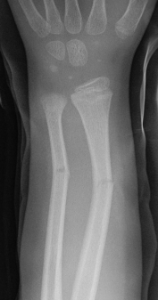
Pediatric fractures can be complete (involving a through-and-through break of the bone) or so-called “greenstick fractures,” a break on one surface of the bone that does not extend to the other side. This configuration occurs in children because, compared to adult bone, pediatric bone tends to be softer and more prone incompletely breakage (much like the branches of a living tree, hence the name “greenstick”).

Galeazzi Fracture
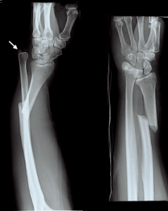
A radial shaft fracture with distal radial ulnar joint (DRUJ) instability is known by its eponym, the “Galeazzi fracture.” A patient with a Galeazzi fracture will present not only with pain in the forearm where the radius is broken, but also swelling, tenderness, and pain at the wrist where there is a dislocation of the distal radioulnar joint.
Ulnar shaft fractures are most often caused by a direct blow to the border of the ulna on the medial forearm. It can occur when the ulnar border is exposed, such as upon raising the arm in defense (the so-called “night stick fracture”). The patient will usually experience pain and show signs of point tenderness in the ulnar region of the forearm. Swelling will likely be present in the forearm.

Monteggia Fractures
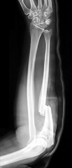
“Monteggia fractures” are injuries to the proximal third of the ulna associated with a dislocation of the head of the radius. It is rare for a fracture to the proximal one-third of the ulna to occur alone. As a result, it is always imperative to do a full work-up of both the ulna and radius to assess whether the radius, especially the radial head, is injured.
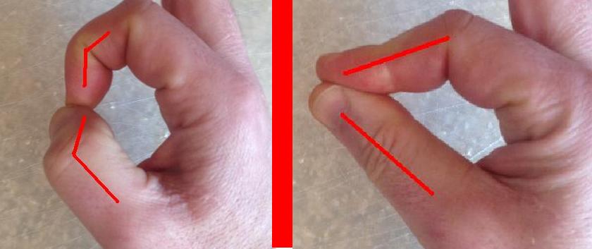
Damage to the posterior interosseous nerve may occur with a Monteggia fracture; it is therefore important to assess this nerve by checking the patient’s ability to extend the hand at the wrist.

objective evidence
Patients with forearm fractures should have the following information collected and documented:
- status of the skin and soft tissues
- range of motion of the wrist and elbow
- gross alignment of the limb
- congruity of the elbow and wrist joints on x-ray
- status of nerves: radial (including posterior interosseous), median (including anterior interosseous), and ulnar (Figure 4)
- perfusion status of hand

Epidemiology
Fractures of the ulnar and radial shaft can occur across all age groups but are most common in children. Indeed, mid-shaft forearm fractures are among the most common type of fracture in children. This type of injury is most often the consequence of direct trauma, frequently from a fall on an outstretched hand from a large height.
Other populations at risk are adults or children involved in events such as traffic accidents and sports injuries, commonly from mountain biking and skating. Osteoporosis, mostly occurring in postmenopausal women, is commonly associated with forearm fractures, although fractures in this population are most often present in the distal forearm near the wrist.
Differential diagnosis
A forearm shaft fracture should be readily diagnosed from radiography. As such, there is no “differential” diagnosis list, per se; the question is either about what other injuries may be present, or whether the fracture was caused by some underlying abnormality such as osteoporosis.
Additional injuries that may be seen with Monteggia fractures, for example, include possible olecranon fracture-dislocation; radial head or coronoid fractures, or lateral collateral ligament injury.
Secondary injuries include not only damage to the elbow or wrist, but otherwise more distal unrelated injuries simply sustained at the same time.
Osteoporosis should be considered if the mechanism of injury suggests particularly low energy.
Fractures through a pre-existing lesion (a so-called “pathological fracture,” for example, one occurring in a bone with a tumor or Paget’s disease) are rare but possible.
Red flags
Radiographs that are too narrowly focused, i.e., do not visualize the elbow and wrist, may lead to missed diagnoses involving those excluded regions.
Both bone fractures can be complicated by acute compartment syndrome of the forearm; this is rare but possible. Signs suggesting compartment syndrome are pain on extension of digits and marked edema. The phrase “pain out of proportion to the injury” is difficult to apply when there is a broken bone, but in general, immobilizing a fracture should reduce the patient’s distress; if it does not, compartment syndrome should be explicitly ruled out by measuring compartment pressures.
Pediatric both-bone fractures, though commonly seen after accidental trauma, may also be the product of child abuse. Careful attention should be paid to the history given by the parents, (obtained from each separately, if abuse is suspected).
Treatment options and outcomes
Displaced and unstable ulnar fractures in adults should be treated operatively. Even non-displaced radial fractures are usually treated with surgery. (Pediatric fractures at times can be treated with closed reduction and casting.)
Monteggia fractures require open reduction and internal fixation of the ulnar shaft fracture. If this procedure fails to reduce the radial head, then that too must be opened and reduced.
Most fractures heal if adequately reduced.
Even subtle malalignment can impede pronation and supination.
Most cases of neuropathy (in the absence of compartment syndrome) tend to resolve with rest.
Risk factors and prevention
Beyond giving trite advice (“Don’t drink and drive. Stay out of fights. Don’t fall.”) there is not much physicians can do to prevent these injuries. The key “prevention” step is to prevent complications by not missing associated injuries at the time of initial presentation.
Miscellany
Although two fractures of the forearm are named after Italian surgeons from Milan, their contribution did not reflect a golden age of orthopaedic surgery in that city. In fact, they were not contemporaries: Giovanni Battista Monteggia died in 1815, more than a half-century before Ricardo Galeazzi was born in 1866.
Key terms
greenstick fracture, Galeazzi fracture, Monteggia fracture
Skills
Recognize and describe fracture patterns on plain radiographs. Assess the neurological function with a patient following fracture. Predict the deforming forces that muscles exert on fractures.
