4 Cells of the Nervous System: Glia
Learning Objectives
Become familiar with the classes of glial cells in the nervous system
-
- Astrocytes
- Oligodendrocytes
- Schwann cells
- Microglia
- Ependymal cells
Appreciate the newly-discovered roles of glial cells
- Produce calcium signals when activated by neurotransmitters
- Release gliotransmitters which influence the activity of neurons
Although most of neuroscience is concerned with understanding the functions of neurons, there are other cells in the nervous system that are just as interesting. These cells are grouped together under the umbrella classification of glia. Historically, when these non-neuronal cells were visualized under the microscope, the histologists and anatomists had no idea about their function. They were seen all around the neurons, so the assumption was that these cells were structural elements, a sort of living glue, that held the nervous system together. Today, we know that these glia serve a variety of functions; unfortunately, the misnomer “glia”—derived from the German word for “glue”—is still used to describe these non-neuronal components of the nervous system.
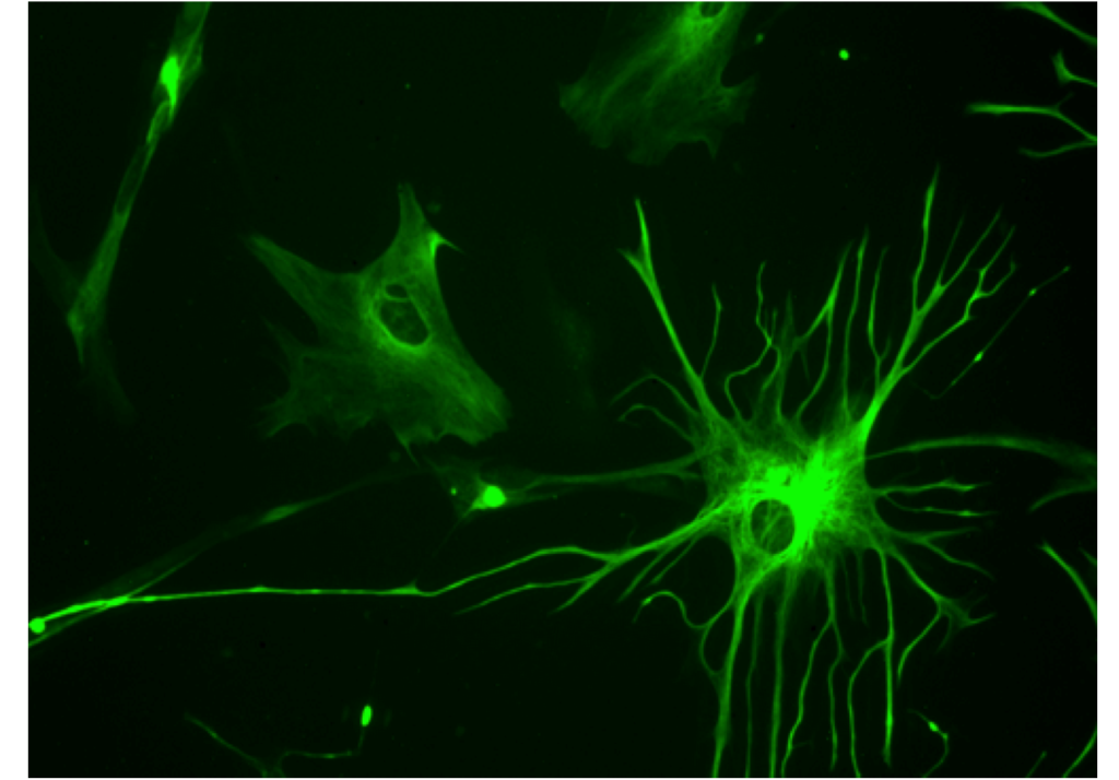
Astrocytes
Astrocytes are named for their characteristic star-shaped morphology. One of the main functions of astrocytes in the brain is to help maintain the blood-brain barrier. At the end of the extensions of the astrocyte are protrusions called “endfeet”. These endfeet are often wrapped around the endothelial cells that surround the blood vessels. The endfeet release important biological compounds that allow the endothelial cells to remain healthy as they function in maintaining the blood-brain barrier. Astrocytes are also very closely associated with synapses.
Astrocytes also synthesize and produce a variety of trophic factors, which are helper molecular signals that serve several different functions. For one, trophic factors signal to neurons that the neuron should continue to live, or that specific synapses should be maintained. They help guide the neurons as they reach out, forming synapses where appropriate.
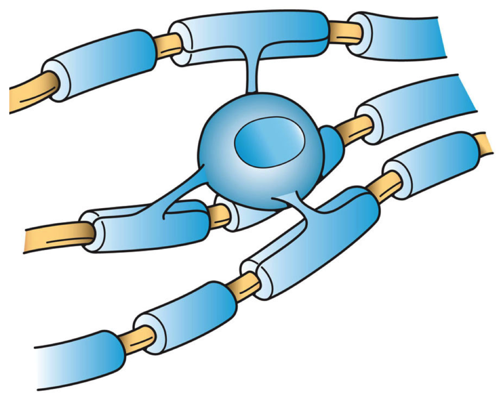
Oligodendrocytes
The main function of the oligodendrocytes is to add a layer of myelin around the axons of nearby neurons in the central nervous system. A single oligodendrocyte is able to myelinate up to 50 segments of axons. As cells that produce myelin, they are responsible for increasing the conduction speed of nearby neurons as they send signals. Oligodendrocytes only exist in the central nervous system.
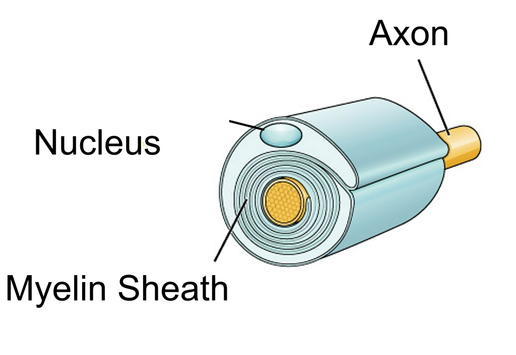
Schwann Cells
Schwann cells can only be found in the peripheral nervous system. The main action of Schwann cells is to provide a section of myelin sheath for peripheral nervous system neurons, and in this way, they function similarly to the oligodendrocytes. Schwann cells produce only a single section of myelin, compared to oligodendrocytes, which myelinate multiple sections. Schwann cells also function in the regeneration of injured axons. When nerves in the peripheral nervous system are damaged after trauma, Schwann cells rapidly mobilize to the site of injury.
Perisynaptic Schwann Cells (PSCs)

There is a subclass of Schwann cells called perisynaptic Schwann cells (PSCs) that cover the presynaptic nerve terminal at the neuromuscular junction. Although they cover and partially wrap around the presynaptic nerve terminal, the nerve terminals are unmyelinated. PSCs function in the peripheral nervous system much like the Astrocytes in the central nervous system.
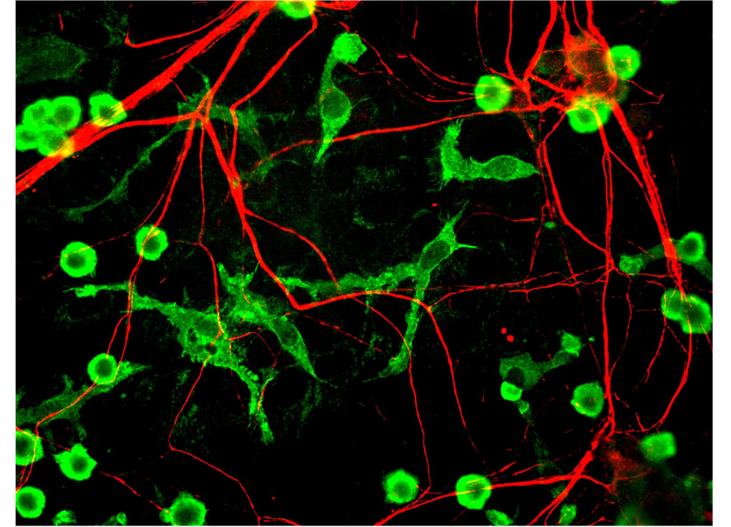
Microglia
Microglia are a bit different from the other glial cell populations. For one, microglia are more immune cells rather than neural. They act as cellular scavengers that travel throughout the brain and spinal cord. It is estimated that microglia make up 10-15% of all cells in the brain.
As immune cells, microglia identify and destroy clumps of proteins, dead/dying cells, or foreign pathogens that enter into the brain. After an injury to the central nervous system, like a traumatic blow to the head, microglia rapidly react to the area of the insult.
Ependymal Cells
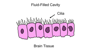
Along the inside of the ventricles are a lining of glia called ependymal cells. These ependymal cells are columnar with small fingerlike extensions called cilia that extend into the ventricles and into the central canal that runs down the inside of the spinal cord. The cilia have motor properties that allow for them to rhythmically beat to create a current in the surrounding fluid.
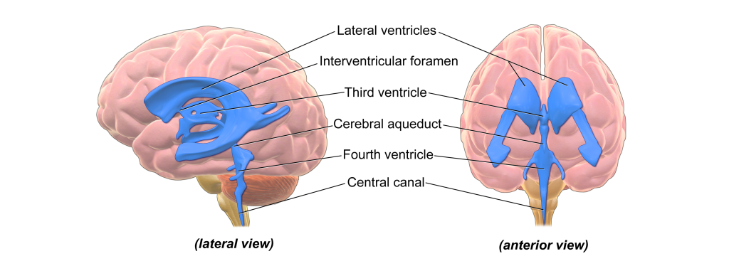
Ependymal cells produce cerebral spinal fluid (CSF). In total, the body can make about half a liter of CSF each day (a little more than two cups.) The ependymal cells are part of a structure called the choroid plexus, the network of blood vessels and cells that form a boundary between the blood and the CSF.
A New appreciation of Glial Cells
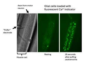
As mentioned briefly in the first chapter, recent discoveries have revealed new, surprising roles for glial cells. Once thought of as primarily supporting the signaling function of neurons (e.g. myelination, metabolic support etc.), we now know that glial cells both receive and send messages to other cells, including neurons. Since their signaling does not happen through electrical means, it was not detected until neuroscientists developed techniques for observing dynamic changes to the concentration of calcium ions (Ca2+) inside cells. These techniques rely on the ability of molecules engineered to indicate the free Ca2+ concentration in their vicinity by altering how they fluoresce when excited by light of specific wavelengths. When these molecules, called fluorophores, were loaded into glial cells, they revealed a big surprise. Glial cells respond to neuronal activity, especially at synapses, by increasing the concentration of Ca2+ in their cytoplasm. This Ca2+ signal is triggered by the binding of neurotransmitter molecules to receptors on the membrane of the glial cell. An example of this is shown below in images collected at a neuromuscular junction (NMJ), in which the glial cells that cover the NMJ were loaded with a fluorescent Ca2+ indicator called Fluo-4. The fluo-4 brightens after oxotremorine was applied to the NMJ via a “puffer pipette”. Oxotremorine activates a type of neurotransmitter receptor called a metabotropic receptor (see “Chapter 12: postsynaptic details” to learn more about these receptors).
In addition to responding to the activity of neurons, glial cells also “talk back” to neurons by releasing their own signaling molecules, referred to as gliotransmitters by analogy to neurotransmitters.
To say that these discoveries were a surprise is an understatement. They challenge the “neuron centric” view of the nervous system that dominated thinking for over a century. The implications of these newly discovered properties of glial cells are still being worked out; however, we already know that they are critical to many of the processes once thought to be exclusively the domain of neurons.
Key Takeaways
- There are multiple different types of glia cells that each have their own functions
- Glial cells have been revealed to receive and transmit signals, resulting in a paradigm shift from a strictly neurocentric view of the nervous system.
Test Yourself!
Attributions
Portions of this chapter were remixed and revised from the following sources:
- Open Neuroscience Initiative by Austin Lim. The original work is licensed under a Creative Commons Attribution-NonCommercial 4.0 International License.
Media Attributions
- Astrocyte
- Oligodendrocyte
- Schwann cell
- PSCs
- Microglia
- Brain Ventricles
- Ependymal Cells
- PSCS fromLizard
Non-neuronal cells of the nervous system.
Histology is the study of biological tissues
A diffusion barrier that prevents some of the substances circulating in the blood to pass to brain tissue.
Junction between neurons or a neuron and a target cell.
Chemicals that promote the growth and development of cells
fatty substance that covers the axons of some neurons
The brain and spinal cord
All nervous system components that are NOT the brain and spinal cord
Cavities within the brain that are filled with cerebral spinal fluid.
column-shaped cells
hairlike vibrating structure found in large numbers on the surface of certain cells that cause currents in the surrounding fluid
a clear fluid formed as a ultra filtrate of blood plasma. CSF is present in both the intracranial and spinal compartments.