23 Brain Anatomy
Learning Objectives
- Know the basic structures of the human brain
- Be able to identify the basic brain structures in multiple views of the brain
- Be familiar with chronic brain trauma
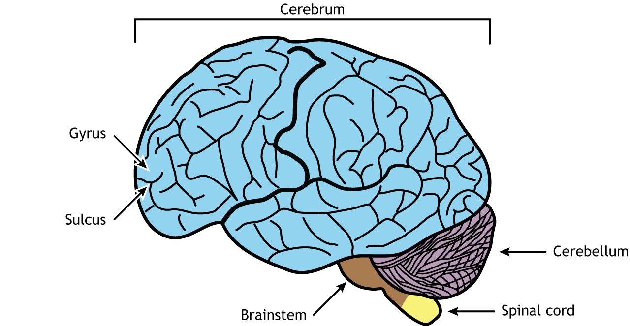
The brain is comprised of the cerebrum, cerebellum, and brainstem. The cerebrum is the most prominent region of the brain. It is divided into left and right hemispheres. The hemispheres have many of the same functions. For example, each perceives touch on one side of the body, but some functions demonstrate laterality, meaning they are primarily controlled on one side of the brain.
Most of what we think of when we imagine the brain is the cerebral cortex in humans. The cortex has many folds to increase the surface area of the brain. The bumps or raised ridges on the outer surface are called gyri (singular gyrus) and the grooved indentations are called sulci (singular sulcus). A large, deep sulcus is sometimes also called a fissure.
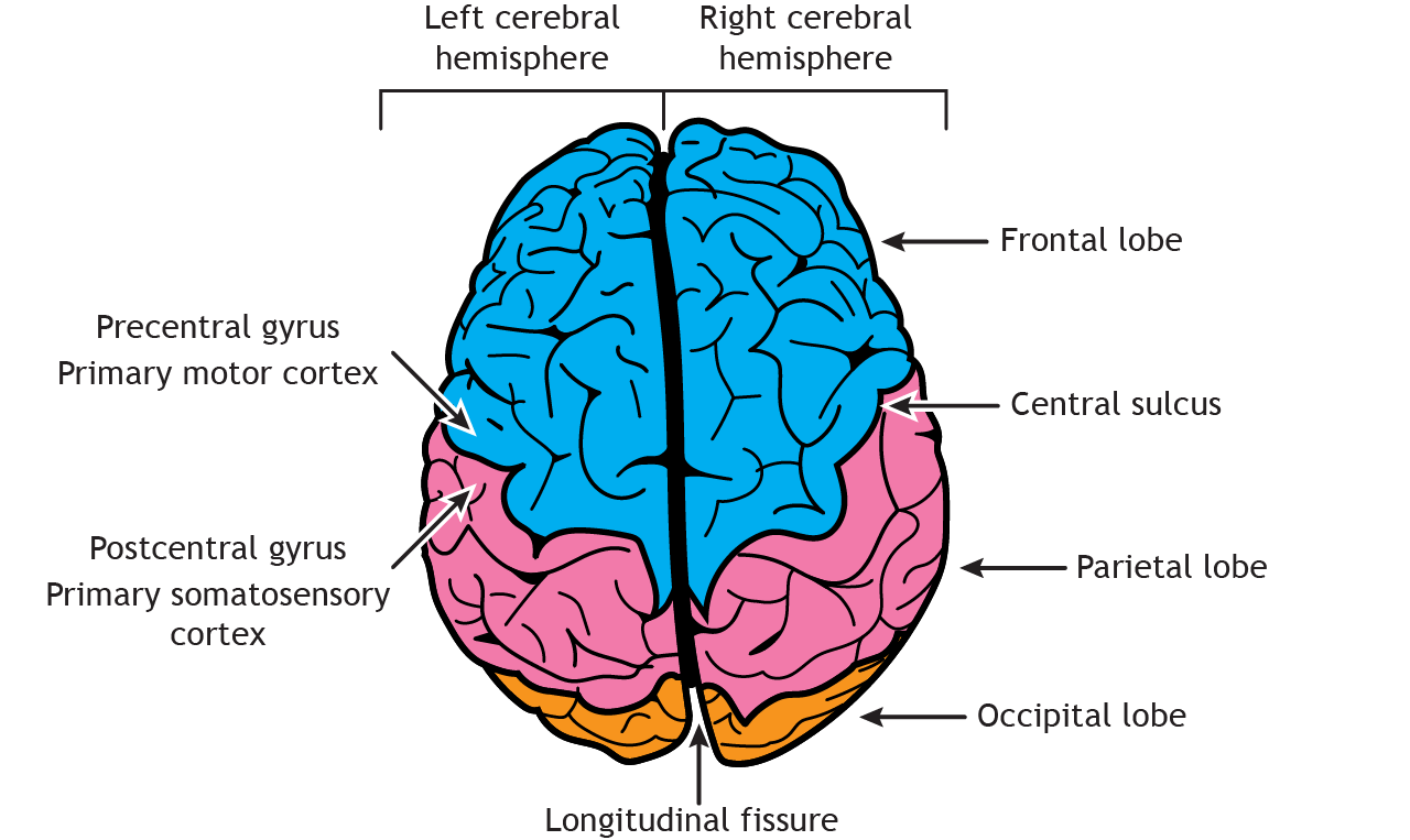
Although each gyrus and sulcus has a name that either identifies its function or location, there are only three sulci that we will introduce here to help orient us around the neuroanatomical features of the cortex. The longitudinal fissure is the most obvious fissure in the brain. It divides the two hemispheres, running along the anterior–posterior axis, visible from a dorsal view of the brain. If you were to cut along the longitudinal fissure completely, you would get two symmetrical portions of brain: the left and right hemispheres.
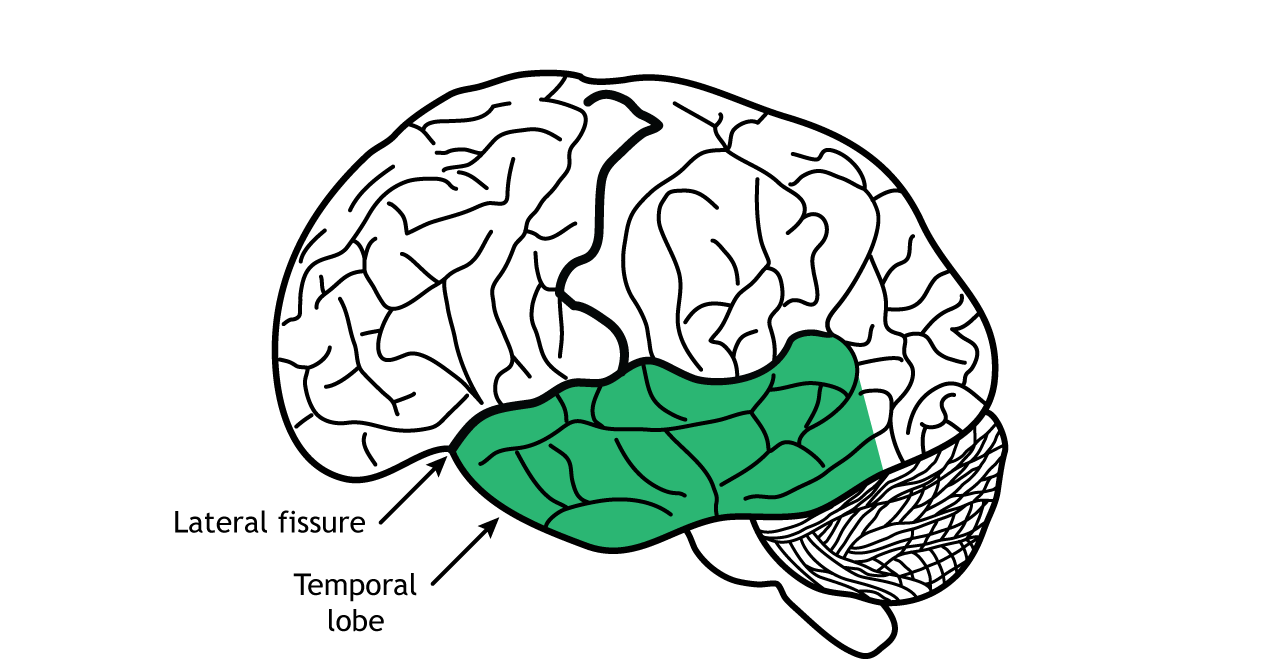
The central sulcus is a large fissure that starts at the dorsal part of the brain at about the halfway point on the anterior–posterior axis. In a sagittal view, the central sulcus runs ventrally about half the length of the brain. The other groove worth noting is the lateral fissure. This one runs roughly along the anterior-to-posterior direction, and curves gently dorsally. Again, in a sagittal view, it is roughly seen in the middle third of the brain in the anterior–posterior axis.
Anatomical Lobes of the Cortex
The cortex is roughly divided into four major lobes, which are named after the bones of the skull that surround each section of brain. The lobes are paired, meaning that the whole brain contains two of each, a left and a right. In general, the structures are roughly symmetrical.
Frontal Lobes
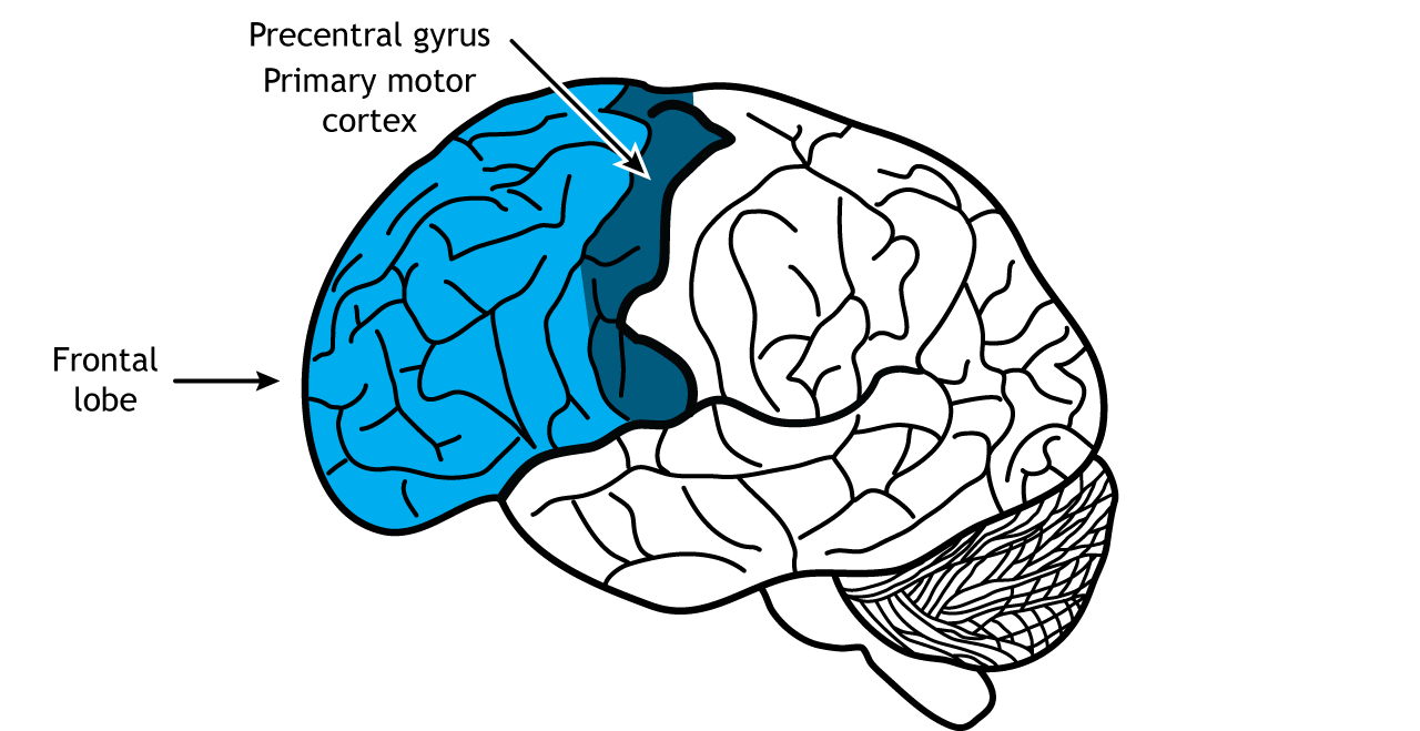
The frontal lobes are the most rostral, located in the front of the brain. The posterior border of the frontal lobe is the central sulcus. Among mammals, it is the largest of the four lobes. The frontal lobes are responsible for higher level executive functions like attention, critical thinking, and impulse control. The frontal lobes allows us to do mental math, to hold a string of letters in our head to be repeated backward, and to suppress socially unacceptable actions. Our personality is influenced by the frontal lobe. An injury to these brain structures can result in a radical change in a person’s behavior.
They are the last brain region to fully develop, not completing development until individuals reach their mid 20s. The frontal lobes are also the location of the primary motor cortex, the region of the brain responsible for planning and executing movement. Specifically, the primary motor cortex is located in the precentral gyrus, which is directly anterior to the central sulcus.
View the frontal lobe using the BrainFacts.org 3D Brain
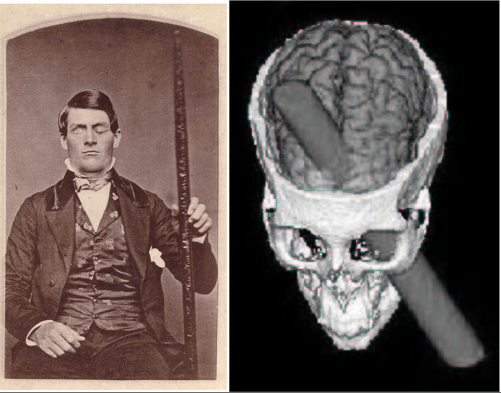
Phineas Gage
Phineas Gage, who was mentioned back in Chapter 1, is one of the most famous case studies in neuroscience.
The mid-1800s saw an expansion of industry in the United States. The most reliable and quickest way to move goods and people was with the railways that were starting to zig-zag across the growing country. The factor slowing down railway expansion was terrain: land had to be relatively flat for the tracks to be laid down. Terraforming mountains was a dangerous ordeal, and in the years before TNT (a relatively safe explosive) was developed, work accidents were a risk that these demolition workers faced.
Phineas Gage was one of those workers. In the green mountain hills of Vermont, Gage was putting explosive powder into a crack in a mountain to clear land for a new railway. As Gage packed the explosive using a three-foot-long metal rod, a spark accidentally ignited the blast prematurely, causing the tamping iron to rocket cleanly through Gage’s skull. Miraculously, Gage survived the blast. Within a month, he had made an almost complete recovery. Gage was talking excitedly with his doctors, he was eating voraciously, and even reported experiencing no pain. While the doctors noticed that his entire frontal lobe had been destroyed, it was his friends who noticed the dramatic change in his personality: whereas he was once a friendly man, adored and respected by his coworkers, the new Gage was irreverent, generally unlikable, and prone to using profanity at the most inappropriate times. The pre-injury Gage was a shrewd businessman who followed through with his plans, but Gage now was unreliable and at times, acted in a more like an animal. Because of the injuries to his frontal lobe, his friends described him as being “no longer Gage”.
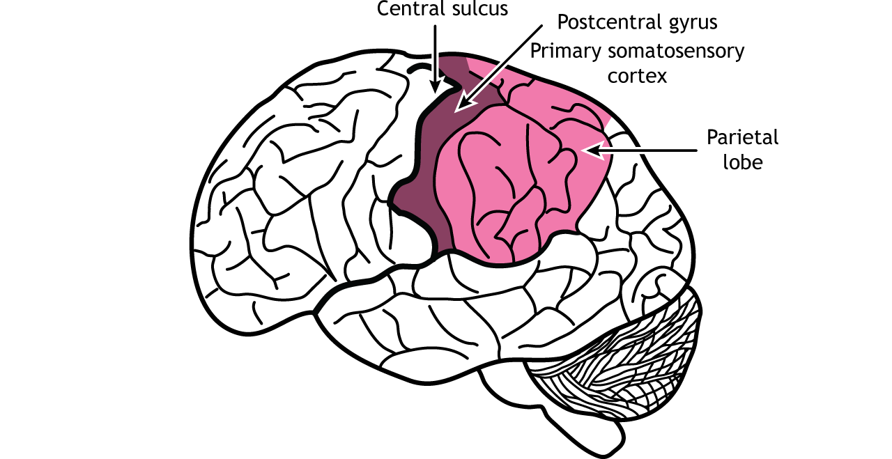
Parietal Lobes
The central sulcus lies caudal to the frontal lobe and divides the frontal lobes from the parietal lobes. The primary somatosensory cortex is located in the postcentral gyrus of the parietal lobe and is responsible for the perception of touch and pain. With our skin, we are able to detect light touch, temperature, pain, vibration, and many other modalities. This ability to sense different tactile properties of things in the world around us with our body is one of the major functions of the parietal lobe. Another closely related sense, proprioception (the ability to identify where parts of your body are located), is also processed by neurons of the parietal lobe. The parietal lobes also perform higher-level visual processing.
View the parietal lobe using the BrainFacts.org 3D Brain
Temporal Lobes
The temporal lobes are located on the side of the brain, separated from the frontal and parietal lobes by the lateral fissure. Like the parietal lobe, the temporal lobe plays a role in sensory processing, specifically with hearing, smell, taste, and higher-level visual processing. The auditory system allows our brain to interpret sound waves. The ability to remember important facts depends on memory-related processes. These functions are carried out in part by a brain structure called the hippocampus, which is buried medially and ventrally in the temporal lobe. The temporal lobe also houses some structures that are important for language.
View the temporal lobe using the BrainFacts.org 3D Brain
Occipital Lobes
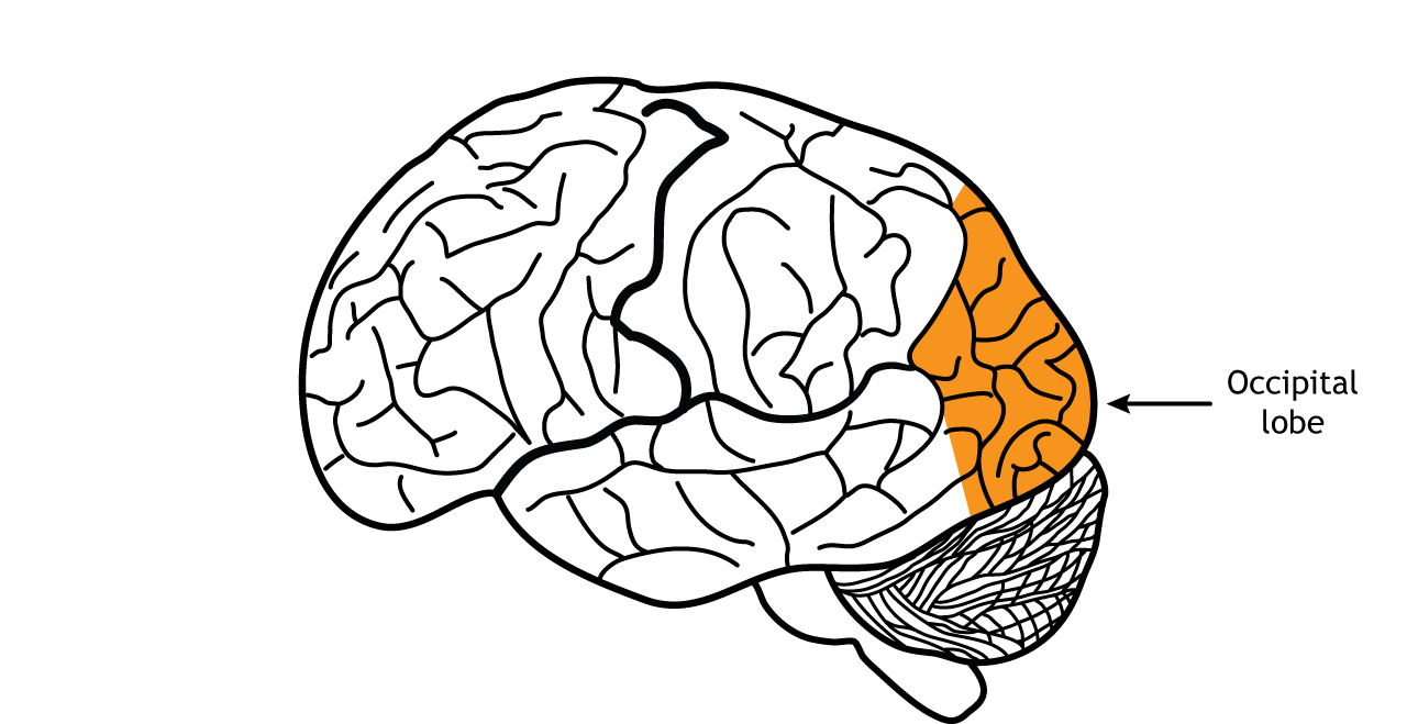
The occipital lobes, the most caudal lobes, are located in the back of the brain. Anatomically, there is not an obvious border that separates the occipital lobe from adjacent areas of the cortex. The occipital lobe is the smallest of the four lobes. The main function of the occipital lobe is for processing of visual stimuli. Our eyes are capable of capturing light and converting that light into signals. The primary visual cortex of the occipital lobe, also called V1, interprets those signals into a representation of the visual world. Other vision-related stimuli, such as objects in motion, object orientation, and color are also processed by neurons in the occipital lobe.
View the occipital lobe using the BrainFacts.org 3D Brain
Non-Cerebral Components
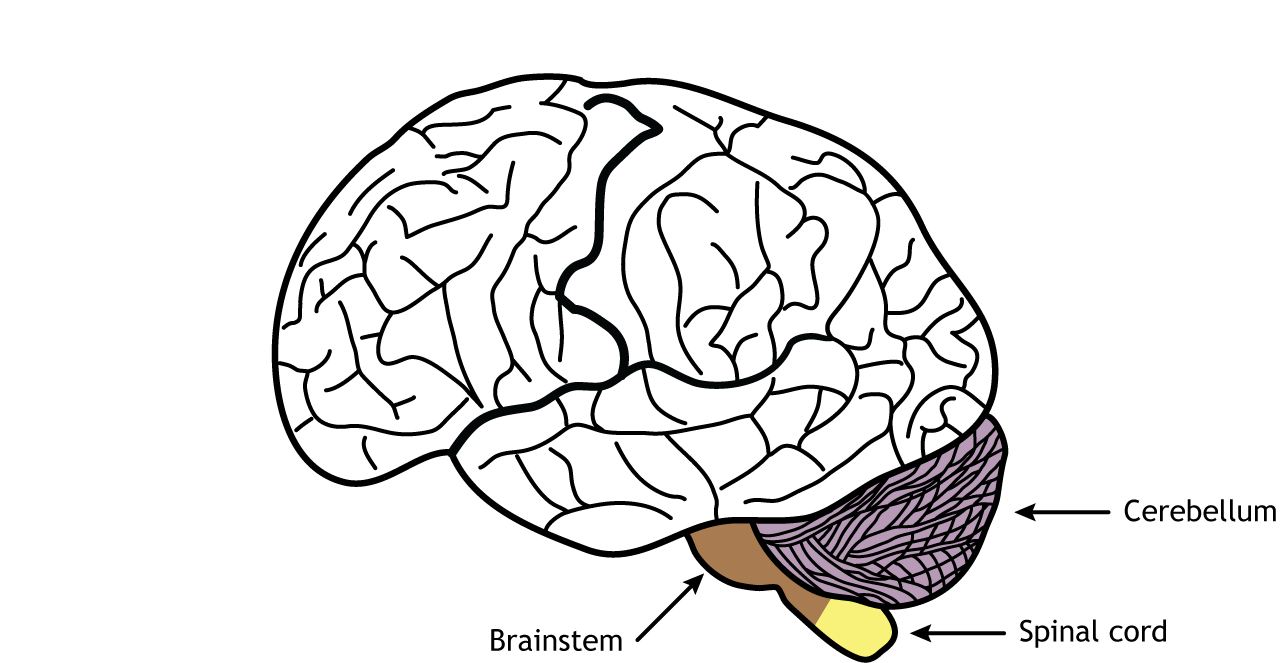
The cerebellum lies inferior to the occipital lobes. The cerebellum is divided into two hemispheres like the cerebral cortex. The cerebellum is best known for its role in regulation and control of movement, but it is also involved in cognitive functions like emotions.
The brainstem is located between the cerebrum and the spinal cord. It is important for regulating critical functions like heart rate, breathing, and sleep. It is also the location of most of the cranial nerves.
The spinal cord, which is part of the central nervous system but not part of the brain, is responsible for receiving sensory information from the body and sending motor information to the body. Involuntary motor reflexes are also a function of the spinal cord, indicating that the spinal cord can process information independently from the brain.
View the brainstem using the BrainFacts.org 3D Brain
View the cerebellum using the BrainFacts.org 3D Brain
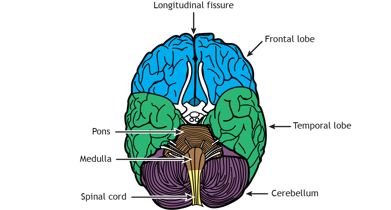
Dorsal View
Viewing the brain from above shows the bilateral symmetry of the left and right cerebral hemispheres, which are separated by the longitudinal fissure. The frontal, parietal, and occipital lobes can be seen. Similar to the lateral view, the central sulcus divides the frontal lobe from the parietal lobe. The precentral gyrus—which is the location of the primary motor cortex—sits rostral to the central sulcus, whereas the postcentral gyrus—which is the location of the primary somatosensory cortex—lies caudal to the central sulcus.
Ventral View
Underneath the brain, the frontal and temporal lobes are visible, as is the cerebellum. Like the dorsal view, the longitudinal fissure divides the cerebrum into right and left hemispheres. The pons and medulla (components of the brain stem) connect the cerebrum to the spinal cord.
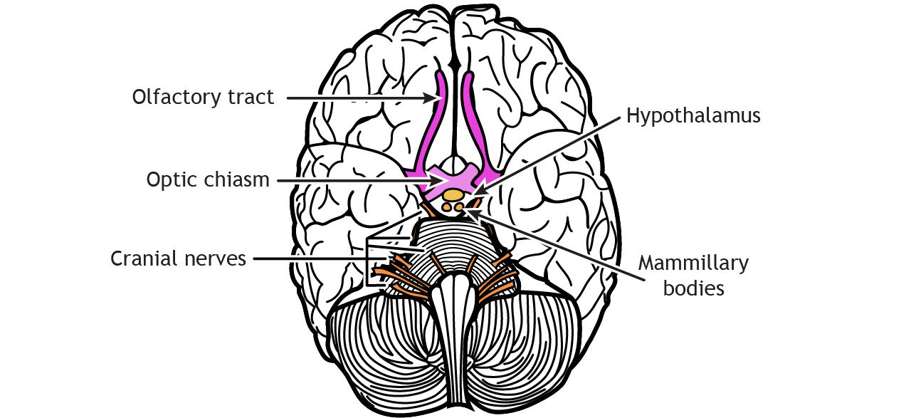
Cranial nerves are also visible on the ventral surface of the brain. The olfactory tract leads out to the olfactory bulb, which connects to the olfactory nerve. The optic tract crosses the midline at the optic chiasm, and then the optic nerve projects to the retina. Other cranial nerves enter or leave the brain at the level of the brainstem. The hypothalamus is located caudal to the pons and the mammillary bodies project out from the hypothalamus.
There are twelve pairs of cranial nerves that are outlined in the table below.
| Cranial Nerve | Type of Nerve | Function |
|---|---|---|
| CN I Olfactory nerve | Sensory | Sense of smell |
| CN II Optic nerve | Sensory | Sense of vision |
| CN III Oculomotor nerve | Motor | Control of extraocular muscles that allow movement of eyeballs; constriction of pupils; changing of lens shape |
| CN IV Trochlear nerve | Motor | Control of the superior oblique muscle of the eye that moves the eyeball down and lateral |
| CN V Trigeminal nerve | Sensory + Motor | Tactile and pain sensory information from the face and mouth; Control of muscles used in chewing |
| CN VI Abducens nerve | Motor | Control of the lateral rectus muscle of the eye that moves the eyeball outward laterally |
| CN VII Facial nerve | Sensory + Motor | Control of the muscles that allow for facial expressions; Taste sensation on the anterior two thirds of the tongue |
| CN VIII Vestibulocochlear nerve | Sensory | Detection of sound information and head positional (vestibular) information |
| CN IX Glossopharyngeal nerve | Sensory + Motor | Detection of somatic sensory in the middle ear and posterior third of the tongue; Taste sensation on the posterior third of the tongue; Controls the stylopharyngeal muscle that allows swallowing |
| CN X Vagus nerve | Sensory + Motor | Control of the internal organs by autonomic nervous system using parasympathetic activity |
| CN XI Accessory nerve | Motor | Control of the sternocleidomastoid and trapezius muscles of the neck and shoulders |
| CN XII Hypoglossal nerve | Motor | Control of the muscles of the tongue |
Mid-Sagittal View
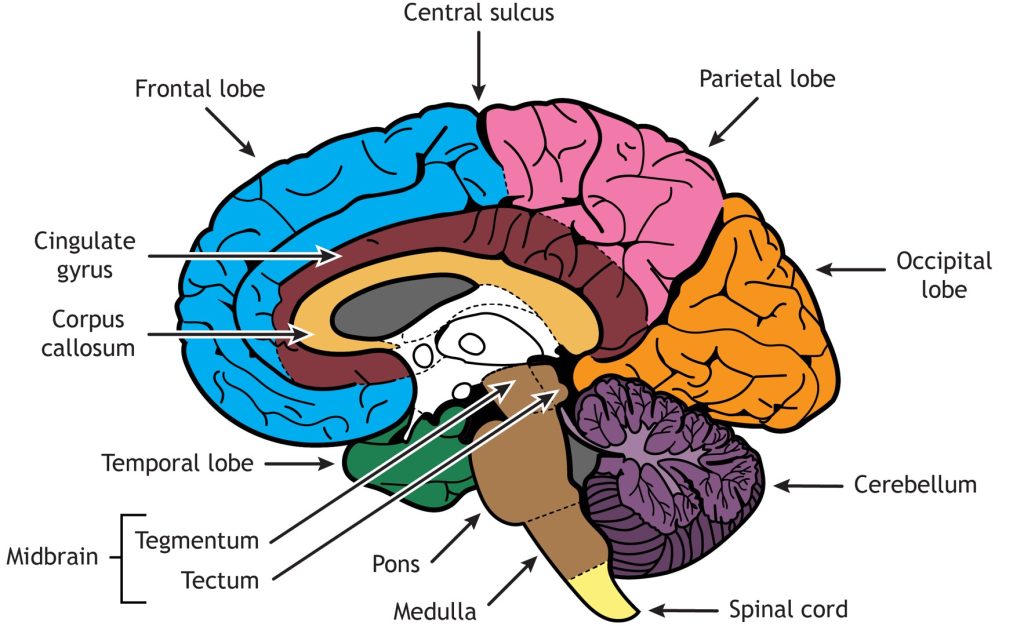
A mid-sagittal section slices the brain through the longitudinal fissure and separates the right hemisphere from the left. It also reveals more structures. In a mid-sagittal view, all four cortical lobes are visible. The frontal lobe is separated from the parietal lobe by the central sulcus, the occipital lobe is in the posterior region of the brain, and the temporal lobe can be seen behind the brainstem. The cerebellum, pons, medulla, and spinal cord are seen caudal to the cerebrum, but in this view, the midbrain—which is made up of two regions: the tegmentum and tectum—is also visible superior to the pons. The corpus callosum is located in the center of the cerebrum and is a white matter bundle made up of axons crossing from one hemisphere to the other. Surrounding the corpus callosum is the cingulate gyrus, a region important for emotion.
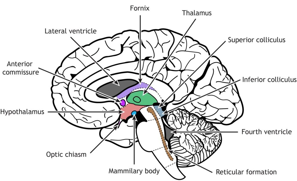
The diencephalon region of the brain consists of the region around the thalamus and hypothalamus. It is located inferior to the fornix and lateral ventricle, posterior to the anterior commissure, and superior to the brainstem. The fornix is a nerve fiber bundle containing primarily output from the hippocampus. The anterior commissure sits above the hypothalamus and is a white matter tract (like the corpus callosum) that allows information to cross from one hemisphere to the other. The thalamus is best known for its role as a relay and processing location for the sensory and motor systems. The hypothalamus has a variety of functions including control of stress and the “fight or flight” response of the autonomic nervous system, reproduction, sleep, thirst, hunger, and other homeostatic functions. The mamillary bodies sit in the posterior part of the hypothalamus and are important for memory. The optic nerves from the retina cross at the optic chiasm and then the optic tracts continue back into the diencephalon.
In the brainstem, the tectum of the midbrain consists of the superior and inferior colliculi, which are important for vision and hearing, respectively. Finally, the reticular formation is located throughout the brainstem. Networks within the reticular formation are important for regulating sleep and consciousness, pain, and motor control.
Coronal Section View
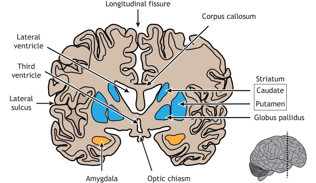
Coronal sections of the brain allow deep tissue structures to be visible. A cut through the anterior portion of the temporal lobe shows the amygdala, a region important for emotion, located in the medial temporal lobe. The regions of the basal ganglia are also visible: the striatum (which consists of the caudate and the putamen) and the globus pallidus. The basal ganglia has multiple functions, but is best known for its role in regulation of movement. The lateral ventricle sits medial to the basal ganglia and below the corpus callosum. The third ventricle is located in the middle of the brain, inferior to the lateral ventricle. The longitudinal fissure separates the left and right cerebral hemispheres and the lateral sulcus is the border between the frontal and temporal lobes.
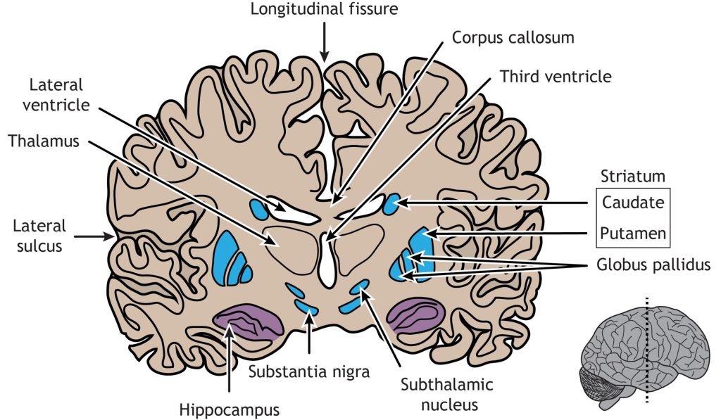
A coronal section taken closer to the central sulcus will make the hippocampus visible. The hippocampus is known for its role in memory and spatial awareness. At this location, the basal ganglia is more defined; the caudate and putamen are still present, but the two separate regions of the globus pallidus (the internal and external segments) can be seen, as well as the subthalamic nucleus and the substantia nigra. The thalamus is located on either side of the third ventricle. The corpus callosum is superior to the lateral ventricle. The cerebrum is divided in half by the longitudinal fissure and the lateral sulcus separates the temporal lobe from the frontal and parietal lobes.
Chronic Brain Injury
The modern human brain faces conditions that are unprecedented in its evolutionary history. Early humans, like other animals, evolved in environments where any physical harm to the brain was typically accompanied by damage to the scalp (the skin covering the skull). For instance, a blow to the head would impact both the brain and the scalp, triggering pain receptors in the skin to warn against repeating the harmful action. Because this warning system was sufficient, the brain itself did not evolve its own pain receptors—they were unnecessary.
However, modern inventions have disrupted this natural synergy between the brain and the scalp, exposing the brain to harm without the same immediate feedback. Three such inventions stand out.
- Boxing Gloves.
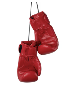 The invention of boxing gloves allowed people to punch each other in the head repeatedly without causing severe damage to the scalp or hands. While this innovation protected the external surface, it did not protect the brain. The brain, with a jelly-like consistency in its living state, is highly sensitive to rapid changes in acceleration. When a person is struck in the head, the brain “sloshes” inside the skull, colliding with the opposite side of the cranium. This motion damages delicate brain cells and disrupts networks involved in memory, decision-making, and emotional regulation.
Boxing gloves inadvertently mask the extent of brain damage. Since symptoms develop over years, boxers often continue fighting without realizing the long-term consequences. By the 1920s, the term “punch drunk” described symptoms seen in boxers, including memory loss, slurred speech, cognitive decline, mood disturbances, and movement disorders like Parkinsonism. These symptoms, caused by cumulative brain trauma, are severe and irreversible by the time they appear.
- Helmets
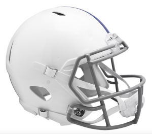 Helmets were another invention intended to protect the head, especially in activities like warfare, motor racing, and American football. While effective at preventing skull fractures, helmets do little to prevent the internal damage caused by repetitive impacts. This issue is particularly evident in football players, where the cumulative effect of non-concussive hits over years can be devastating.
Even with advanced helmets, linemen and other players experience frequent low-level impacts during games. These repeated blows can lead to chronic traumatic encephalopathy (CTE), a condition characterized by pathological changes in the brain. Dr. Bennet Omalu, a neurologist, was among the first to document CTE in football players and to challenge the NFL on the dangers of repeated head trauma. His efforts are highlighted in the 2015 film Concussion.
- Hypersonic Fighter Jets

The development of hypersonic fighter jets introduced a new source of brain trauma. Fighter pilots, particularly those in elite training programs like TOP GUN, experience extreme accelerative forces during complex maneuvers and abrupt deceleration when landing on aircraft carriers. Over years, these forces repeatedly displace the brain within the skull, leading to neurological problems similar to those seen in boxers and football players.
Recently, the U.S. Navy initiated a confidential study to better understand the extent of brain damage among fighter pilots. This research aims to address the long-term effects of repeated exposure to these forces and develop strategies to mitigate the risks.
In each case—boxing gloves, helmets, and hypersonic jets—human innovation inadvertently created scenarios where the brain sustains damage without immediate warning. These examples highlight the need for better awareness and preventive measures to protect the brain from the cumulative effects of modern activities.
Key Takeaways
- The four lobes of the cerebral cortex each have specific functions
- The cerebral cortex has gyri and sulci to increase the surface area
- The cerebral cortex, underlying structures, cerebellum, brainstem, and spinal cord form the central nervous system
- Cranial nerves are visible on the ventral surface of the brain
Test Yourself!
Attributions
Portions of this chapter were remixed and revised from the following sources:
- Foundations of Neuroscience by Casey Henley. The original work is licensed under a Creative Commons Attribution-NonCommercial-ShareAlike 4.0 International License
- Open Neuroscience Initiative by Austin Lim. The original work is licensed under a Creative Commons Attribution-NonCommercial 4.0 International License.
Media Attributions
- ExternalBrainRegions-1024×530-1
- FrontalLobe-1024×527-1
- Phineas Gage and his injury
- ParietalLobe
- TemporalLobe
- OccipitalLobe
- Hindbrain
- Dorsal-Surface-of-Brain
- Ventral-Surface-Lobes
- Ventral-Surface-Cranial-Nerves
- Sagittal section of brain structures
- sagittal 2
- Amygdala and Basal Ganglia
- Hippocampus and Basal Ganglia
- Screenshot 2024-12-10 at 1.02.25 PM
- Screenshot 2024-12-10 at 1.09.34 PM
- Screenshot 2024-12-10 at 1.18.57 PM
a raised bump on the surface of the brain
a groove on the surface of the brain
Toward the front of the brain or the top of the spinal cord
in front of; toward the face
Toward the back of the brain or the bottom of the spinal cord
Below; toward the feet
The brain and spinal cord