Biology of sensory perception
4 Olfaction: the Sense of Smell
RETROnasal AND ORTHONASAL SMELL
Smell plays a key role in flavour perception — before, during, and after ingestion of food. When you smell a food item, the odorant first goes through your nose before being registered in the brain. Odorants bind to the olfactory epithelium in the nasal cavity. This type of smell is called orthonasal smell.
As you can see in Figure 2, the mouth and nasal cavity are connected, allowing odorants to travel from the mouth to the nasal cavity. This type of smell is called retronasal smell. It travels from the mouth to the oral cavity through the retronasal passage to the olfactory epithelium. This happens when the food is chewed and swallowed.
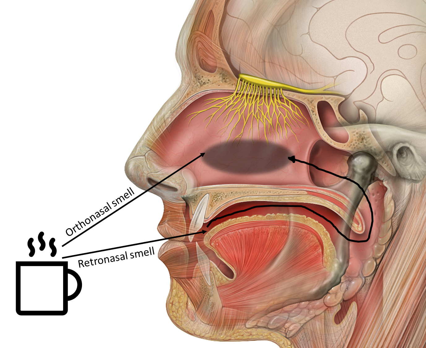
Figure 2. The anatomy of the olfactory organ. Adapted from: “Human Biology fig. 1.45 – Olfactory receptors – no labels” at AnatomyTOOL.org by Patrick J. Lynch [1]. License: Creative Commons Attribution.
biology of smell
The sense of smell works through the olfactory system. It begins when a smell in the air enters the nose and reaches the olfactory epithelium.
The size of the olfactory epithelium and the number of sensory neurons is different in each species. In humans, this area is only about 9 to 10 square centimeters. The smell receptors are found in tiny hair-like parts called cilia, which are part of olfactory neurons. These neurons have two ends: one end receives the smell by binding odorants, and the other sends the signal to the brain’s olfactory bulb. Each neuron has only one kind of smell receptor, but each receptor can detect many different smells.
For example, receptor A can detect smells 1 and 2, receptor B can detect smells 2 and 3, and receptor C can detect smells 1 and 3. This means that the smell is recognised by the brain through the combination of receptors that are activated. This system is called combinatorial coding of olfaction.
The olfactory epithelium is covered with a thin layer of mucus. This mucus layer protects the surface and breaks down complex smells into simpler chemicals.
olfactory bulb
The olfactory bulb receives the information from the olfactory sensory neurons. Figure 3 shows the anatomy of the olfactory organ. The olfactory bulb is a part of the brain that is only separated by the cribriform plate from the nasal cavity. Resulting in a very quick transport of olfactory information. Much faster compared to taste, for example.
Inside the olfactory bulb, the axons from sensory neurons connect to an area called a glomerulus. Neurons that have the same type of smell receptor all send their axons to the same one or two glomeruli. This means that thousands of similar neurons can send signals to just one glomerulus.
Odour is multidimensional: the quality and intensity of a chemical interact. When we change the brightness of a colour, like making green lighter or darker, it still stays green. But with smells, changing the amount (more or less) can actually change the way the smell is experienced. In this way, concentration can directly affect the quality of the sensation.
Given that humans have at least 320 different odour receptor types, and that both the odorant and its concentration are important, this leads to endlessly different odorant composition and intensity, yielding countless numbers of smells. This is called olfactory diversity.
NEURAL PROCESSING OF smell
From the olfactory bulb, the smell information goes straight to the olfactory cortex. The olfactory cortex is made up of several parts of the brain that work together to process olfactory information, so to understand smells.
One cortical area, the cortical amygdala, influences emotional responses to smell. The second olfactory area, the orbitofrontal cortex, is involved in the identification of odours and the reward value of odours and tastes. The entorhinal cortex, another olfactory cortical area, projects to the hippocampus, which is implicated in olfactory memory.
One part of the olfactory brain, called the cortical amygdala, helps control emotional response to smells. Another part, the orbitofrontal cortex, helps recognise different smells by identifying them, and to give a reward value to odours and tastes. A third part, the entorhinal cortex, sends smell information to the hippocampus, which is important for remembering smells.
The ability to notice and name smells uses higher parts of the brain. These are areas that help us think, make decisions, remember things, and understand what we sense. They are found in the cerebral cortex, which is the outer layer of the brain. Because the olfactory bulb connects directly to brain areas that handle emotions and memory, smells often have a strong emotional effect.
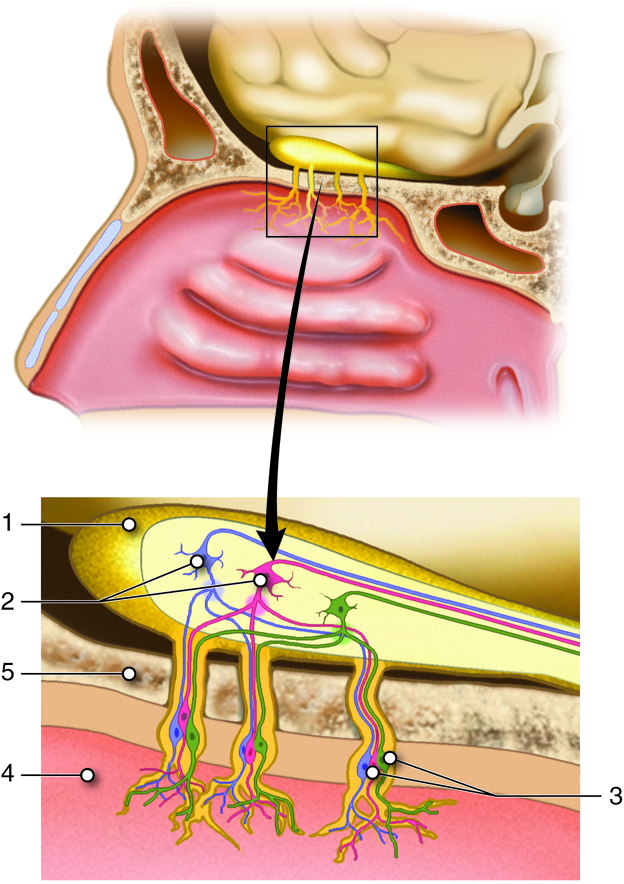
Figure 3. Anatomy of the Structures Involved in Smell (Olfaction).
The olfactory bulb (1) holds mitral cells (2) that receive information from the olfactory cells (3). The place where axons from the olfactory cells come together is the glomerulus. The olfactory cells are found within the nasal epithelium (4) and pass their information through the cribriform plate (5) of the ethmoid bone. From: “Cenveo – Drawing Anatomy of the Structures Involved in Smell (Olfaction) – Numbered labels” at AnatomyTOOL.org by Cenveo. License: Creative Commons Attribution
- ‘Human Biology’, https://textbookequity.org/Textbooks/HumanBiologyCK12.pdf. ↵
compound that gives a smell. It can be water soluble, fat soluble or volatile.
The specialized epithelial tissue inside the nasal cavity that is involved in smell. It lies in the top of the nasal cavity and contains odor receptors.
The nasal cavity is a large, air-filled space above and behind the nose in the middle of the face.
Smell that occurs through the nose.
Smell that occurs through the mouth during food ingestion.
The olfactory sensory system is the sense of smell. Including both the smell coming from the nasal and oral cavities.
microscopic hair-like structure that contains G-protein coupled receptors on which odorants bind.
The olfactory system uses combinations of receptors to detect a wide range of odors, reducing the need for a specific receptor for each odor.
Olfactory Bulb (OB) is the part in the brain that sends information regarding smell (olfactory information) to be processed further.
Nerve cell.
The cribriform plate is a part of the ethmoid bone located at the base of the skull. It forms the roof of the nasal cavity.
An axon or nerve fiber is a long, slender part of a nerve cell, or neuron, that conducts electrical impulses known as action potentials.
Place in the Olfactory bulb where axons from the olfactory cells come together.
The phenomenon that odor receptor cells give a different response based on odorant intensity and smell composition.
The Olfactory Cortex is the portion of the cerebral cortex concerned with the sense of smell. It is part of the Cerebrum.
The areas of the brain that process and register incoming sensory information and make possible the conscious awareness of the world.
The amygdala is the main processing center for emotions. It also links emotions to many other brain abilities, especially memories, learning and the senses.