Before we learn about the medications that are used to treat analgesic and musculoskeletal conditions in our patients, we must first review the physiology of pain and the anatomy of the musculoskeletal system.
Review of the Anatomy and Physiology of Pain
Pain occurs when there is tissue damage in the body. Tissue damage activates pain receptors of peripheral nerves. Nociceptors, the nerve endings that respond to painful stimuli, are located in arterial walls, joint surfaces, muscle fascia, periosteum, skin, and soft tissue. Nociceptors are barely present in most internal organs.[1]
The cause of tissue damage may be physical (e.g., heat, cold, pressure, stretch, spasm, and ischemia) or chemical (pain-producing substances are released into the extracellular fluid surrounding the nerve fibers that carry the pain signal). These pain-producing substances activate pain receptors, increase the sensitivity of pain receptors, or stimulate the release of inflammatory substances (e.g., prostaglandins).[2]
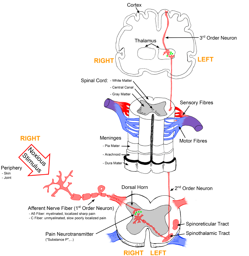
For a person to feel pain, the signal from the nociceptors in peripheral tissues must be transmitted to the spinal cord and then to the hypothalamus and cerebral cortex of the brain. The signal is transmitted to the brain by two types of nerve cells (A-delta and C fibers). The dorsal horn of the spinal cord is the relay station for information from these fibers. In the brain the thalamus is the relay station for incoming sensory stimuli, including pain. From the thalamus the pain messages are relayed to the cerebral cortex where they are perceived.[3] See Figure 10.1[4] for an illustration of how the pain signal is transmitted from peripheral tissues to the spinal cord and then to the brain.

Endogenous Analgesia
The CNS has its own endogenous analgesia system for relieving pain. The CNS suppresses pain signals from peripheral nerves. Opioid peptides interact with opioid receptors to inhibit perception and transmission of pain signals. These opioid peptides are endorphins, enkephalins, and dynorphins.[5]
Visit the YouTube video below for more information about how pain relievers work.
Review of Anatomy and Physiology of the Musculoskeletal System
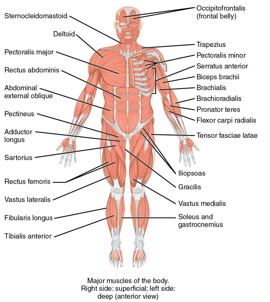
In the musculoskeletal system, the muscular and skeletal systems work together to support and move the body. The bones of the skeletal system serve to protect the body’s organs, support the weight of the body, and give the body shape. The muscles of the muscular system attach to these bones, pulling on them to allow for movement of the body.[7] See Figure 10.2[8] for an illustration of the musculoskeletal system.

Muscles
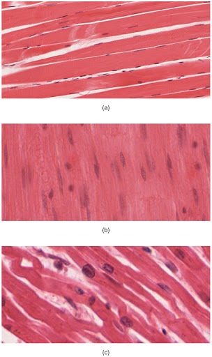
The body contains three types of muscle tissue: skeletal muscle, smooth muscle, and cardiac muscle. See Figure 10.3[9] for images of different types of muscle.

Skeletal muscle is voluntary and striated. These are the muscles that attach to bones and control conscious movement. Smooth muscle is involuntary and nonstriated. It is found in the hollow organs of the body, such as the stomach, intestines, and around blood vessels. Cardiac muscle is involuntary and striated. It is found only in the heart and is specialized to help pump blood throughout the body.[10]
When a muscle fiber receives a signal from the nervous system, myosin filaments are stimulated, pulling actin filaments closer together. This shortens sarcomeres within a fiber, causing it to contract.[11]
- . Frandsen, G., & Pennington, S. (2018). Abrams’ clinical drug: Rationales for nursing practice (11th ed.). pp. 305, 310, 952-953, 959-960. Wolters Kluwer. ↵
- . Frandsen, G., & Pennington, S. (2018). Abrams’ clinical drug: Rationales for nursing practice (11th ed.). pp. 305, 310, 952-953, 959-960. Wolters Kluwer. ↵
- . Frandsen, G., & Pennington, S. (2018). Abrams’ clinical drug: Rationales for nursing practice (11th ed.). pp. 305, 310, 952-953, 959-960. Wolters Kluwer. ↵
- “Sketch colored final.png” by Bettina Guebeli is licensed under CC BY-SA 4.0 ↵
- . Frandsen, G., & Pennington, S. (2018). Abrams’ clinical drug: Rationales for nursing practice (11th ed.). pp. 305, 310, 952-953, 959-960. Wolters Kluwer. ↵
- Ted-Ed. (2012, June 26). How do pain relievers work? - George Zaidan [Video]. YouTube. https://youtu.be/9mcuIc5O-DE ↵
- Khan Academy. (n.d.). The musculoskeletal system review. https://www.khanacademy.org/science/high-school-biology/hs-human-body-systems/hs-the-musculoskeletal-system/a/hs-the-musculoskeletal-system-review ↵
- This image is a derivative of “1105 Anterior and Posterior Views of Muscles.jpg” by CFCF licensed under CC BY 4.0 ↵
- “414 Skeletal Smooth Cardiac.jpg” by OpenStax College is licensed under CC BY 4.0 ↵
- Khan Academy. (n.d.). The musculoskeletal system review. https://www.khanacademy.org/science/high-school-biology/hs-human-body-systems/hs-the-musculoskeletal-system/a/hs-the-musculoskeletal-system-review ↵
- Khan Academy. (n.d.). The musculoskeletal system review. https://www.khanacademy.org/science/high-school-biology/hs-human-body-systems/hs-the-musculoskeletal-system/a/hs-the-musculoskeletal-system-review ↵
Nerve endings that selectively respond to painful stimuli and send pain signals to the brain and spinal cord.
Produced in nearly all cells and are part of the body’s way of dealing with injury and illness. Prostaglandins act as signals to control several different processes depending on the part of the body in which they are made. Prostaglandins are made at the sites of tissue damage or infection, where they cause inflammation, pain, and fever as part of the healing process.
