ULTRASOUND FINDINGS WITH ABNORMAL PREGNANCIES
| ANEMBRYONIC PREGNANCY
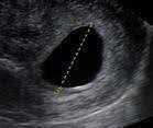
Empty GS without fetal pole. Need three views (L, W, H) to calculate MSD. An empty GS with a MSD of ≥25mm is diagnostic of anembryonic pregnancy. Early pregnancy loss occurs in approximately 10-20% of clinically recognized pregnancies. |
 |
| ECTOPIC PREGNANCY
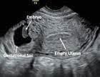
Note this GS with fetal pole is not intrauterine (no endometrial stripe seen in the same plane). Ectopic pregnancy occurs in 0.25% pregnant people seeking abortion (vs. 1-2 % of all pregnancies) (Duncan 2022, Panelli 2015). |
 |
| FREE FLUID IN CUL-DE-SAC
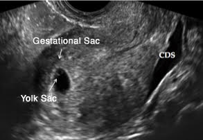
Seen best in a longitudinal view of the uterus. Note the presence of anechoic (dark) fluid in the posterior cul-de-sac. This finding may be consistent with blood from an ectopic pregnancy, ruptured ovarian cyst, or uterine perforation. |
 |
| GESTATIONAL TROPHOBLASTIC DISEASE (GTD or MOLAR PREGNANCY)
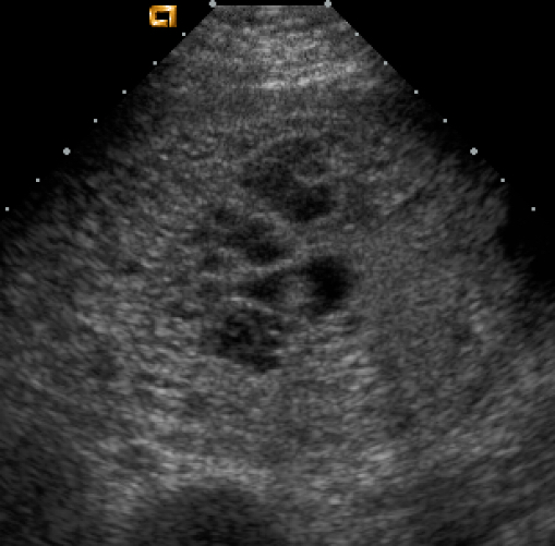
Image of complete mole (no embryo), showing cystic intrauterine mass with no distinct GS, YS, or fetal pole. Often has a swiss cheese, snowstorm, or moth-eaten appearance on US. Consider referral for higher level of care > 12-week size (by exam or US) due to increased bleeding risk. GTD occurs in approximately 0.1% of pregnancies and should be followed carefully to confirm appropriate treatment and resolution (Horowitz 2021, Soper 2021). |
 |
|
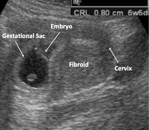
FIBROID UTERUS Uterine fibroids are a common benign pelvic tumor that may enlarge or distort the cervix or uterine cavity, presenting technical difficulty for aspiration. US can help identify size, location, and orientation to the pregnancy. |
 |
|
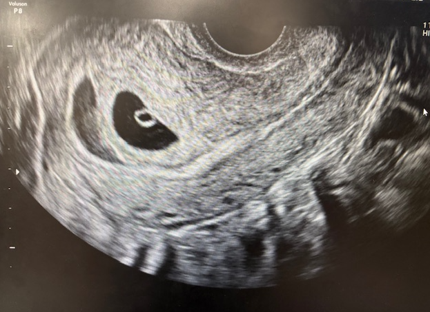
The sonolucent area adjacent to GS represents a fluid collection between the chorion and the uterine wall. This may be seen in the setting of vaginal bleeding or as an incidental finding and is associated with a higher risk of EPL (Qin 2022). |
 |
|
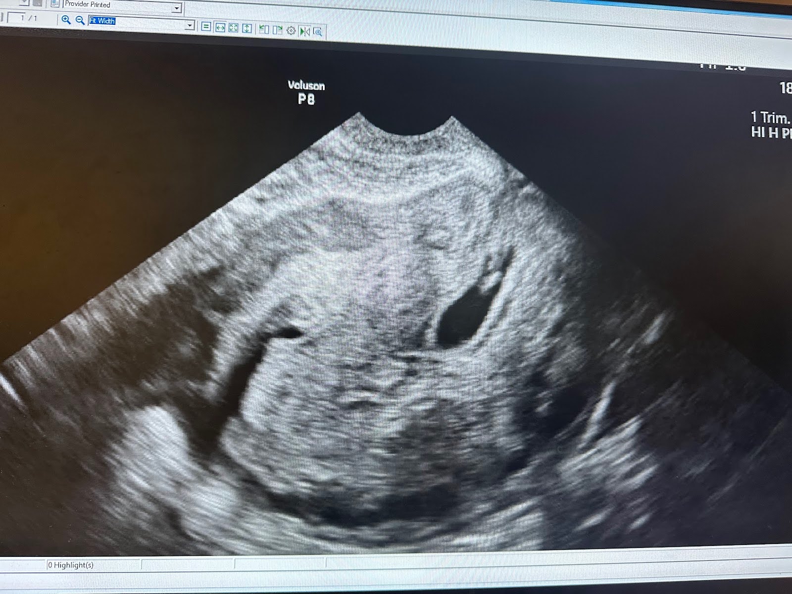
CESAREAN SCAR ECTOPIC PREGNANCY (CSEP) Variant of uterine ectopic pregnancy with full or partial implantation of pregnancy in scar from previous cesarean section. Carries significant risk of morbidity and mortality and may require higher level of care for surgical management (SMFM 2022). |
 |