Meiosis and Gametogenesis
This chapter will focus on the events of meiosis and the process of gametogenesis, or the production of sperm and egg. Before we move into the events of meiosis, we will first review the events of mitosis so that we can draw comparisons between the two processes.
Diploid vs Haploid
Nearly all the cells in the human body contain one maternal and one paternal copy of each of our 23 chromosomes. When we talk about the events of mitosis and meiosis, the number of unique chromosomes a species has is given a placeholder (n) to keep things simple as we track the cell division events in mitosis and meiosis. A cell that has two of each chromosome is diploid [from the Greek for double], which we write as 2n. A cell that has only one of each chromosome is haploid [from the Greek for single], which we write as n.
The cells that make up our body’s organ systems are diploid (2n) and we call them somatic [from the Greek for body] cells. The only haploid cells in the human body are the gametes [Latin and Greek origins meaning husband or wife] – male sperm and female oocytes (eggs). When a haploid sperm cell (n) combines with a haploid oocyte (n), the resulting cell is diploid (2n) and contains the correct number of chromosomes to make a human embryo.
Important Note: In the first book in this book series, Anatomy and Physiology I: An Interactive Histology Atlas, we discuss the events of mitosis in detail. The following section will highlight the major events for the purpose of distinguishing between mitosis and meiosis. For a more detailed explanation of mitosis, please visit this link: https://pressbooks.pub/aandp1histologyatlasandworkbook/chapter/mitosis-and-the-cell-cycle/
Review of Mitosis
Mitosis and cytokinesis are the final events in the cell cycle and correspond to division of the nuclear (mitosis) and cytoplasmic (cytokinesis) parts of the cell. The final events of mitosis and cytokinesis occur simultaneously. Mitosis is used to replicate diploid (2n) cells, producing two daughter cells that are identical to each other and identical to the parent cell.
A somatic cell spends most of its time in what is called interphase. During interphase the cell carries out normal functions, grows, and replicates its DNA. When stimulated to divide, the cell then leaves interphase, undergoes mitosis and cytokinesis resulting in two genetically identical daughter cells.
Mitosis is nuclear division that separates the replicated chromosomes resulting in two nuclei. During mitosis (prophase, metaphase, telophase, and anaphase) the chromatin (DNA wrapped around protein) condenses into tight structures that you can visually see (chromosomes). Cytokinesis is the division of the cytoplasm resulting in two new cells. The pinching of the membrane that occurs during cytokinesis can be visualized as a structure we call the cleavage furrow.
A simplified abbreviation for the phases of mitosis is PMAT (prophase, metaphase, anaphase, and telophase). However, prophase represents a wide set of events and should be separated into early prophase and late prophase/prometaphase. Stained cells in the various phases of mitosis typically have the following visual characteristics, which are highlighted in Figure 1:
Early Prophase – Chromatin begins to condense into distinct chromosomes, each one formed by a pair of sister chromatids attached by a centromere. The nuclear envelope is disintegrating but still typically visible.
Late Prophase/Prometaphase – Chromatin is fully condensed into distinct chromosomes, nuclear envelope is no longer visible, spindle apparatus (mitotic spindle) is forming at the poles of the cell.
Metaphase – Each of the chromosomes are aligned on the equator of the cell (metaphase plate), an imaginary line between the two mitotic spindles.
Anaphase – The two sister chromatids of each chromosome are being separated, and pulled to the opposite sides of the cell (poles) and resemble the shape of a “V”.
Telophase – Two groups of chromatids are localized at the poles. By the very end of telophase, the nuclear envelope will be reformed, but this process is not as commonly seen. It is more common to find the nuclear envelope missing.
It is important to note that cytokinesis occurs simultaneously with telophase, however the names describe different events. Cytokinesis represents cytoplasmic events and is not considered part of mitosis. Stained cells undergoing cytokinesis will show pinching/narrowing of the plasma membrane due to formation of the cleavage furrow.
Figure 1: Phases of the whitefish blastula cell cycle, with and without illustration overlay
Overview of Meiosis
In order for gametes (sperm and oocyte) to be haploid cells, they must be produced by a different cell division process. This process, meiosis, shares some similarities with mitosis, but contains two sequential division events instead of one. Figure 2 illustrates the events of meiosis as they compare with the events of mitosis. Like mitosis, meiosis begins following DNA replication. Also like mitosis, each division event goes through prophase, metaphase, anaphase and telophase/cytokinesis. The major difference occurs in how the chromosomes line up in metaphase to be separated.
Before we move into comparing the specific events in mitosis and meiosis, we need to clarify some terminology about the DNA strands. When the DNA is replicated, two identical copies are produced. When condensed, this pair of identical DNA strands (chromosomes) are called sister chromatids and are attached to each other by a centromere.
Each diploid cell contains a maternal and paternal chromosome for each of our 23 chromosomes, which we call homologous chromosomes. Following DNA replication, each homologous chromosome is made of two sister chromatids attached at the centromere.
Figure 2: Cellular events of mitosis (left) and meiosis (right)
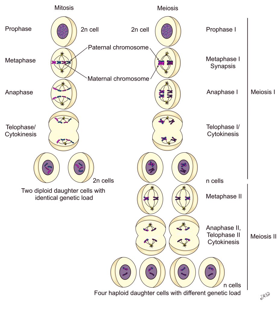
In Figure 2, the maternal sister chromatid is shown in pink and the paternal sister chromatid is shown in blue. For simplicity, Figure 2 only shows two chromosomes, one lighter set (light pink and light blue) and one darker set (dark pink and dark blue). The maternal and paternal strands for a specific chromosome contain the same genes for a particular trait, but each can have slight variations in DNA sequence between the maternal and paternal strands. This maternal and paternal set of the same chromosome (light set = light pink maternal and light blue paternal) are called homologous chromosomes. To give a more specific example, homologous chromosome number 1 contains the maternal copy and paternal copy of chromosome 1.
In mitosis, each chromosome, containing a pair of identical sister chromatids, lines up separately on the metaphase plate. This allows each cell to receive one maternal and one paternal copy of each chromosome (diploid 2n). However, the goal of meiosis is to produce a haploid (n) gamete. To be haploid, the gamete must get a paternal or maternal copy of each chromosome, but not both.
The first division event, meiosis I, reduces the cell’s chromosome number from diploid (2n) to haploid (n). During prophase I the homologous chromosomes (maternal sister chromatid + paternal sister chromatid) pair up with each other in a process called synapsis to form a 4-strand unit called a tetrad. While joined together in prophase I, the maternal and paternal strands exchange small sections of their DNA strands in a process called crossing-over. This genetic recombination increases the genetic diversity of resulting offspring. The 23 tetrads line up on the metaphase plate in metaphase I. In anaphase I in Figure 2, note that the once solid pink or blue strands (metaphase I), now contain small sections of the opposite color. In anaphase I the homologous chromosomes are separated, with the two sister chromatids still attached at the centromere. The product of meiosis I are two haploid (n) daughter cells that are genetically distinct from each other and from their diploid parent cell.
Events in the second division event, meiosis II, match the events of mitosis. Like mitosis, in meiosis II sister chromatids are separated into individual daughter cells. Unlike mitosis, which results in 2 genetically identical diploid (2n) daughter cells, meiosis results in 4 genetically distinct/unique haploid (n) daughter cells.
Gametogenesis
Gamete production, gametogenesis, and hormone production take place in the primary sex organ for males and females. Spermatogenesis, production of sperm cells, takes place in the testis of the male. In spermatogenesis the events of meiosis result in 4 sperm per parent cell.
Oogenesis, production of oocytes, takes place in the ovary of the female. In oogenesis, the events of meiosis are modified. Two nuclear division events (meiosis I and II occur), but the cytokinesis events occur unequally so that one daughter cell receives nearly all cytoplasmic contents. The other three “cells”, which lack cytoplasmic contents are called polar bodies. Therefore, in oogenesis, the events of meiosis result in one functional oocyte per parent cell plus 3 polar bodies. The polar bodies are reabsorbed by the body.
The events of spermatogenesis and oogenesis will be discussed in detail in the following chapters:
Chapter illustrations by:
Juan Manuel Ramiro Diaz, PhD
a cell that posesses two copies of each chromosome (2n)
a cell that posesses one copy of each chromosome (n)
