Mitosis and the Cell Cycle
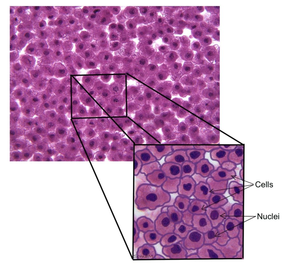
To identify cells in the various phases of the cell cycle, we will be using fixed slides of white fish blastula. A blastula is a sphere of cells produced during the development of an embryo by repeated mitosis and cleavage of a fertilized egg. The following sections will illustrate the phases of mitosis as seen in the whitefish blastula (a good example of animal cells).
Figure 1: Whitefish blastula sections under different magnification power – 4x (left), 10x (center), and 40x (right) objective lenses.

Introduction to the cell cycle
The cell cycle is a series of events that takes place in a cell as it grows and divides. A cell spends most of its time in what is called interphase. During Interphase the cell carries out normal functions, grows, and replicates its DNA. When stimulated to divide, the cell then leaves interphase, undergoes mitosis and cytokinesis resulting in two genetically identical daughter cells.
Mitosis is nuclear division that separates the replicated chromosomes resulting in two nuclei. During mitosis (prophase, metaphase, telophase, and anaphase) the chromatin (DNA wrapped around protein) condenses into tight structures that you can visually see (chromosomes). Cytokinesis is the division of the cytoplasm resulting in two new cells. The pinching of the membrane that occurs during cytokinesis can be visualized as structures we call cleavage furrows.
The next sections will highlight the two major parts/phases of the cell cycle: interphase and mitotic (M) phase. The cell spends nearly all of its life in interphase, followed the division events of the mitotic (M) phase.
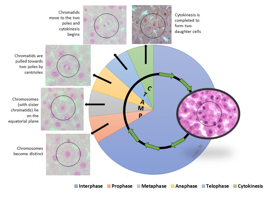
Figure 2: Phases of the cell cycle
Interphase
Interphase is divided into 3 distinct phases:
- G1 phase – normal cell growth and cell functions
- S phase – DNA synthesis/replication
- G2 phase – growth and organelle replication in preparation for division
However, using a compound light microscope, it is difficult to visualize the separate events of interphase. Stained cells in interphase will typically have the following visual characteristics:
- Clear nuclear envelope
- Darkly staining nucleolus
- Uniform appearance to chromatin staining
Figure 3: Illustrated overlay of microscope image showing cells in interphase.
Mitotic (m) phase
The mitotic (M) phase consists of two distinct but concurrent/simultaneous events: mitosis and cytokinesis. Mitosis represents the events involved in division of the nucleus, while cytokinesis represents the division of the cytoplasm. Cytokinesis occurs simultaneously with the latter phases of mitosis (late anaphase and telophase).
Mitosis is a continuous process, where the cell moves smoothly from one phase to another without pausing. We must remember that the cell we observe has been preserved in that phase because of the tissue preparation technique. The cell has been essentially frozen in that moment in time.
A simplified abbreviation for the phases of mitosis is PMAT (prophase, metaphase, anaphase, and telophase). However, prophase represents a wide set of events and should be separated into early prophase and late prophase/prometaphase. Stained cells in the various phases of mitosis typically have the following visual characteristics:
Early Prophase – Chromatin begins to condense into distinct chromosomes, each one formed by a pair of sister chromatids attached by a centromere. The nuclear envelope is disintegrating but still typically visible.
Late Prophase/Prometaphase – Chromatin is fully condensed into distinct chromosomes, nuclear envelope is no longer visible, spindle apparatus (mitotic spindle) is forming at the poles of the cell
Metaphase – Each of the chromosomes are aligned on the equator of the cell (metaphase plate), an imaginary line between the two mitotic spindles
Anaphase – The two sister chromatids of each chromosome are being separated, and pulled to the opposite sides of the cell (poles) and resemble the shape of a “V”
Telophase – Two groups of chromatids are localized at the poles. By the very end of telophase, the nuclear envelope will be reformed, but this process is not as commonly seen. It is more common to find the nuclear envelope missing.
It is important to note that cytokinesis occurs simultaneously with telophase, however the names describe different events. Cytokinesis represents cytoplasmic events, and is not considered part of mitosis. Stained cells undergoing cytokinesis will show pinching/narrowing of the plasma membrane due to formation of the cleavage furrow.
Figure 4: Phases of the whitefish blastula cell cycle, with and without illustration overlay
Check out our YouTube video on how to properly use the microscope as we examine the whitefish blastula slide: YouTube Video – Whitefish Blastula Slide
Chapter Illustrations By:
Juan Manuel Ramiro-Diaz, Ph.D.
Soma Mukhopadhyay, Ph.D.
a blastula is a sphere of cells produced during the development of a white fish embryo by repeated mitosis and cleavage of a fertilized egg
During Interphase the cell carries out normal functions, grows, and replicates its DNA
nuclear division that separates the replicated chromosomes resulting in two nuclei
DNA wrapped around histone proteins
tightly coiled, condensed DNA wrapped around proteins
occurring with mitosis, cytokinesis is the division of the cytoplasm resulting in two new cells
pinching or narrowing of the plasma membrane during cytokinesis
occurring with or simultaneous
