UNIT OBJECTIVES
Become aware of the contributions of neuroscience to adolescent research.
Distinguish between various areas of the brain and their functions.
Understand the concept of Executive Function.
Recognize implications of brain plasticity research.
Identify contemporary methods of brain research.
Recognize present-day impediments to human neuroscience research.
NEUROTRANSMITTERS
The human brain weighs about three pounds. It is perhaps the most complex structure in the known universe. Although it has been dissected for at least 2500 years of recorded history, most of its functioning remains a secret. It has only been recently that new techniques have been developed to directly study the brain.
We know that the brain communicates with itself and other parts of the body through neurons, which are special cells. These cells fire or are released by chemicals called neurotransmitters. This is how information is transmitted. We have about 87 billion neurons in our adult brain. The way that they could link up is uncountable.
Neurons tend to be specific. This means that a neuron fires for only one particular neurotransmitter. It will usually not fire when exposed to a different neurotransmitter. Some neurotransmitters act as helpers and speed up the firing process. Some act as gatekeepers and slow down or inhibit firing and transmission.
There are over 100 different neurotransmitters that have been identified. However, neuroscientists believe that there are likely many more. Some are very common and are located throughout the brain. Others have very specialized functioning.
Certain areas of the brain may be rich in specific receptors for certain types of neurotransmitters. For example, the frontal lobes, described later in this unit, are rich in norepinephrine receptors. Therefore, the neurons will fire only when they encounter norepinephrine and not other neurotransmitters.
Certain neurotransmitters are located throughout the brain. For example, GABA (abbreviated for Gamma-aminobutyric acid) is a neurotransmitter that inhibits or slows reactions. It is located in almost all brain areas. Glutamate is a neurotransmitter that speeds up reactions. It also is located across the brain in every area.
Some neurotransmitters have one function in one portion of the brain and a different one in another. Dopamine is associated with reward and motivation in the brain’s frontal areas and movement in the midbrain. These two tasks are entirely different and rely on different brain circuits. Dopamine neurons are also critical in the visual system, where they play a still different role.
Furthermore, neurons may have subtypes. GABA has at least six subtypes that have different roles. Dopamine has at least five subtypes. Often these work in coordination so that when one subtype of receptor fires, another is inhibited.
BRAIN ANATOMY
There are many ways to subdivide the brain. Many clinicians and researchers divide the brain into three essential parts: the brain stem, cerebellum, and cerebral hemispheres. Each piece has a specialized function. However, it is a mistake to believe that each part exists in isolation. The entire brain works together to make us who we are.
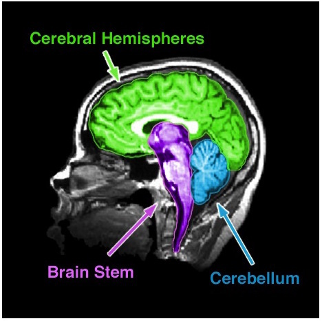
An MRI of the human brain show three clear structures: the cerebral hemispheres, brain stem, and cerebellum. Photo by National Institute of Mental Health.
The brain stem is the most fundamental part of the brain. Its functions include many that keep us alive, including regulating our breathing, heart rate, and temperature. If it is severely damaged, a person will require life support. The brain stem includes several structures: the medulla, pons, midbrain, and diencephalon. The latter consists of the thalamus and hypothalamus, which regulate growth and are especially important at puberty.
The cerebellum is critical for coordination and posture. It is the densest structure in the brain, with hundreds of millions of synaptic connections. However, its exact function in these areas is unclear. More recently, researchers realize that it has a role in language and other cognitive abilities.
Cerebral Hemispheres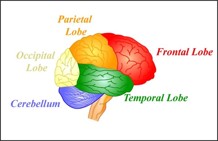 The four lobes of the brain and the cerebellum. [image: MIT OpenCourseWare, https://goo.gl/rwuevt, cc by-nc-sa 2.0, https://goo.gl/toc0zf]
At the top of the brain is the cerebral cortex. It has two halves, called hemispheres. The cerebral cortex is where our thinking occurs. Scientists usually believe this is where we get our sense of consciousness. Under the cerebral cortex are the basal ganglia, amygdala, and hippocampus. These structures are responsible for mood, emotions, and bodily regulation.
The two cerebral hemispheres can be subdivided into four lobes. The occipital lobe is responsible for vision. The temporal lobes process vision and are involved in hearing, memory, and integrating vision and sound. Finally, the parietal lobe houses the somatosensory cortex, which response to bodily sensations. This lobe is also involved in visual attention and decoding patterns, such as facial expressions.
The frontal lobes contain the motor cortex, which is involved in planning and movement. It also includes areas for the processing of judgment and decision-making. The frontal lobes are also engaged with abstract thinking. The human frontal lobe is more prominent than in other mammals. Damage to the frontal lobes can have a lifetime effect on attention and concentration. An injury to the forehead may injure this area. This is one reason why it is so important to wear helmets while bicycling and in other sports activities, even those that seem fairly low risk.
The frontal lobes are where much of the task of executive function occurs. Executive function is the ability to plan and purposefully coordinate activities and monitor your progress towards them. Skills of self-control, paying attention, and memory are involved. Much of this activity occurs in a more specific area called the prefrontal cortex, which is part of the frontal lobes.
The subcortical structures are underneath the cortex. The basal ganglia are involved in voluntary movement (movement you control). The amygdala and hippocampus are part of the limbic system. The limbic system is involved in emotions. The hippocampus also has a vital role in memory. If it is substantially damaged, a person has problems forming new memories.
The two cerebral hemispheres are connected by nerves called the corpus callosum. Some functions of the brain occur in both the left and right hemispheres. For example, both hemispheres are responsible for sensory and motor functions. However, the sensory and motor cortices have an opposite-side representation. The left cerebral hemisphere is responsible for most movements and sensations on the right side of the body. The right cerebral hemisphere is primarily responsible for most movements on the left side of the body.
Some functions are more located in one hemisphere or another. Language, for example, tends to be processed in the left hemisphere of right-handed people. However, for left-handed people, this is not always true.
On the other hand, some spatial skills like responding to faces are processed in the other hemisphere. The entire brain works as a unit. When people say they are “left-brained” or “right-brained,” they are not really correct. Everyone uses their entire brain all of the time.
“When I was 13, I banged up my frontal lobes,” said Andrew, a college freshman. “Or that’s what they tell me. It wasn’t fun.
“I fell off a bike while going down a hill. I was wearing a helmet, but somehow I hit a tree. I was being crazy, I guess. Without the helmet, I probably would have been killed… I’m not sure what happened after that. I wasn’t knocked out. I went to the emergency room and had a couple of stitches and some tests. That’s all I remember. It’s a blur, really.
“I thought I was okay after a while, but I felt really ‘spacey.’ Things seemed like I wasn’t underwater. That’s the only way I can describe it. I don’t remember a lot about it.
“This happened on the weekend, a Saturday. By Monday, I thought I was fine, maybe a headache and some soreness. I went back to school. But it was really hard concentrating. I couldn’t read anything, or really, couldn’t remember what I read was the problem. I wrote something down, and it didn’t make sense.
“I told everyone at home I was okay. I went to my room and hung out alone. But my family knew I was having problems. I was forgetting things, yelling at them, and just had a hard time. I also started getting sad for no reason. Even happy things made me cry.
“After a couple of days, my dad called the doctor. I remember screaming at him not to. But he did. The doctor saw me and said everything was probably fine. Somewhere along the line, they did some more tests. But he recommended that I talk to a psychologist who treats head injuries.
“Sure. Okay. It seemed really strange because I didn’t think of myself as having a head injury, but I went. I did what she told me. It was helpful and gave me time to heal.
“It took a few months before I felt normal again. The psychologist said I hit my frontal lobes. That might explain why it was so hard to concentrate.”
Studying the Brain
Efforts to study a living human brain can be risky. So far, there is no easy and completely safe way to insert monitors or brain probes without potentially disrupting brain processes. We, therefore, have to use other techniques.
One way that we study the brain is in animals. Some researchers remove or damage parts of the brain in animals such as rats. This is called lesioning. If the animal’s behavior changes, we infer that the removed structure is essential for that behavior.
Researchers can also genetically “knock out” or remove the function of specific aspects of animals’ brains. This enables us to see the effects of particular areas or neurotransmitters on behavior. We cannot perform these experiments on humans because they would not be ethical.
Because of our ethical concerns, lesions or damage of human brains is studied only when they occur in patient populations. Examples are patients who have lost a brain region due to a stroke or other injury or have had surgical removal of a structure to treat a particular disease. We infer brain function by measuring changes in the patients’ behavior before and after the lesions.
Brain waves detected through EEGs, discussed in Unit 1, have been a valuable research tool. They furnish a great deal of information about changes in brain functioning. Yet, they do not provide a good location of what is happening. They can usually tell us only how fast information is being processed. This is important for many questions. However, EEGs are not very good at locating where the processing is occurring.
The real revolution that made knowledge about the brain more possible was with the advent of sophisticated neuroimaging. These techniques are based on the biological fact that the brain uses oxygen and glucose (a type of sugar that the brain uses for fuel) delivered through the blood. Thus, devices that can measure the brain’s oxygen or glucose allow us to measure the brain as processing information. Computer programs that analyze this data allow us to visually reconstruct what is occurring.
Thanks to this technology, we can observe the brain and see it change while performing specific psychological tasks such as math problems or identifying faces. This gives researchers an idea of which brain areas are important for which types of functions.
The Adolescent Brain
Until recently, it was believed that the brain was fixed from early childhood onward. It was thought that no more brain growth was possible past the ages of about four or five.
More recent research indicates that is not the case. Moreover, the wiring of the brain can change through experiences. Major rewiring may and does take place during the adolescent years. This is one more event that makes this period so critical.
Marian Diamond and her colleagues showed that brains of any age could change. Furthermore, the environment is essential to the development of phenomena, which is called neural plasticity.
Neuroimaging techniques have allowed behavior scientists to study the brain changes associated with puberty. Magnetic resonance imaging (MRI) enables researchers to see structures of the brain in greater detail. fMRI, a related technique, shows more minor detail but often is a better way to see the brain “while in process” or working. These two methods can show how puberty and adolescence affect both the brain’s structure and function. In general, brain scans have shown that with the onset of puberty, there is an activation of the brain’s limbic system or emotional areas. There is also a decrease in the frontal regions of the brain. This suggests that some of the stereotyped emotionality of adolescence may be due to a change in brain balance. The emotional areas become more dominant. The parts of the brain that inhibit behaviors and encourage reflection are actually suppressed. It may take several years for all areas of the brain to catch up.
The effects of specific hormones released during puberty can also be studied with neuroimaging. An exciting research area is that hormones released in puberty seem to reorganize adolescent behavior by changing brain structure and function. For example, reward circuitry appears to undergo significant change, as does the capacity to respond to social situations.
Testosterone tends to thicken the cortex of males but does not have this effect on females. A thicker cortex is generally associated with a more neutral activity. Impact on individuals who do not identify as binary is not known but is presently being investigated.
Greater levels of testosterone are associated with more reward-oriented behavior and risk-taking. Testosterone also seems to decrease the assessment of risk. The prefrontal lobes, which influence Executive Function, lag other areas in the brain during adolescence. This may help account for some impulsive behavior of adolescents. This effect tends to be most pronounced when adolescents are in groups.
In general, puberty is associated with gender-specific changes in brain function. There is some evidence that males experience an enlarging of the left or dominant hemisphere during puberty. In contrast, females experience an increase of the right. The pattern of individuals who identify as nonbinary is not known at this time.
The ability to respond to nonverbal communications is enhanced during puberty. People who show deficits in this ability may actually be at risk for social problems or criminal behavior. Perhaps just as important, the ability to respond to ambiguous or complex nonverbal communication is increased. Whether this relates to the development of hormones is not clear.
Substantial research has helped reduce risky behavior by teaching adolescents to reflect on peer influences while in groups. These have been effective in some instances. Moreover, when they have been implemented, they have shown that it is possible to prevent many factors that lead to unwanted teen behavior. Likely, such methods will also be implemented with the assistance of neuroimaging in the future.
CRITICAL THINKING
Why is it a myth that we only use 10% of our brain?
Why might the effects of a brain injury be worse on adults than on adolescents?
If science progress to the point where we can observe people’s thoughts, what ethical problems are we likely to encounter?
Are methods to enhance or “hack” the brain likely to succeed? Why or why not? Are they ethical?
Do you think that the use of social media has an effect on the rewiring of the brain? Could this effect be both negative and positive?
