2
Learning Objectives
Upon completion of this module, participants will be able to:
- Identify and describe three basic brain structures and functions
- Describe the functions of the peripheral nervous system
- Explain the purpose and process of neurotransmission
- Identify and describe three neurotransmitters
- Differentiate between neuroplasticity and neurogenesis.
Brain fun facts… Did you know? (Northwestern Medicine, 2022; Brain facts, 2018)
- The brain continues to develop up until around age 25
- There are approximately 86 billion neurons (cells) in the brain
- The brain is made up of about 75% water
- It is thought that a yawn works to send more oxygen to the brain, therefore working to cool it down and wake it up
- The brain is believed to generate up to 50,000 thoughts per day
- Thoughts in the brain can travel up to an impressive 268 miles per hour
- You can’t tickle yourself because your brain distinguished between unexpected external touch and your own touch.
- Anomia is the technical word for tip-of-the-tongue syndrome when you can almost remember a word, but it just won’t quite come to you.
The Nervous System
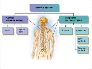
I like to think of the nervous system as an overarching command center consisting of components: the central and peripheral nervous systems (See figure 1). The nervous system controls almost everything we experience including movement, balance, coordination, thoughts, feelings, memory, and breathing.
Figure 1: Nervous System Overview

(Source: Nervous system structure, Pinterest)
The Central Nervous System
The central nervous system (CNS) includes the brain and spinal cord. Both the brain and spinal cord play a role in how we experience anxiety (i.e., our thoughts, memories, reactions, sensations etc.). Our brain is the body’s most complex organ, and we can think of it as a conductor. Like a conductor, it provides instruction and coordination of system functions. The brain contains numerous structures which means it must work in coordination with other parts of the nervous system. The brain plays a role in such things as cognition (planning, problem-solving), emotion (fear, anxiety, happiness), and movement (walking, balance). The spinal cord’s primary role is to facilitate communication between the body and brain via nerves and cells.
Brain Basics
The brain is a complex and impressive organ. Brain development takes place overtime and begins in utero and into adulthood. Development continues until approximately 25 years of age. While the brain stops growing around the age of 25, it continues to develop new neurons throughout life. This process is known as neurogenesis. It is easy to get neurogenesis confused with neuroplasticity. Although related, “neuroplasticity and neurogenesis are two different concepts. Neuroplasticity is the ability of the brain to form new connections and pathways and change how its circuits are wired; neurogenesis is the even more amazing ability of the brain to grow new neurons” (Bergland, 2017). Neurogenesis on the other hand, uses brain-derived neurotrophic factor (BDNF) to assist in the production of new neurons. It is known that BDNF plays an important role in modifying/changing fear memories (Wehrenberg, 2018).
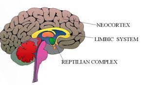
You can think of brain development as taking a bottom-up approach. The brain contains three stages of development: reptilian brain, mid-brain, and neocortex (See figure 2). These three areas do not work independent of one another. Rather, they are interconnected and communicate to fulfill various body functions. (Note: You may also see these parts of the brain being referred to as the hindbrain, midbrain, and forebrain).
Reptilian brain: The reptilian brain is considered the oldest part of the brain. Therefore, many refer to it as the “old brain”. This part of the brain contains the brainstem and cerebellum. The reptilian brain is responsible for such things as balance, body temperature, heart rate, and breathing. It makes sense that this is the first part of the brain to develop as it provides us with basic life sustaining needs.
Midbrain: The mid-brain, also known as the “limbic brain” is responsible for our emotions, memory, and motivation. There are several important structures within the limbic brain including the amygdala, thalamus, hypothalamus, and hippocampus (Wehrenberg, 2018).
Neocortex: The neocortex also known as the “new brain”, is the largest and most developed part of the brain. The neocortex is divided into two hemispheres: right and left hemispheres. While both hemispheres are structurally identical, they provide different functions. It is also true, that people typically have a dominant hemisphere. The left hemisphere is primarily responsible for language (thinking in words), logic, linear thinking, and sequencing. The right brain on the other hand is known for imagination, intuition, creativity, daydreaming, and feelings visualization (Cherry, 2020).
The neocortex also contains a total of four lobes: the frontal, parietal, occipital, and temporal lobes. For this chapter, we will specifically be looking at the frontal lobe. The frontal lobe, also referred to as the prefrontal cortex (PFC), plays a significant role in anxiety and is located towards the front of the brain near the forehead. The PFC is responsible for several cognitive functions. For example, the PFC guides a person in how they might think through and prioritize daily responsibilities. This is linked to logic/reasoning and problem solving. In a nutshell, the PFC is responsible for learning, perceiving, planning, and logic/reasoning. One additional important function of the PFC is to attach meaning to your memories. The functions of the PFC are critical to understanding anxiety as well as how to change one’s reactions that lead to feelings of anxiety.
Click the video link below to learn about the four lobes.
https://www.youtube.com/watch?v=Vy8EvyQoQIE
Figure 2: Brain Areas of Development

(Source: Three brains, Flickr)
In addition to these three areas of the brain, it is also important to highlight the brainstem. The brainstem is located at the base of the brain and acts like a hitch thus connecting brain to the spinal cord. It basically is responsible for regulating many of the body’s automatic functions such as breathing and heart rate and does so in concert with the peripheral nervous system.
The Limbic System
While the limbic system is part of the midbrain, it plays an important role in anxiety. As indicated earlier, it is made up of several parts including the amygdala, thalamus, hippocampus, and hypothalamus. Understanding the function of each part is important in making sense of anxiety and the stress or fear response. See table 1 for a brief description of each limbic system part and their associated function.
Click the video link below to learn about the limbic system.
Table 1: Limbic System Parts and Functions
| PART | FUNCTION |
| Thalamus | The thalamus sits on top of the brainstem. Its primary function is that of a router. It receives information from your senses, sorts out that information and decides where to send it within the brain. Typically sends information to the limbic system and/or the cortex. Sensory information routed through the thalamus tend to be sights and sounds. Smell is the only sense that goes directly to the amygdala. |
| Amygdala | It is most known for processing fear and detecting threats. It acts as a guard and prepares us to protect ourselves by activating other brain/body parts. The amygdala helps process and regulate both positive and negative emotion. It works closely with the hippocampus as it attaches emotion to memories. Stimulation of the amygdala can lead to a stress response (fight, flight, or freeze) which is linked to anxiety and fear. More recently, research has identified an additional stress response known as fawning (i.e., people pleasing, difficulty saying no, neglecting one’s own needs). |
| Hypothalamus | The hypothalamus’s primary role is to keep the body stable and in balance and it does so with the release of hormones (i.e., stress hormones and adrenaline). It does this through the autonomic functions of the nervous system. It acts as a switchboard between the endocrine and nervous system. It assists in regulating temperature, hunger, sex drive, and sleep. |
| Hippocampus | The hippocampus has many functions but is most known for its role in learning and memory. This is where memories are processed, organized, retrieved, and stored. Specifically aids in the development of long-term memories. It takes data and facts and sends it to the thinking part of the brain- the cortex. |
The Peripheral Nervous System
The peripheral nervous system (PNS) functions take place outside of the brain and spinal cord. The primary role of the PNS is to communicate with the CNS and other body parts via nerves. The PNS’s communication highway allows the CNS to receive and send messages to the body including the skin and other organs (Mcleod, 2021). The PNS has two additional systems within it: somatic and autonomic nervous systems.
Somatic nervous system: This system communicates by giving and taking in information using two different types of neurons: motor and sensory neurons. Sensory neurons take in information via our senses and relays that information to the CNS. Motor neurons transmit information from the brain and spinal cord to muscle fibers in the body. This process also assists in movement. Both types of neurons guide our CNS to take some form of action (Mcleod, 2021)
Autonomic nervous system: This system automatically activates/regulates bodily functions including breathing, heart rate, blood pressure, and digestion. Again, these bodily functions occur automatically and involuntarily. For example, we don’t have to consciously remember to breathe minute to minute as our body does it naturally. There are two subsystems within the autonomic nervous system: the sympathetic and parasympathetic nervous systems. The sympathetic nervous system, (SNS) engages in a stress response and prepares the body for “fight or flight” which is an involuntarily response to threat and/or danger. When activated, the body responds by experiencing increased heart and breathing rates, increased alertness, and increased blood flow to muscles. During this time, a person may experience a “rush” of adrenalin. In contrast, the parasympathetic nervous system (PSNS), known as “rest and digest” and the “freeze” response. During this involuntary resting period, heart and breathing rates begin to slow, pupils begin to constrict, and the body begins to calm thus returning to a normal baseline resting rate (Waxenbaum, et. al., 2021). The PSNS plays a critical role in regulating and treating anxiety.
In a nutshell, the peripheral nervous system:
- Regulates involuntary bodily functions such as the fight, flight, and freeze responses.
- Fosters the communication process thus allowing the brain and spinal cord to receive and send information to other parts of the body
- Delivers sensory and motor information to and from the CNS
- Connects the CNS other parts of the body including the skin and other organs
Click the video link below to learn more about the peripheral nervous system.
https://www.youtube.com/watch?v=jaWrMYChc5A
Additional Parts of the Central Nervous System to Note
Hypothalamic-pituitary-adrenal (HPA) axis. When discussing the stress/fear response, it would be neglectful not to address the hypothalamic-pituitary-adrenal (HPA) axis. The HPA axis is a central part of the body’s stress response system. As stated in the name, it is made up of three parts (the hypothalamus, pituitary gland, and the adrenal glands) all of which support the stress response. It plays an important adaptive function for short-term stress management but can also become dysregulated. Dysregulation can occur when a person is exposed to and experiences chronic stress (Cleveland Clinic, 2024).
In a nutshell, the primary role of the HPA axis is to coordinate with other parts of the nervous system to release hormones including cortisol so that the body will have enough energy to protect and defend itself from possible harm. Remember the sympathetic nervous system and its role if fight, flight, or freeze? The sympathetic nervous system is also part of the stress response.
Chronic dysregulation of the HPA axis can fuel a negative feedback loop. According to the Cleveland Clinic (2024), “The HPA axis is meant to have a fine-tuned negative feedback loop: the cortisol in your body then triggers your hypothalamus to stop making corticotrophin-releasing hormone (CRH), ending the stress response. But experiencing frequent or intense stress and other issues can cause dysfunction with your HPA axis.” Simply stated, cortisol plays a major role by communicating to the brain to halt the stress response. Because chronic stress can deplete cortisol, it creates the challenge of shutting off/down the stress response (Wehrenberg, 2018). Chronic exposure to stress can result in several issues including mental health related challenges (i.e., anxiety, depression).
Basal Ganglia. The basal ganglia (BG) are a complex group of nuclei located under the cortex deep within the brains cerebral two hemispheres. They are involved in several motor and non-motor functions many of which are connected to the limbic system and the prefrontal cortex. These functions are primarily related to movement/motor control, emotions including arousal, behaviors, executive functioning/decision making, reward/reinforcement, and the formation of habits (Simonyan, 2019). While the BG serve many functions, they often work in concert with other areas of the brain thus adding to their complexity.
Insula. The insula, also known as the insular cortex, is located deep within the brain and separates the temporal lobe from the parietal and frontal lobes. It is complex in that it works in concert with other parts of the brain including the pre-frontal cortex and the amygdala. While the insula is responsible for several functions, for the purposes of this text, we will focus on the functions of decision-making and sensorimotor (the coordinator and interpretation of bodily sensations). Thus, the insula collects information on sensations being experienced by the body, attaches an interpretation to that experience, which influences decision-making (Wehrenberg, 2018).
The Brain and Communication
For our body to experience such things as movement, thought, emotion, and behavior, our cells must communicate with one another. They communicate via a complex network of cells called neurons. Believe it or not, there are between 86-100 billion neurons in our brain (Brain facts, 2018)! Neurons communicate back and forth via electrical and chemical impulses. More specifically, they use special messengers known as neurotransmitters.
There are several types of neurotransmitters, and each type dictates the type of message that is sent and received. Each neurotransmitter functions as excitatory, inhibitory, or a combination of both (See Table 3). Excitatory neurons excite the neuron and are more likely to trigger an action potential where inhibitory neurons have a calming effect and are less likely to fire an action potential (Cherry, 2021).
Before jumping to the table that identifies and describes the various neurotransmitters, I want to introduce you to another important protein/molecule in the brain. Brain-derived neurotrophic factor (BDNF) is produced in the brain and plays a critical role in the growth, maintenance, and survival of neurons. Specifically, BDNF has an influence on cognitive and emotional functions in the brain. It modulates the activity of specific neurotransmitters, including serotonin, dopamine, and glutamate, which are important for mood regulation and brain function (Bathina, 2015). Research has found that altered levels of BDNF have been linked to mental health conditions such as depression, anxiety, and schizophrenia. Higher levels of BDNF are often associated with better mental health and cognitive function (Lin, 2020).
Table 3: Neurotransmitters
| Neurotransmitter | Role in the Body |
| Acetylcholine | A neurotransmitter used by the spinal cord neurons to control muscles. It also aides in regulating memory, learning and neuroplasticity. It works in both the central and peripheral nervous systems. In most instances, acetylcholine is excitatory. |
| Dopamine | Dopamine has multiple functions depending where in the brain it is activated including movement, motivation, and pleasure. Considered to be both excitatory and inhibitory. |
| GABA (gamma-aminobutyric acid) | The major inhibitory neurotransmitter in the brain. Known for producing a calming effect. |
| Glutamate | The most common excitatory neurotransmitter in the brain. Plays a major role in learning and memory. |
| Glycine | A neurotransmitter used mainly by neurons in the spinal cord. It plays a role in sensory and motor function. It typically acts as an an inhibitory neurotransmitter. |
| Norepinephrine | Norepinephrine acts as both a neurotransmitter and a hormone. In the peripheral nervous system, it is part of the flight/flight/freeze response. It plays a critical role in regulating attention, arousal, stress reactions, and cognition. Norepinephrine is usually excitatory, but is inhibitory in a few brain areas. |
| Serotonin | A neurotransmitter involved in many functions including mood, appetite, pain, and sleep patterns. Serotonin is inhibitory. |
(Source: Major Neurotransmitters, NIH publication 00-4871)
Problems in Neurotransmission
As with all forms of communication, there can be a breakdown, and this is also true with neurotransmission. It is a breakdown in neurotransmission that can influence a person’s health and mental health. What are some problems that might arise if there is a communication challenge? First, neurons may not produce enough of a specific type of neurotransmitter (i.e., dopamine). Second, absorption of a specific transmitter may occur quickly or too slowly. Third, too much of a specific neurotransmitter may be released. Last, certain enzymes may work to deactivate neurons (Cherry, 2021).
Here are a couple of examples of how a breakdown in communication can impact both health and mental health. In the case of Parkinson’s disease, neurons responsible for producing dopamine slowly begin to breakdown and/or die. This leads to an array of symptoms which can include trembling and hallucinations. In terms of mental health, an issue with the reuptake of serotonin that occurs in the synaptic cleft that can lead to symptoms of depression and anxiety.
Neuron Anatomy
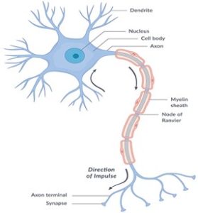
While neurons are quite small, they play a critical role in communication. To better understand neuronal communication, let’s take a closer look at the anatomy of the neuron. First, there are several important parts that make up a neuron all of which participate in the process of communication. These parts are dendrites, nucleus, some, axon, myelin sheaths, and axon terminals (See figure 4).
Figure 4: Neuron Anatomy

(Source: What are the parts of the nervous system? NICHD)
In breaking down communication, let’s look at the process of neurotransmission (also known as synaptic transmission). The space between which two neurons communicate is the synaptic cleft. It is in this space where chemical messengers are transmitted. These chemical messengers are known as neurotransmitters. The process of communication from one neuron (presynaptic neuron) to another (postsynaptic neuron) is triggered by an electrical impulse. This action is known as an action potential. In brief, a chemical message is sent from a presynaptic neuron to a postsynaptic neuron (NIH, 2018).
Click the link below to watch a video illustration of neural communication.
https://www.youtube.com/watch?v=WhowH0kb7n0
Role of Medication
While we will not do a deep dive into medication and their relationship to neurotransmission, I do want to provide you with a few helpful and important pieces of information. More specifically, I want to address the role medications (aka psychotropic medications) play in neurotransmission and their effects on mental health.
Medications that specifically target neurotransmitters act as either an agonist or antagonist. Agonist’s primary function is to increase the impact or effect of certain neurotransmitters. In contrast, antagonists block the impact or effect of certain neurotransmitters. Furthermore, these medications all have what is called a mechanism of action (MOA). For example, some MOA’s work to mimic a specific neurotransmitter while others work with various synaptic receptors to block or assist in the reuptake process (Cherry, 2021).
Conclusion
Developing foundational knowledge of the nervous system is critical to understanding the relationship between anxiety and the brain. This requires an understanding of the structure and function of the central and peripheral nervous systems. Furthermore, understanding the role of neurotransmitters and how cells communicate will provide further understanding of anxiety development and exacerbation.
Learning Activities
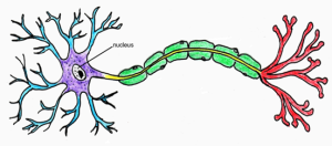
- Using the neuron image below, label all parts (soma, dendrites, axon, myelin sheath, axon terminals and dendrites).

(Source: Neuron, Clipartlibrary)
2. Video challenge. It is not uncommon for those in the helping profession (i.e., psychotherapists, counselors, etc.) to provide psychoeducation to clients. For example, you may share with a client how neurons communicate and when such communication is disrupted, how it may lead to the experience of negative emotions such as anxiety or depression. In this video challenge, record yourself explaining the process of neural communication as if you were talking to a client. Perhaps be creative and think of a metaphor as way to describe this communication. The video should be 2-3 minutes in length.
3. Describe your understanding of the limbic system. For example, how would you define this system? What are the functions of this system?
References
Bathina, S., & Das. U. (2015). Brain-derived neurotrophic factor and its clinical implications. Arch Med Sci. 11(6), 1164-1178. https://www.ncbi.nlm.nih.gov/pmc/articles/PMC4697050/
Bergland, C. (2017). How do neuroplasticity and neurogenesis rewire your brain? Psychology Today. Retrieved from https://www.psychologytoday.com/us/blog/the-athletes-way/201702/how-do-neuroplasticity-and-neurogenesis-rewire-your-brain
Brain facts (2018). How many neurons are in the brain? Retrieved from https://www.brainfacts.org/in-the-lab/meet-the-researcher/2018/how-many-neurons-are-in-the-brain-120418
Cherry, K. (2021, July 8). The Role of Neurotransmitters. Verywell mind. https://www.verywellmind.com/what-is-a-neurotransmitter-2795394
Cherry, K. (2020, April 19th). Left Brain vs. Right Brain Dominance: Is the analytical-creative separation true or false? Verywell mind, https://www.verywellmind.com/left-brain-vs-right-brain-2795005
Cleveland Clinic (2024). Hypothalamic-pituitary-adrenal (HPA) axis. Retrieved from https://my.clevelandclinic.org/health/body/hypothalamic-pituitary-adrenal-hpa-axis
Lin, C., & Huang, T. (2020). Brain-derived neurotrophic factor and mental disorders. Bio Med J. 43(2). 134-142. https://www.ncbi.nlm.nih.gov/pmc/articles/PMC7283564/
Mcleod, S. (2021, April 23). Peripheral Nervous System: Definition, Parts and Function. Simple Psychology. https://www.simplypsychology.org/peripheral-nervous-system.html
National Institute of Health (2018). What are the parts of the nervous system? https://www.nichd.nih.gov/health/topics/neuro/conditioninfo/parts
Northwestern Medicine (2022). 11 Facts about your brain. https://www.nm.org/healthbeat/healthy-tips/11-fun-facts-about-your-brain
Pittman, C.M., & Karle, E.M. (2015). Rewiring the anxious brain. How to use the neuroscience of fear to end anxiety, panic and worry. New harbinger publications.
Waxenbaum, J.A., Reddy, V., Varacallo, M. (2021). Anatomy, Autonomic Nervous System. https://www.ncbi.nlm.nih.gov/books/NBK539845/
Wehrenberg, M. (2018). The 10 best-ever anxiety management techniques (2nd ed). Norton and company.
Images
Nervous system image https://www.pinterest.com/pin/440508407284810726/
Neuron image https://clipart-library.com/neuron-cliparts.html
Three brains (image) https://www.flickr.com/photos/caseorganic/5229979702
What are the parts of the nervous system (image)?
https://www.nichd.nih.gov/health/topics/neuro/conditioninfo/parts
