61
DNA is the genetic material passed from parent to offspring for all life on Earth. The three letters “DNA” have now become associated with crime solving, paternity testing, human identification, and genetic testing. DNA can be retrieved from hair, blood, or saliva. With the exception of identical twins, each person’s DNA is unique and it is possible to detect differences between human beings on the basis of their unique DNA sequence.
DNA analysis has many practical applications beyond forensics and paternity testing. DNA testing is used for tracing genealogy and identifying pathogens. In the medical field, DNA is used in diagnostics, new vaccine development, and cancer therapy. It is now possible to determine predisposition to many diseases by analyzing genes.
In the 1950s, Francis Crick and James Watson worked together to determine the structure of DNA at the University of Cambridge, England. Other scientists like Linus Pauling and Maurice Wilkins were also actively exploring this field. Pauling had discovered the secondary structure of proteins using X-ray crystallography. In Wilkins’ lab, researcher Rosalind Franklin was using X-ray diffraction methods to understand the structure of DNA. Watson and Crick were able to piece together the puzzle of the DNA molecule on the basis of Franklin’s data because Crick had also studied X-ray diffraction (Figure 1).

Unfortunately, Watson and Crick gained access to Franklin’s data without her knowledge or approval. In 1962, James Watson, Francis Crick, and Maurice Wilkins were awarded the Nobel Prize in Medicine. Unfortunately, by then Franklin had died (of ovarian cancer, likely caused by exposure to X-rays), and Nobel prizes are not awarded posthumously (after death). This is actually a really interesting story of “sexism in the sciences” – there’s a movie called “The Secret of Photo 51” that you can find on YouTube if you’re interested.
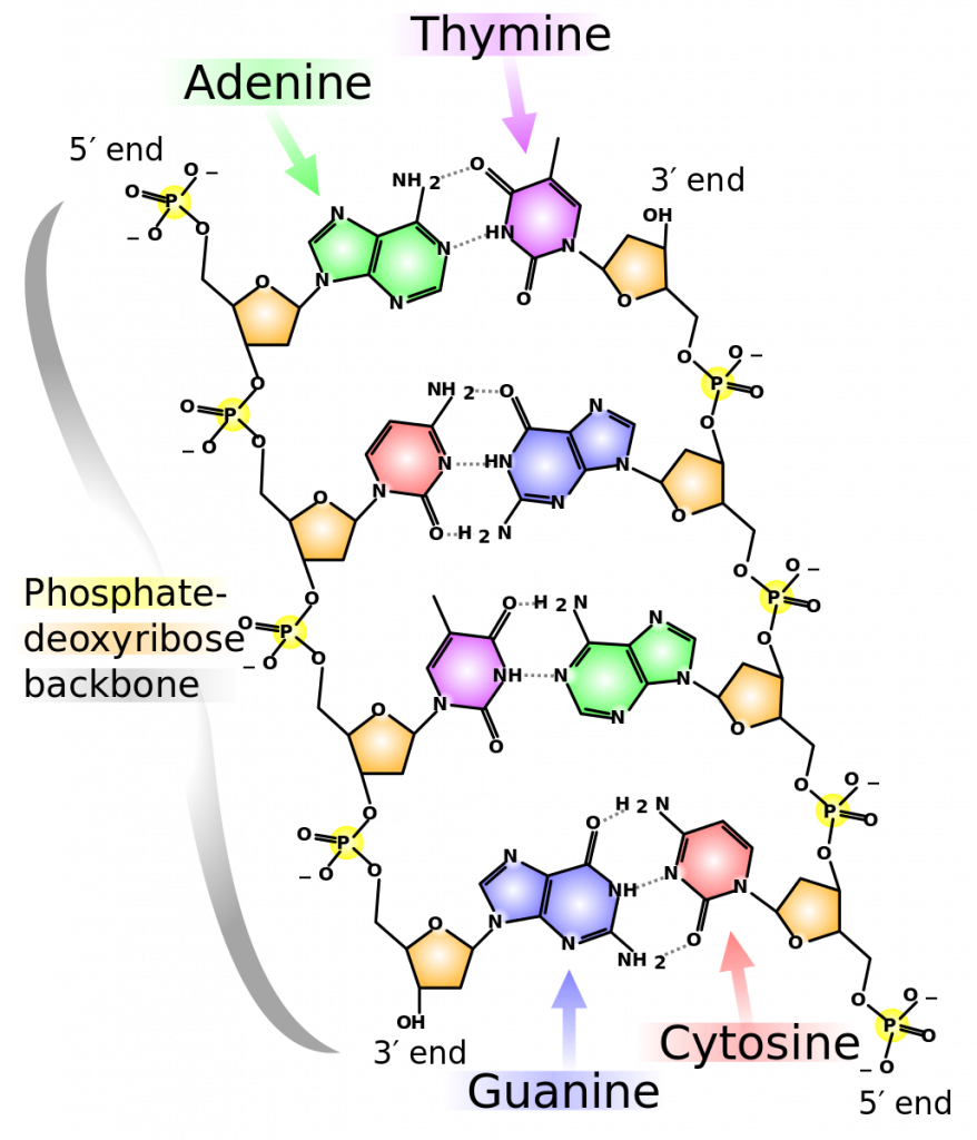
Based on Rosalind Franklin’s X-ray diffraction photograph, and work by other scientists, Watson and Crick proposed that DNA is made up of two strands of nucleotidesthat are twisted around each other to form a right-handed helix. The nucleotides are joined together in a chain by covalent bonds. Scientists already knew that nucleotides contain the same three important components: a nitrogenous base, a deoxyribose(5-carbon sugar), and a phosphate group (Figure 1). The nucleotide is named depending on the nitrogenous base: adenine (A), thymine (T), cytosine (C), and guanine (G).
Watson and Crick’s model proposed that the two strands of nucleotides interact through base pairing between the nucleotides: A pairs with T and G pairs with C. Adenine and thymine are complementary base pairs, and cytosine and guanine are also complementary base pairs. The base pairs are stabilized by hydrogen bonds (a weak type of bond that forms between partially positive and partially negative atoms). Adenine and thymine form two hydrogen bonds and cytosine and guanine form three hydrogen bonds.

The two strands of a DNA double-helix are anti-parallel in nature; that is, the phosphate end of one strand points in one direction, while the sugar end of the other strand points in that direction. The sugar and phosphate of the nucleotides form the backbone of the structure, while the nitrogenous bases are stacked inside.
References
Unless otherwise noted, images on this page are licensed under CC-BY 4.0 by OpenStax.
OpenStax, Biology. OpenStax CNX. December 21, 2017 http://cnx.org/contents/s8Hh0oOc@9.10:QhGQhr4x@6/Biological-Molecules
