27
We have already mentioned that vesicles can bud from the ER, but where do the vesicles go? Before reaching their final destination, the lipids or proteins within the transport vesicles need to be sorted, packaged, and tagged so that they wind up in the right place. The sorting, tagging, packaging, and distribution of lipids and proteins take place in the Golgi apparatus(also called the Golgi body), a series of flattened membranous sacs (Figure 1).

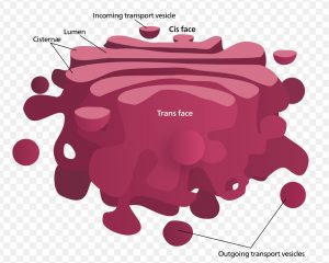
The Golgi apparatus has a receiving face near the endoplasmic reticulum (the cis face) and a releasing face on the side away from the ER, toward the cell membrane (the trans face) (Figure 2). The transport vesicles that form from the ER travel to the receiving face, fuse with it, and empty their contents into the lumen (empty space inside) of the Golgi apparatus. As the proteins and lipids travel through the Golgi, they undergo further modifications. The most frequent modification is the addition of short chains of sugar molecules. The newly modified proteins and lipids are then tagged with small molecular groups to enable them to be routed to their proper destinations.
Finally, the modified and tagged proteins are packaged into vesicles that bud from the opposite face of the Golgi. While some of these vesicles, transport vesicles, deposit their contents into other parts of the cell where they will be used, others, secretory vesicles, fuse with the plasma membrane and release their contents outside the cell.
The amount of Golgi in different cell types again illustrates that form follows function within cells. Cells that engage in a great deal of secretory activity (such as cells of the salivary glands that secrete digestive enzymes or cells of the immune system that secrete antibodies) have an abundant number of Golgi.
In plant cells, the Golgi has an additional role of synthesizing polysaccharides, some of which are incorporated into the cell wall and some of which are used in other parts of the cell.
References
Unless otherwise noted, images on this page are licensed under CC-BY 4.0 by OpenStax.
Text adapted from: OpenStax, Concepts of Biology. OpenStax CNX. May 18, 2016 http://cnx.org/contents/b3c1e1d2-839c-42b0-a314-e119a8aafbdd@9.10

-en.svg)