59
ACR – Chest – Acute Chest Pain – Suspected Pulmonary Embolism
Case 1
Pulmonary Embolism
Chest X-ray
Clinical:
History – This 40 year old female had returned to Canada from China on a long trans-Pacific flight the day before. She presented to the ER with chest pain and shortness of breath. She was on oral birth control. No other medications.
Symptoms – The patient complained of a moderate, dull, achy, left chest pain and mild shortness of breath. It came on about 2 hours after landing in the airport.
Physical – A pleuritic rub was heard in the left chest. No other abnormalities.
Laboratory – The patient’s pregnancy test was negative. Her D-Dimer was elevated. Nil else.
DDx:
Pulmonary Thromboembolism
Pneumothorax
Pneumonia
Pleurisy
Imaging Recommendation
ACR – Chest – Acute Chest Pain – Suspected Pulmonary Embolism, Variant 1
Chest X-ray
Chest CT Angiography
Imaging Assessment
Findings:
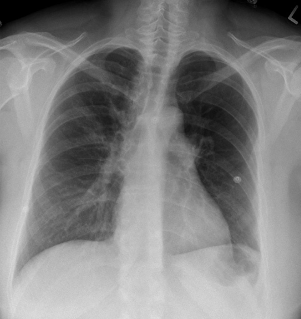
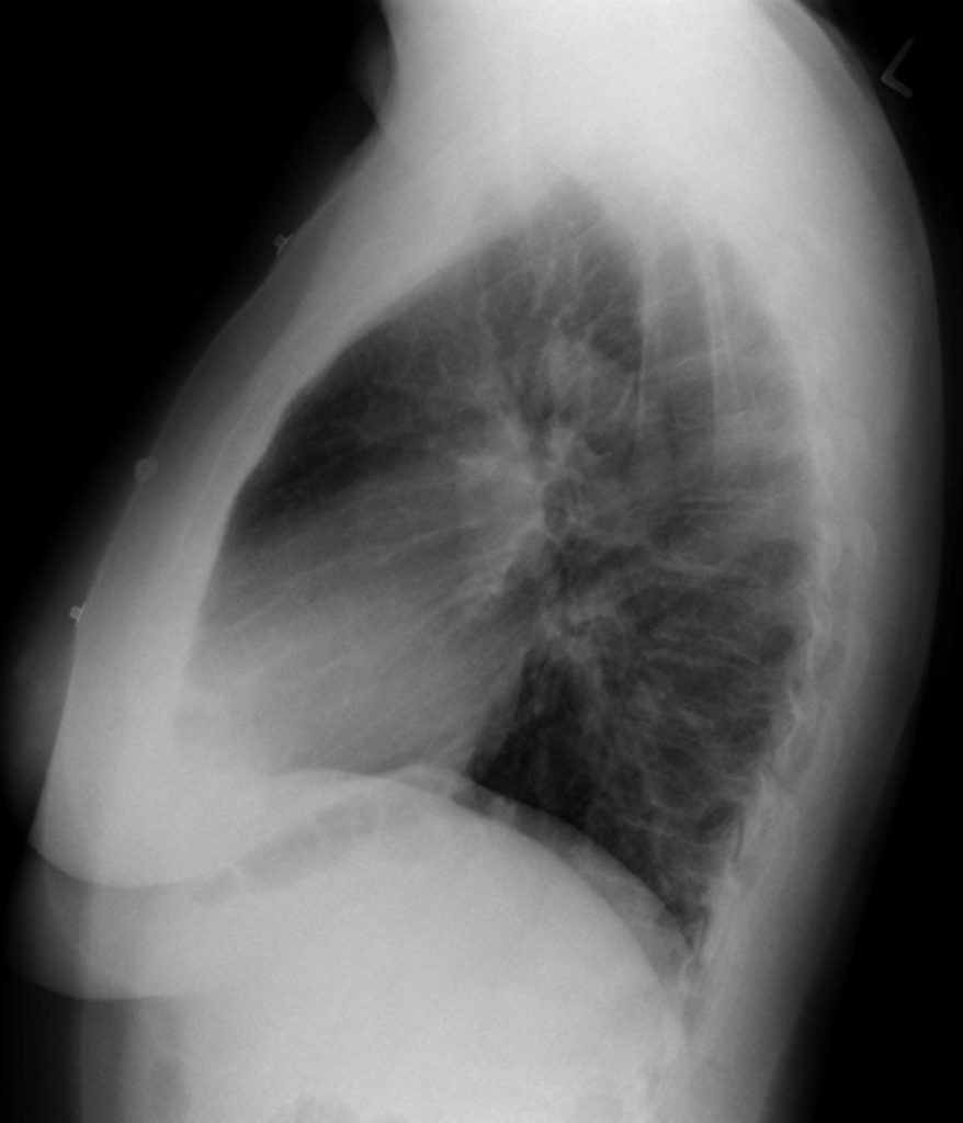
The cardiac silhouette was not enlarged. The left pulmonary artery was prominent and seemed to end abruptly just downstream of the left hilum. There was a paucity of vessels in the left lung.
Interpretation:
The radiographs demonstrated the, “Westermark sign” and a prominent knuckle of the left pulmonary artery. This suggested an abrupt cut-off of the pulmonary artery secondary to a thromboembolism. CT PE imaging was recommended.
Diagnosis:
High Suspicion for Pulmonary Thromboembolism. CT PE study was recommended.
Pathology:
Venous thrombus, formed in the peripheral venous system, leg, pelvis, arm, embolizes to the pulmonary arterial circulation.
Discussion:
- Over 90% of pulmonary emboli (PE) develop from thrombi in the deep veins of the leg, especially above the level of the popliteal veins. There are often concomitant associations of, recent surgery, prolonged bed rest, cancer, venous foreign body, clotting disorder, and oral contraceptives.
- Although conventional chest radiographs are frequently normal in patients with PE, they can demonstrate nonspecific findings, such as subsegmental atelectasis or small pleural effusions.
- Chest x-ray findings suggestive of PE are very rare i.e. Westermark’s sign, Hampton’s hump, vessel paucity.
- CT pulmonary angiography (CT-PA), the best test for the detection of PE (CT-PE), is made possible by the rapid acquisition of spiral CT images (one breath hold) combined with thin CT slices and rapid bolus injection of intravenous, iodinated, contrast that produces maximal opacification of the pulmonary arteries with little or no motion artifact.
- Another benefit of CT-PE studies is the ability to diagnose other diseases the may simulate PE i.e. aortic dissection, pleural effusion, etc.
- CT-PE has a sensitivity in excess of 90% and has replaced the use of V/Q scans in patients with chronic obstructive pulmonary disease or with an abnormal chest radiograph, in whom, a Nuclear Medicine V/Q scan is known to be less sensitive.
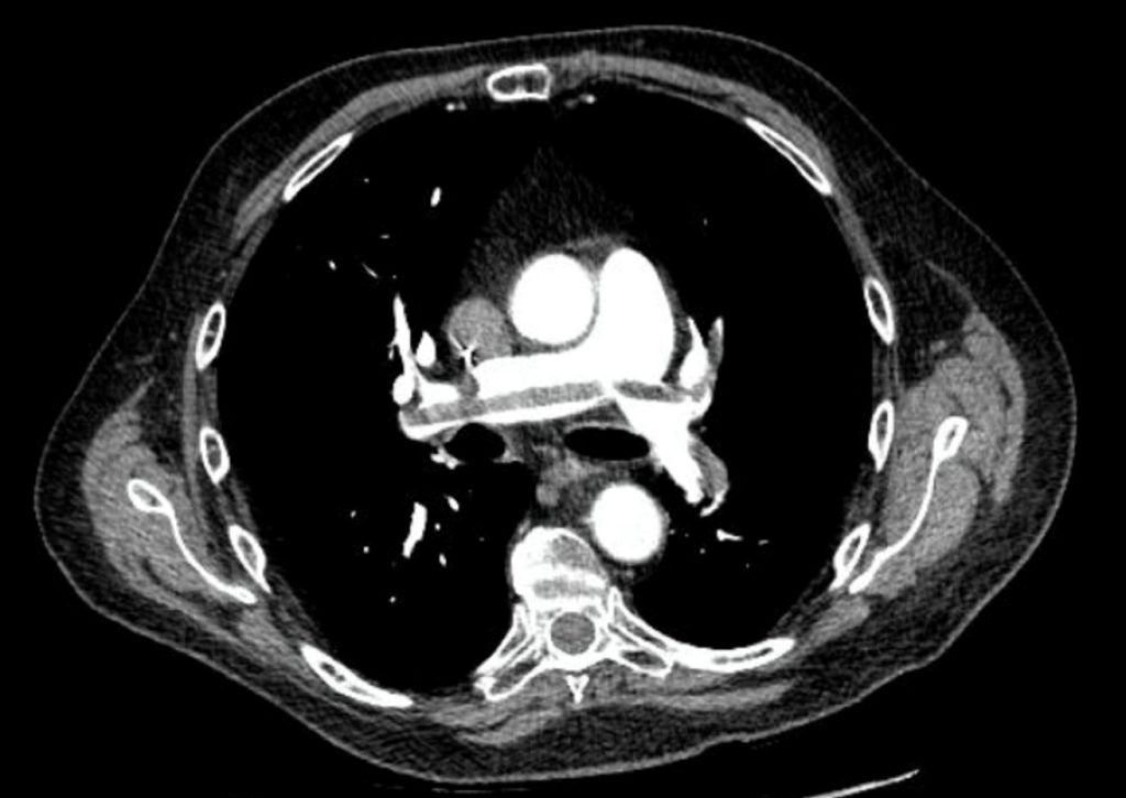
- On CT-PE, acute pulmonary emboli appear as partial or complete filling defects located within the contrast-enhanced lumina of the pulmonary arteries. (See below, Case 2 and 3)
X-ray findings may include:
- May infrequently manifest one of the “classic” findings for pulmonary embolism:
- Wedge-shaped peripheral air-space disease (Hampton hump)
- Focal oligemia (Westermark sign).
- A prominent central pulmonary artery (knuckle sign).
- Pleural effusion.
- No abnormality, the most common finding.
Case 2
Pulmonary Embolism
Chest X-ray and CT
Clinical:
History – This 65 year old male was receiving chemotherapy for colon cancer. He was admitted to the hospital for management of weight loss and swollen legs. He complained of mild shortness of breath.
Symptoms – He had swollen edematous legs.
Physical – No other abnormalities on the chest examination. The legs were swollen with pitting edema.
Laboratory – Hypo-albuminia and mild anemia were noted. His D-Dimer was elevated.
Imaging Assessment
Findings, X-rays:
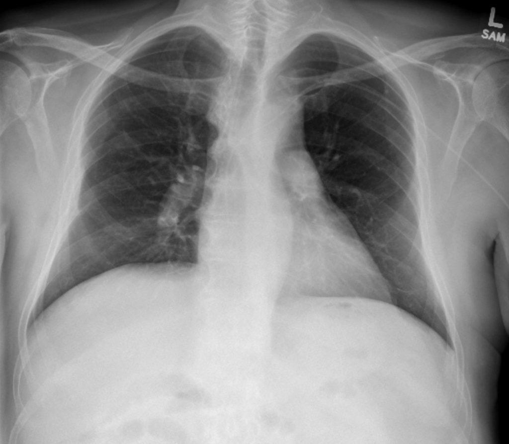
The cardiac silhouette was not enlarged. The central pulmonary arteries may have been mildly enlarged but this was thought to be due to the result of poor inspiratory effort leading to crowding of hilar anatomy.
Interpretation:
Normal chest x-ray. Due to the patient’s signs and symptoms CT-PE imaging was recommended.
Diagnosis:
Clinically, High Suspicion for Pulmonary Thromboembolism.
Findings CT:
There was extensive pulmonary thromboembolism. There was a saddle embolus, with a long segment of thrombus straddling the main pulmonary artery bifurcation. This suggested a large peripheral venous clot burden prior to embolization. Given the patient’s leg swelling, consideration should be given to the possibility of extensive leg and pelvic venous thrombosis.
Pathology:
Venous thrombus, formed in the peripheral venous system, leg, pelvis, arm, embolized to the pulmonary arterial circulation.
Case 3
Pulmonary Embolism
Chest X-ray, Nuclear Medicine, and CT
Clinical:
History – This 25 year old female was pregnant. She was admitted to the hospital after she attended the ER for the management of moderate shortness of breath.
Symptoms – Shortness of breath.
Physical – No other abnormalities on the chest examination. Findings consistent with a 20 week gestation were note. No leg symptoms
Laboratory – Her D-Dimer was elevated.
Diagnosis: – Suspected Pulmonary Thromboembolism.
Imaging Recommendation
ACR – Chest – Acute Chest Pain – Suspected Pulmonary Embolism, Variant 2
Chest X-ray
Lower Limb Venous Doppler Ultrasound
Perfusion Nuclear Medicine Lung Scan
CT Angiography
Imaging Recommendation: Pulmonary Thromboembolism, Variant 2
Chest CT, selected image
Imaging Assessment
Findings, X-rays:
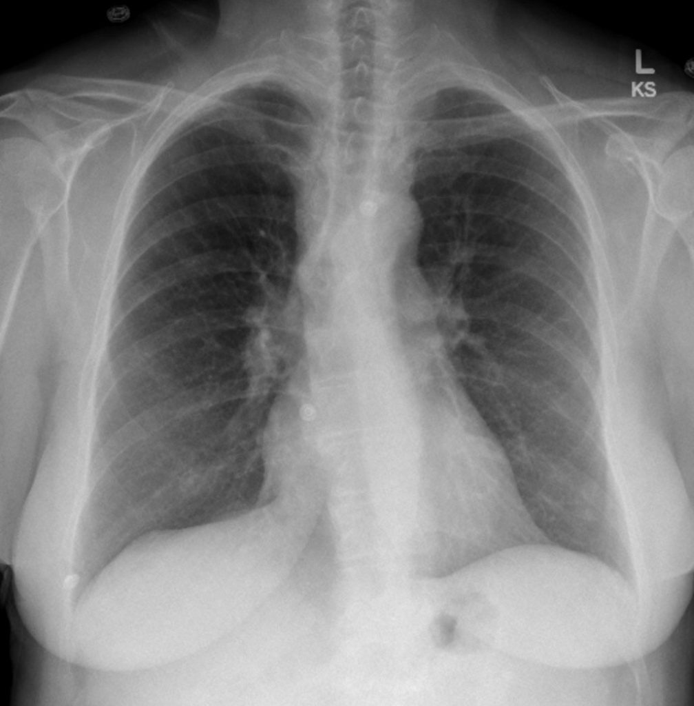
The cardiac silhouette was not enlarged. There was significant peripheral vascular deficiency in the lungs.
Interpretation:
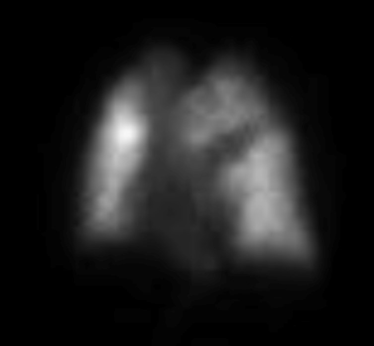
Due to the patient’s signs and symptoms in a pregnant patient perfusion Nuclear Medicine imaging only was recommended.
Diagnosis:
Clinically High Suspicion for Pulmonary Thromboembolism.
Findings Nuclear Medicine:
There were areas of wedge-shaped lack of perfusion on the perfusion Nuclear Medicine scan. High probability of Pulmonary Embolism. Leg Doppler Ultrasound was recommended.
Doppler US Findings:
Negative bilateral leg Doppler Ultrasound. CT PE recommended.
Findings CT:
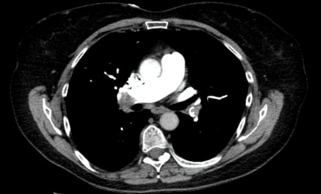
There was extensive central pulmonary thromboembolism in both lungs.
Pathology:
Venous thrombus, formed in the peripheral venous system, legs or pelvis, embolized to the pulmonary arterial circulation.
Attributions
Figure 9.26A PA Chest x-ray, Pulmonary Embolism by Dr. Brent Burbridge MD, FRCPC, University Medical Imaging Consultants, College of Medicine, University of Saskatchewan is used under a CC-BY-NC-SA 4.0 license.
Figure 9.26B Lateral Chest x-ray, Pulmonary Embolism by Dr. Brent Burbridge MD, FRCPC, University Medical Imaging Consultants, College of Medicine, University of Saskatchewan is used under a CC-BY-NC-SA 4.0 license.
Figure 9.27A PA Chest x-ray, Pulmonary Embolism by Dr. Brent Burbridge MD, FRCPC, University Medical Imaging Consultants, College of Medicine, University of Saskatchewan is used under a CC-BY-NC-SA 4.0 license.
Figure 9.27B CT of the Chest, Pulmonary Embolism by Dr. Brent Burbridge MD, FRCPC, University Medical Imaging Consultants, College of Medicine, University of Saskatchewan is used under a CC-BY-NC-SA 4.0 license.
Figure 9.28 PA Chest x-ray, Pulmonary Embolism by Dr. Brent Burbridge MD, FRCPC, University Medical Imaging Consultants, College of Medicine, University of Saskatchewan is used under a CC-BY-NC-SA 4.0 license.
Figure 9.29 Nuclear Medicine Scan of Chest. Pulmonary Embolism by Dr. Brent Burbridge MD, FRCPC, University Medical Imaging Consultants, College of Medicine, University of Saskatchewan is used under a CC-BY-NC-SA 4.0 license.
Figure 9.30 CT Scan of the Chest, Pulmonary Embolism by Dr. Brent Burbridge MD, FRCPC, University Medical Imaging Consultants, College of Medicine, University of Saskatchewan is used under a CC-BY-NC-SA 4.0 license.
