107
ACR – Urologic – Indeterminate Renal Mass
Case
Renal Cell Carcinoma
Clinical:
History – A mass in the vicinity and probably involving the left kidney was incidentally discovered during an abdominal ultrasound for right upper quadrant pain.
Symptoms – None
Physical – None
Laboratory – Microscopic hematuria was detected.
DDx:
Adrenal mass
Renal mass
Splenic mass
Adenopathy
Imaging Recommendation
ACR – Urologic – Indeterminate Renal Mass, Variant 1
CT Abdomen


Imaging Assessment
Findings:
Ultrasound
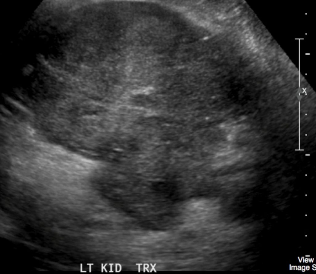
An inhomogeneous mass was seen in the vicinity of the pancreatic tail and left kidney. The organ of origin was not easily determined. No other findings. CT was recommended.
Computed Tomography
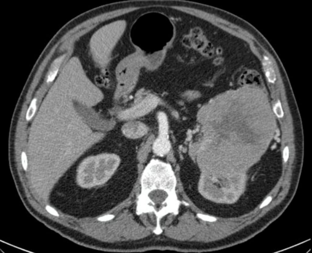
The large, exophytic, mass arose from the cranial left kidney. It was of inhomogeneous density with some low density in the centre. The mass poorly contrast enhanced. No lymphadenopathy. The left renal vein was normal in appearance.
Interpretation:
Renal mass – High suspicion for renal carcinoma.
Diagnosis:
Renal Cell Carcinoma (Chromophobe subtype) – Biopsy proven
Discussion:
An indeterminate renal mass is one that cannot be diagnosed confidently as benign or malignant at the time it is discovered. Renal masses are increasingly detected in asymptomatic individuals as incidental findings. Computed tomography (CT), ultrasonography (US), and magnetic resonance imaging (MRI) of renal masses with fast-scan techniques and intravenous (IV) gadolinium are the mainstays of evaluation.
Dual-energy CT, contrast-enhanced US, positron emission tomography PET/CT, and percutaneous biopsy are all technologies that are gaining traction in the characterization of the indeterminate renal mass.
Computed Tomography
CT is the most utilized imaging technique for evaluating the indeterminate renal mass, playing an important role in the characterization of both solid and cystic lesions. Although the majority of lesions are characterized on initial imaging, one definition for the indeterminate renal mass is a lesion containing areas that measure 20–70 Hounsfield units (HU) on non-contract imaging.
Homogeneous lesions measuring <20 HU or >70 HU can be considered benign, whereas lesions either entirely or partially within the 20–70 HU range should be considered indeterminate and warrant further evaluation.
For those lesions requiring further evaluation, enhancement after IV contrast is key in determining if a renal mass warrants treatment. Enhancing solid renal masses or enhancing components in cystic masses indicates a vascularized mass and, therefore, a possible malignancy. The degree of renal enhancement is dependent on many factors, including the amount and rate of contrast material injection, the timing of contrast-enhanced imaging, and the intrinsic characteristics of both the mass and the adjacent renal parenchyma.
Proper characterization of a renal mass includes at least 3 phases: non-contract imaging followed by contrast-enhanced imaging in corticomedullary and nephrographic phases.
Attributions
Figure 16.2A Transverse ultrasound of the left kidney mass by Dr. Brent Burbridge MD, FRCPC, University Medical Imaging Consultants, College of Medicine, University of Saskatchewan is used under a CC-BY-NC-SA 4.0 license.
Figure 16.2B Axial CT of the abdomen revealing a renal tumor by Dr. Brent Burbridge MD, FRCPC, University Medical Imaging Consultants, College of Medicine, University of Saskatchewan is used under a CC-BY-NC-SA 4.0 license.
