71
ACR – Women’s Imaging – Clinically Suspected Adnexal/Pelvic Mass/Uterus
Case 1
Uterine Leiomyoma (Fibroid)
Clinical:
History – Annual physical examination.
Symptoms – The patient reported a vague sense of pelvic fullness over the last six months, but no other symptoms.
Physical – The uterus was felt to be globally enlarged and had a discernible, separate, mass in the fundus. Nil else.
DDx:
Uterine Mass
Adnexal Mass
Peritoneal Mass
Imaging Recommendation
ACR – Women’s Imaging – Clinically Suspected Adnexal/Pelvic Mass/Uterus, Variant 1
Pelvic Ultrasound
ODIN Link for Leiomyoma images (Pelvic Ultrasound), Figure 11.1A and B: https://mistr.usask.ca/odin/?caseID=20170502170438155
Imaging Assessment
Findings:
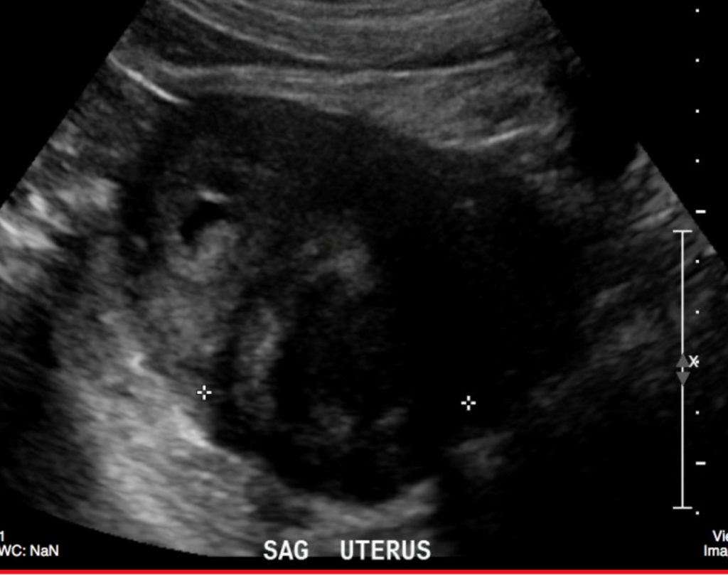
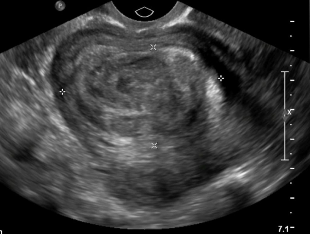
There was a large, homogeneous mass in the uterus that was of low echogenicity. It caused shadowing of the ultrasound beam. Some fluid was seen in the endometrial cavity. The ovaries were normal.
Interpretation:
Probable Leiomyoma of the Uterus
Diagnosis:
Uterine Leiomyoma (Fibroid)
Discussion:
Suspected uterine and adnexal masses are a common problem clinically, and pelvic ultrasonography (US), specifically endovaginal US, is the first-line imaging modality for assessing them. Its findings, however, should be correlated with the physical examination, history, and laboratory tests. Morphological analysis of adnexal masses with US can help narrow the differential diagnosis.
Recent studies have shown that US (transvaginal plus color Doppler) may discriminate benign from malignant lesions with a sensitivity of 99.1 % and a specificity of 85.9%. However, US is not always reliable for triage of patients to surgery.
Transabdominal sonography (TAS) and transvaginal sonography (TVS) are complementary. In some facilities, patients are scanned by both techniques, but most recent literature regarding adnexal mass US refers to TVS.
- Leiomyomas are benign smooth muscle tumors of the uterus that occur in up to 50% of women over the age of 30.
- Although most women with fibroids are asymptomatic, fibroids can cause pain, infertility, menorrhagia, and urinary or bowel symptoms if they grow large enough.
- US (transabdominal and transvaginal) is the imaging study of choice in evaluating uterine fibroids.
- Magnetic resonance imaging (MRI) is used mainly to evaluate complicated cases or in surgical planning.
Imaging findings may include
- US (transabdominal and transvaginal) is the imaging study of choice in evaluating uterine fibroids.
- Uterine leiomyomas on US are heterogeneously hypoechoic, solid masses, meaning they may display some areas that contain many echoes and others that demonstrate few echoes.
- It is important to characterize the location of fibroids as submucosal (which has implications for bleeding and infertility), myometrial (most common), or serosal ( can be pedunculated).
Case 2
Simple Ovarian Cyst
Clinical:
History – Annual physical examination.
Symptoms – No symptoms.
Physical – The right ovary was globally enlarged and had a soft compressible feel on pelvic examination. Nil else.
DDx:
Ovarian Mass
Ovarian Cyst
Pelvic Adenopathy
Imaging Recommendation
ACR – Women’s Imaging – Clinically Suspected Adnexal/Pelvic Mass/Uterus, Variant 1
Ultrasound
ODIN Link for Ovarian Cyst images (Ultrasound), Figure 11.2A and B – https://mistr.usask.ca/odin/?caseID=20170123215147808
Imaging Assessment
Findings:
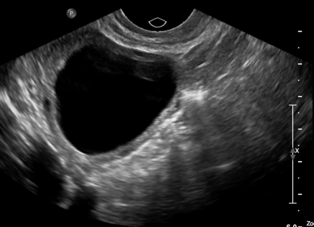
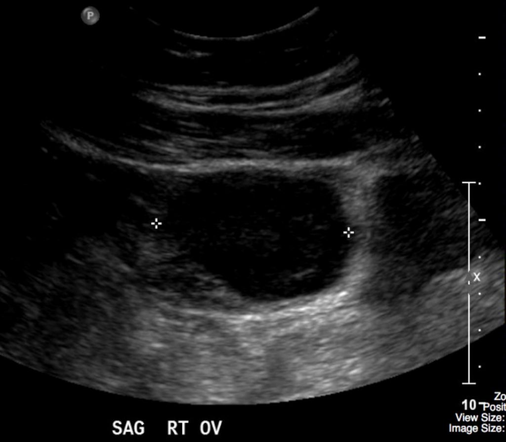
The images revealed a simple cyst in the right ovary with no nodules in the cyst wall or abnormal Doppler signal.
Interpretation:
Simple cyst of the right ovary.
Diagnosis:
Simple Ovarian Cyst
Discussion:
Suspected uterine and adnexal masses are a common problem clinically, and pelvic sonography (US), specifically endovaginal US, is the first-line imaging modality for assessing them. Its findings, however, should be correlated with the history, clinical, and laboratory findings. Morphological analysis of adnexal masses with US can help narrow the differential diagnosis.
Recent studies have shown that US (transvaginal plus color Doppler) may discriminate benign from malignant lesions with a sensitivity of 99.1 % and a specificity of 85.9%. However, US is not always reliable for triaging patients for surgery. Accurate staging is best achieved with MRI.
If one of the non-dominant follicles fills with fluid and does not rupture, a follicular cyst is formed. Follicular cysts are unilateral, usually asymptomatic, and usually involute during the next menstrual cycle or two. Follicular cysts and corpus luteum cysts are called functional cysts of the ovary because they occur as a result of the hormonal stimuli associated with ovulation. Both are characteristically well-defined, thin-walled, anechoic structures with homogeneous internal fluid. They may contain echogenic material if hemorrhage occurs into the “cyst”.
Case 3
Ovarian Dermoid
Clinical:
History: An ultrasound, performed for pelvic pain, incidentally detected an ovarian mass.
Symptoms: None
Physical Examination: Trans-abdominal and bi-manual pelvic examination detected a solid mass in the left adnexa.
DDx:
Ovarian Tumor
Ovarian Cyst
Pelvic Adenopathy
Imaging Recommendation
ACR – Women’s Imaging – Clinically Suspected Adnexal/Pelvic Mass, Variant 1
Ultrasound
ODIN Link for Ovarian Dermoid images (Ultrasound and CT), Figure 11.3A and B: https://mistr.usask.ca/odin/?caseID=20170123215550360
Imaging Assessment
Ultrasound
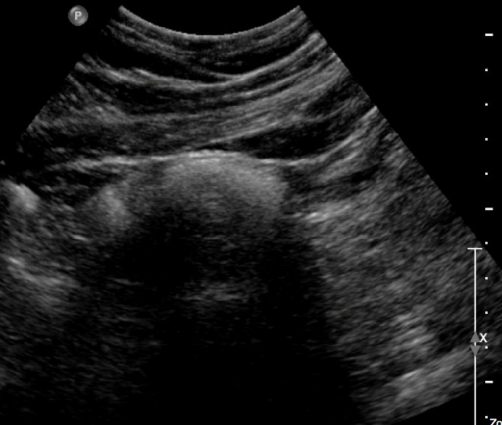
Ultrasound detected a left adnexal mass that demonstrated increased echogenicity and shadowing of the ultrasound beam. No focal calcification detected.
Computed Tomography
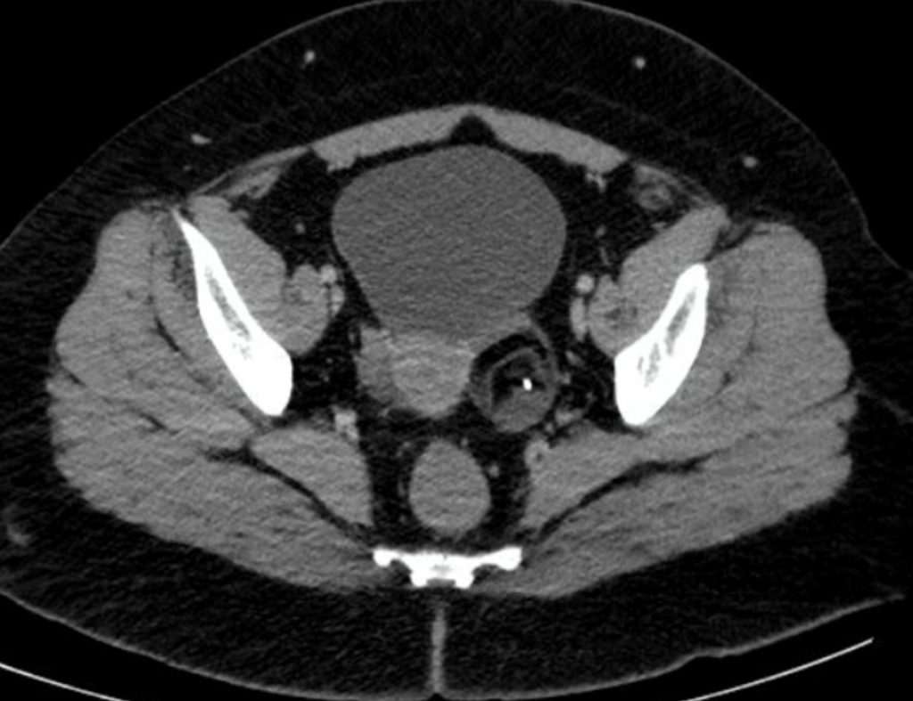
CT revealed a mass containing, fat, soft tissue, and calcification in the left adnexa.
Diagnosis: Ovarian Dermoid Tumor
Diagnosis:
Ovarian Dermoid (left)
Discussion:
Dermoid cysts are mature teratomas composed of cells from all three germ layers – ectoderm, mesoderm, and endoderm – although the ectodermal elements (hair, bone) predominate. They are most commonly found in women of reproductive age, are bilateral in up to 25% of cases, and may serve as a lead point for ovarian torsion.
Imaging findings of a dermoid cyst may include:
- An adnexal mass is present.
- Normal ovarian tissue may be present.
- Dermoid cysts have a variety of echo textures on ultrasound due to the complex tissue present.
- There may be echogenic tissue consistent with fat.
- There may be dense calcifications due to teeth or bone in the mass.
- There may be a complex dot – dash pattern due to layered hair.
- A dermoid plug is a peripheral echogenic nodule – Rokitansky nodule.
- The total size of the mass may not be visible due to shadowing from calcification or fat – the tip of the iceberg sign.
- Fluid – fluid levels may be seen.
Attributions
Figure 11.1A Sagittal Ultrasound of the Uterus with a uterine mass by Dr. Brent Burbridge MD, FRCPC, University Medical Imaging Consultants, College of Medicine, University of Saskatchewan is used under a CC-BY-NC-SA 4.0 license.
Figure 11.1B Transverse Ultrasound of the Uterus with a mass by Dr. Brent Burbridge MD, FRCPC, University Medical Imaging Consultants, College of Medicine, University of Saskatchewan is used under a CC-BY-NC-SA 4.0 license.
Figure 11.2A Pelvic ultrasound in a female with an ovarian cyst by Dr. Brent Burbridge MD, FRCPC, University Medical Imaging Consultants, College of Medicine, University of Saskatchewan is used under a CC-BY-NC-SA 4.0 license.
Figure 11.2B Sagittal ultrasound of a right ovarian cyst by Dr. Brent Burbridge MD, FRCPC, University Medical Imaging Consultants, College of Medicine, University of Saskatchewan is used under a CC-BY-NC-SA 4.0 license.
Figure 11.3A Ultrasound of female uterus, echogenic mass with acoustic shadowing by Dr. Brent Burbridge MD, FRCPC, University Medical Imaging Consultants, College of Medicine, University of Saskatchewan is used under a CC-BY-NC-SA 4.0 license.
Figure 11.3B CT Scan of female pelvis, left adnexal mass with bone, fat and soft tissue by Dr. Brent Burbridge MD, FRCPC, University Medical Imaging Consultants, College of Medicine, University of Saskatchewan is used under a CC-BY-NC-SA 4.0 license.
