80
Case
Epiglottitis
Clinical:
History – Sore throat and drooling.
Symptoms– This 5 year old reported severely sore throat and difficulty swallowing.
Physical– The child was sitting up on the stretcher drooling and was showing signs of mild respiratory distress with inspiratory stridor and grunting. Fever, with temperature of 40.5C.
DDx:
Tonsillitis
Pharyngitis
Epiglottitis
Imaging Recommendation
Upright, lateral, soft-tissue x-ray of the pharynx

Imaging Assessment
Findings:
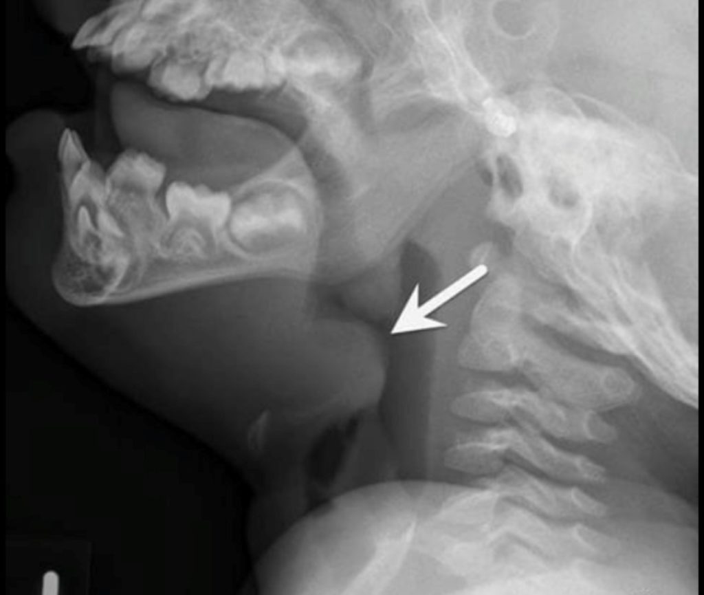
The epiglottis and the aryepiglottic folds were severely swollen creating a thumb-like projection that was partially occluding the trachea. The adenoid tissue was normal for a child this age.
Interpretation:
Epiglottitis
Diagnosis:
Epiglottitis
Discussion:
Acute bacterial epiglottitis can be a life-threatening medical emergency, leading to airway obstruction caused by infection, with edema of the epiglottis and aryepiglottic folds.
The most frequent causative organism had previously been Haemophilus influenzae type B, but introduction of the vaccine in 1985 has led to a marked decrease in the number of cases of epiglottitis. Other, rarer, causative may include: Pneumococcus, Streptococcus group A, viral infection such as herpes simplex 1, parainfluenza, thermal injury, or angioneurotic edema.
Epiglottitis typically has a peak incidence from about 3 to 6 years of age. Clinically epiglottitis resembles croup, but the clinician should think of epiglottitis if the child cannot breath unless sitting up, if what appears to be croup seems to be worsening, or the child cannot swallow saliva and drools. Cough is rare. The classical triad of epiglottitis is drooling, severe dysphagia, and respiratory distress with inspiratory stridor.
The imaging study of choice is the lateral neck radiograph, which should be exposed in the upright position only as the supine position may precipitate airway occlusion. The patient should be accompanied everywhere by someone experienced in endotracheal intubation. Imaging studies are not always necessary for the diagnosis and may be falsely negative in early stages.
Imaging findings may include:
- Enlargement of the epiglottis.
- There is thickening of the aryepiglottic folds (which is an important component of airway obstruction and the true cause of stridor).
- Sometimes circumferential narrowing of the subglottic portion of the trachea is seen during inspiration.
Treatment of Epiglottitis
- Securing the airway, which may require intubation or emergency tracheostomy;
- Start empiric antibiotic therapy;
- Corticosteroids may be added to minimize swelling of the epiglottis.
Attributions
Figure 12.7 Lateral x-ray of the neck with an arrow pointing to the enlarged epiglottis by Dr. Brent Burbridge MD, FRCPC, University Medical Imaging Consultants, College of Medicine, University of Saskatchewan is used under a CC-BY-NC-SA 4.0 license.
