113
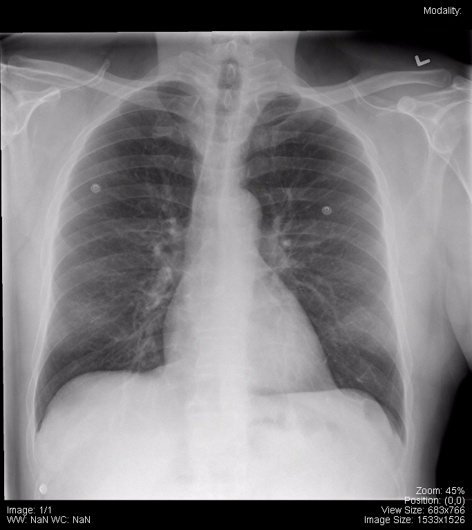
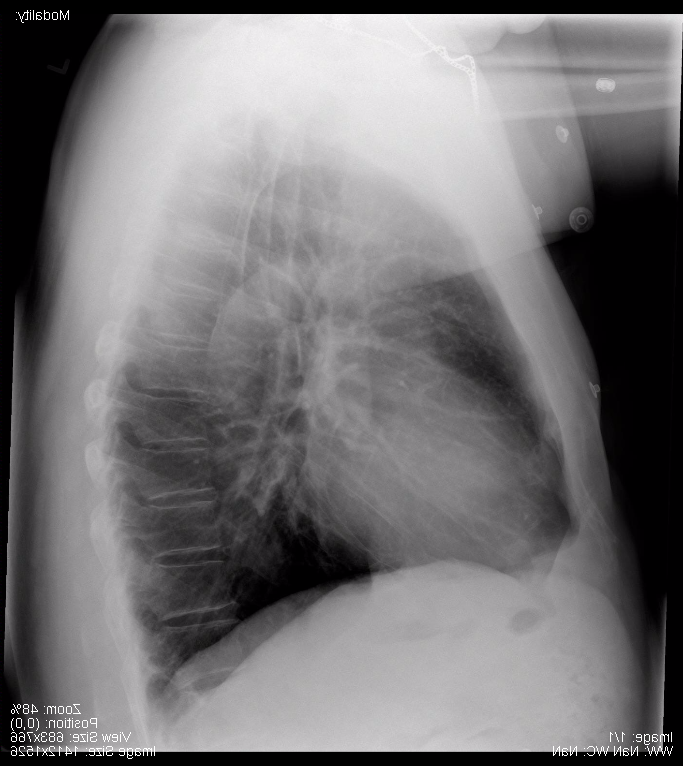
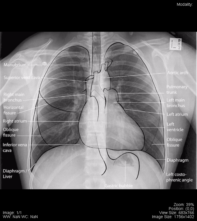
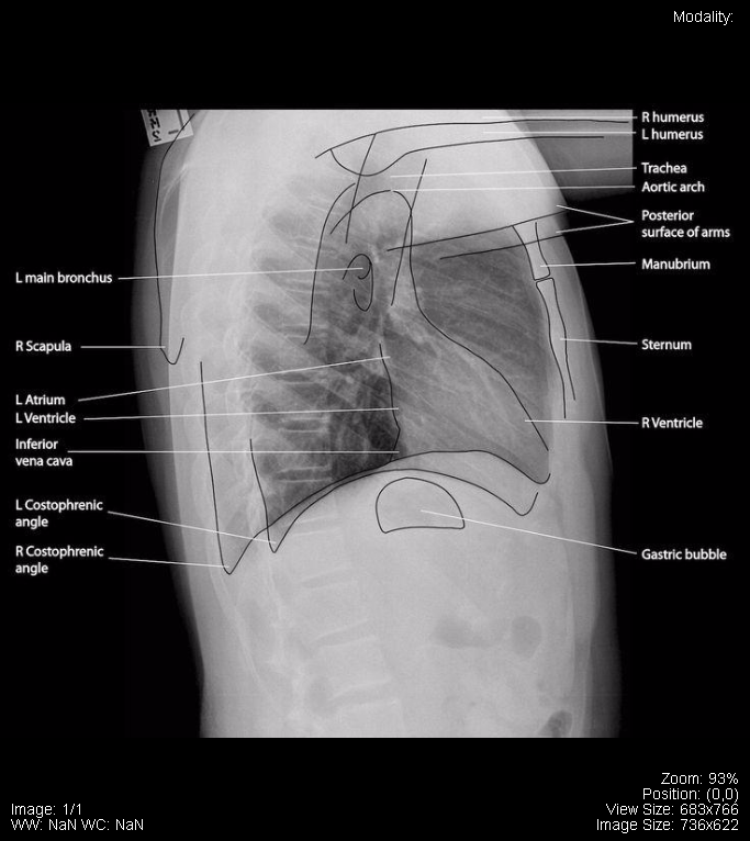
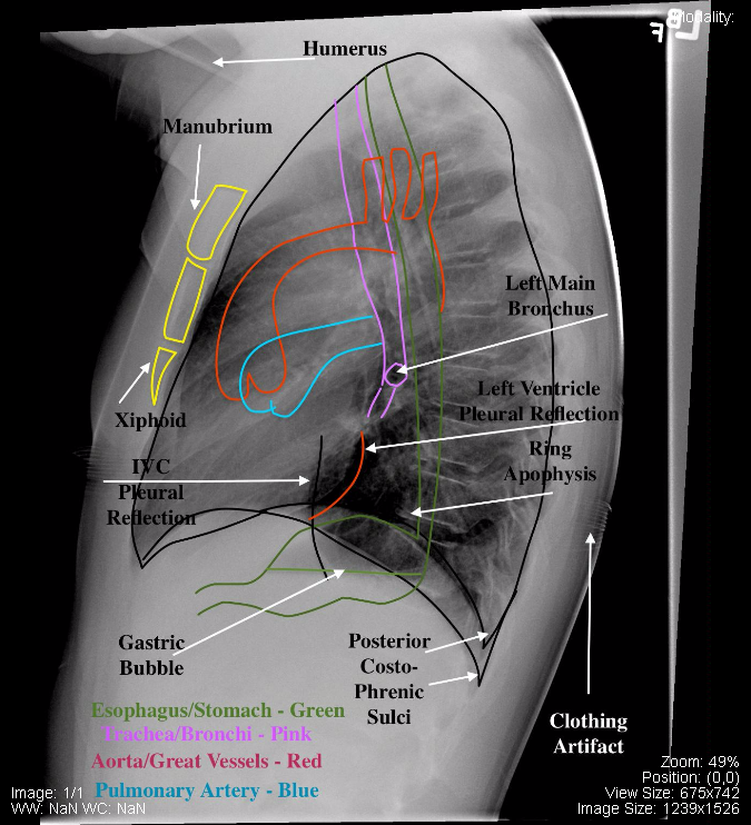
The following are normal PA and Lateral x-ray images of the chest:
The following is a normal AP (Portable) x-ray of the chest:
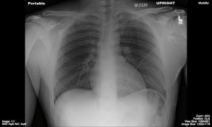
The following are normal pediatric chest x-rays:
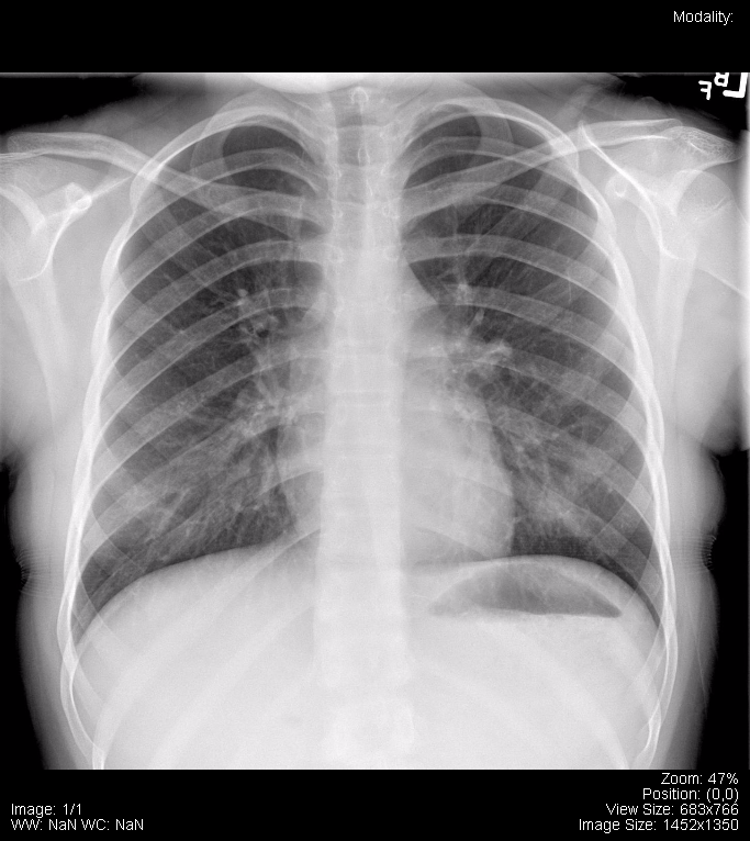
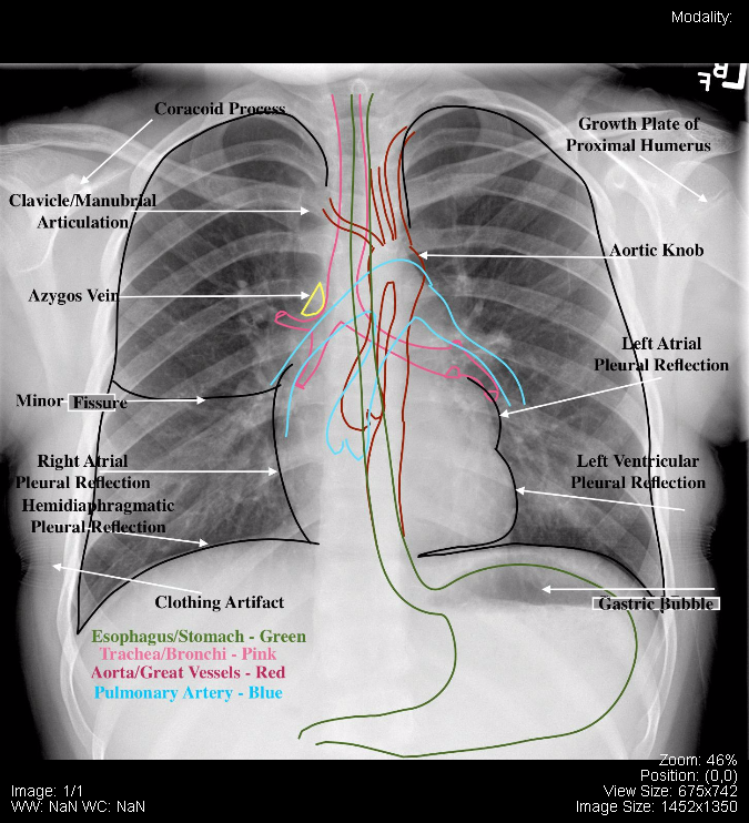
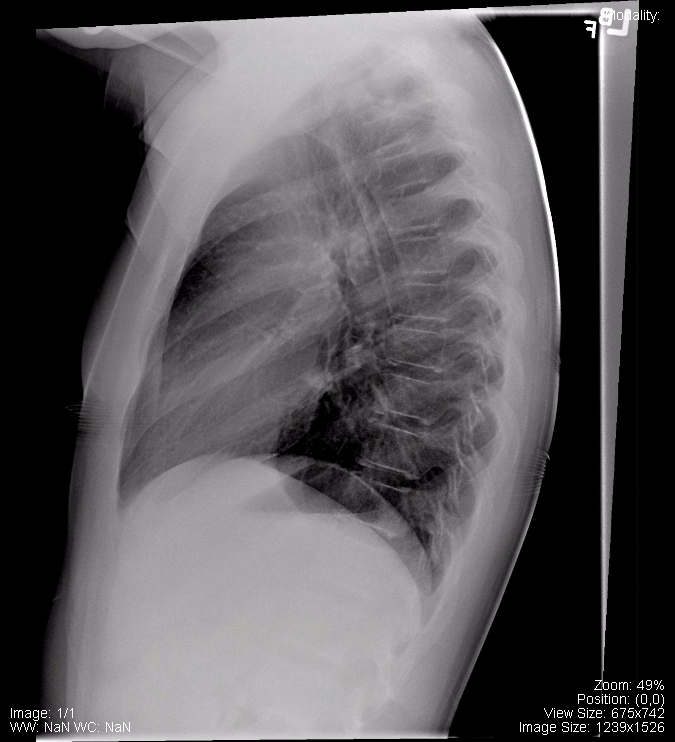
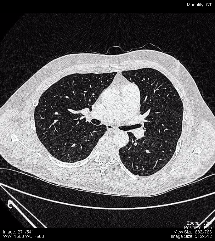
The following are sections from a High Resolution CT (HRCT) of a normal chest:
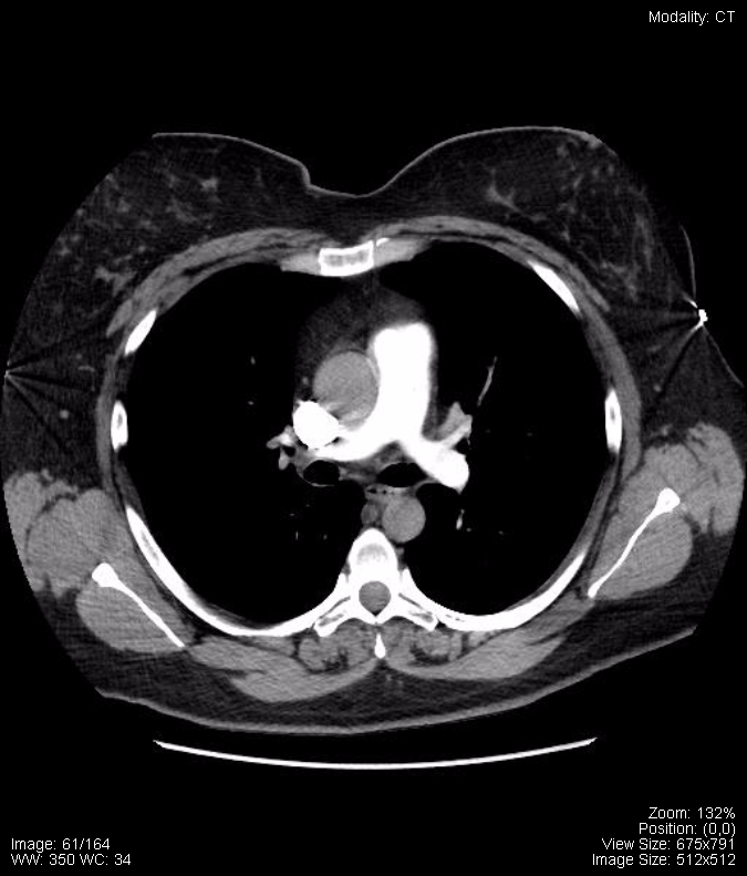
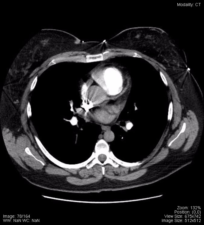
The following is a normal CT Angiography (CTA) of the Pulmonary Arteries:
Attributions
All figures in “Chapter 17: Chest” by Dr. Brent Burbridge MD, FRCPC, University Medical Imaging Consultants, College of Medicine, University of Saskatchewan is used under a CC-BY-NC-SA 4.0 license.
