61
ACR – Gastrointestinal – Right Upper Quadrant Pain
Case
Cholecystitis
Clinical:
History – This 45 year old female presented to the emergency room with moderate to severe right upper quadrant pain. She had noted vague pain in the right upper quadrant when eating for the last 4 months.
Symptoms – The patient relates that she had moderate, persistent, right upper quadrant pain.
Physical – The liver was difficult to palpate due to the patient’s large size (Body Mass Index – 30). The patient was tender in the right upper quadrant and there was a positive Murphy’s sign. She had a fever of 40C. There was guarding on palpation in the right upper abdomen.
Laboratory – The serum liver enzymes and bilirubin were normal. Her white blood cell count was elevated.
DDx:
Cholecystitis
Choledocholithiasis
Liver abscess
Diverticulitis
Colitis
Imaging Recommendation
ACR – Gastrointestinal – Right Upper Quadrant Pain – Suspected Cholecystitis, Variant 1
Ultrasound Abdomen

Imaging Assessment
Findings:
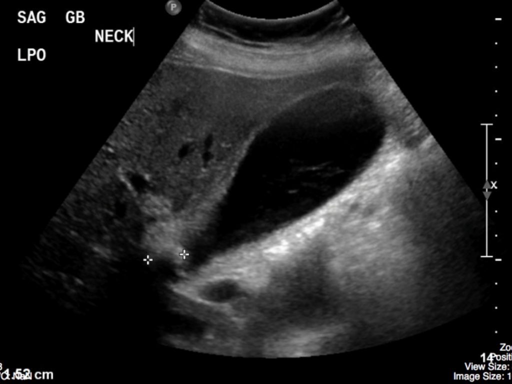
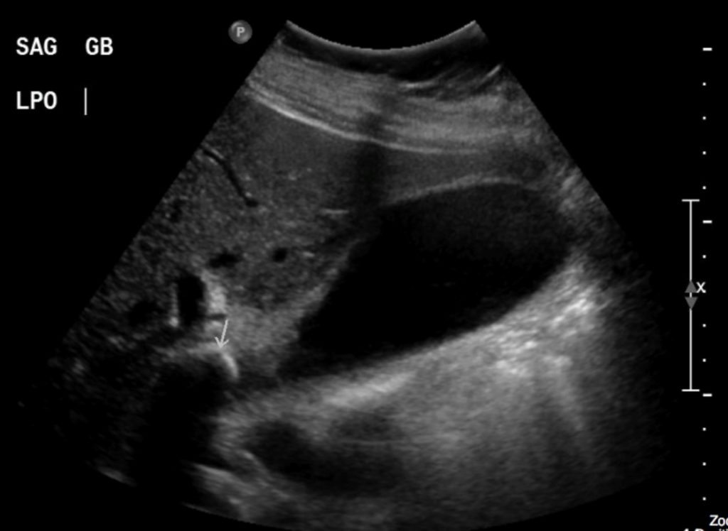
The gallbladder was distended. The gallbladder was very tender on ultrasound probe pressure (Sonographic Murphy’s Sign). There was a large gallstone seen in the neck of the gallbladder. The gallbladder wall was mildly thickened and there was a small amount of fluid adjacent to the gallbladder.
Interpretation:
The findings were in keeping with Cholelithiasis and Cholecystitis.
Diagnosis:
Cholelithiasis and Cholecystitis
Discussion:
- Cholelithiasis is estimated to affect more than 20 million Americans. This does not lead to cholecystitis in a large number of individuals. In almost all cases, acute cholecystitis starts with a gallstone impacted in the neck of the gallbladder or cystic duct. The presence of gallstones does not, by itself, mean that the patient’s pain is emanating from the gallbladder, because asymptomatic gallstones are common. Cholecystitis may occur, less commonly, in the absence of stones (acalculous cholecystitis).
X-ray findings may include:
- No abnormalities
- Visible, calculi, may be multiple and faceted but only 15% of biliary calculi have enough calcium within them to be visible on x-rays.
- Gas may be seen in the biliary tree if there has been recent passage of a bile duct stone.
Ultrasound findings may include:
- Gallstones are characteristically echogenic and produce acoustical shadowing because they reflect most of the signal back to the transducer.
- Acoustic shadowing describes a band of reduced echoes behind an echo-dense object (e.g., a gallstone) that reflects most of the sound waves. This finding can have diagnostic value in identifying a calculus, such as in the gallbladder and kidney.
- Gallstones usually fall to the dependent part of the gallbladder, based upon the patient’s position at the time of the scan. Gallbladder calculi will move when the patient is re-positioned. This helps to differentiate gallstones from polyps or tumors.
- Biliary sludge can be found in the lumen of the gallbladder and is an aggregation that may contain cholesterol crystals, bilirubin, and glycoproteins. It is often associated with biliary stasis. Although it may be echogenic, sludge does not produce acoustical shadowing as gallstones do.
Recognizing Acute Cholecystitis on US:
- The presence of gallstones, possibly impacted in the neck of the gallbladder or in the cystic duct.
- Thickening of the gallbladder wall (>3 mm)
- Pericholecystic fluid (fluid around the gallbladder)
- A positive sonographic Murphy sign (Pain that is elicited by compression of the gallbladder with the US probe).
- In the presence of gallstones and gallbladder wall thickening, US has a positive predictive value for acute cholecystitis as high as 94%.
Attributions
Figure 10.1A Sagittal ultrasound of the Gallbladder by Dr. Brent Burbridge MD, FRCPC, University Medical Imaging Consultants, College of Medicine, University of Saskatchewan is used under a CC–BY-NC-SA 4.0 license.
Figure 10.1B Sagittal Ultrasound of the Gallbladder Neck and Calculus by Dr. Brent Burbridge MD, FRCPC, University Medical Imaging Consultants, College of Medicine, University of Saskatchewan is used under a CC–BY-NC-SA 4.0 license.

