97
ACR – MSK – Chronic Extremity Joint Pain
Case
Hip Osteoarthritis.
Clinical:
History – This elderly patient had chronic hip pain, right worse than left.
Symptoms – The chronic pain was limiting the patients activities.
Physical – Limited, painful, range of motion. Limp when walking. Stiffness upon arising from a chair or bed.
DDx:
Degenerative arthritis (Osteoarthritis) of the hip
Imaging Recommendation
ACR – MSK – Chronic Extremity Joint Pain
X-rays

Imaging Assessment
Findings:
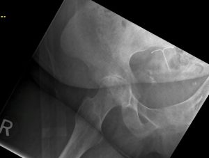
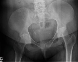
Severe right hip joint narrowing was present. There was moderate hip joint degeneration on the left. Bone density was normal. Small marginal joint osteophytes were seen on the right and there is subchondral sclerosis and subchondral cysts in the right joint space. Incidental note was made of an intra-uterine contraceptive device in this 59 year old female.
Interpretation:
Moderate to severe right hip osteoarthritis
Diagnosis:
Hip osteoarthritis. Treatment planning included, physiotherapy, pain management and eventual planning for a hip replacement.
Discussion:
Osteoarthritis (degenerative) is the most common form of arthritis. It results from intrinsic degeneration of the articular cartilage, mostly from the mechanical stress of excessive wear and tear on weight-bearing joints. It mostly involves the hips, knees, and hands and increases in prevalence with increasing age.
X-ray findings may include:
- Marginal osteophyte formation. A hallmark of hypertrophic arthritis, osseous transformation of cartilaginous excrescences, and metaplasia of synovial lining cells leads to the production these bone protrusions at or near the joint.
- Subchondral sclerosis. This is a reaction of the bone to the mechanical stress to which it is subjected when its protective cartilage has been destroyed.
- Subchondral cysts. As a result of chronic impaction, necrosis of bone, and/or imposition of synovial fluid into the subchondral bone, cysts of varying sizes form in the subchondral bone.
- Narrowing of the joint space. Seen in all forms of arthritis.
Attributions
Figure 14.15A X-ray of the pelvis by Dr. Brent Burbridge MD, FRCPC, University Medical Imaging Consultants, College of Medicine, University of Saskatchewan is used under a CC-BY-NC-SA 4.0 license.
Figure 14.15B X-ray of the femur and hip joint by Dr. Brent Burbridge MD, FRCPC, University Medical Imaging Consultants, College of Medicine, University of Saskatchewan is used under a CC-BY-NC-SA 4.0 license.

