98
ACR – MSK – Chronic Extremity Joint Pain
Case
Rheumatoid Arthritis
Clinical:
History – This patient had a long history of polyarthritis.
Symptoms – Painful deformed joints.
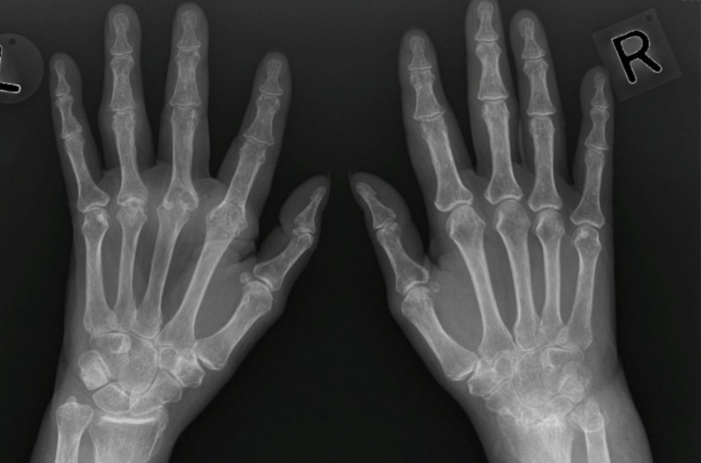
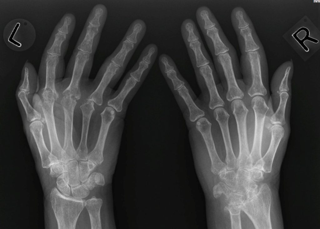
Physical – There were deformed joints in the hands and feet that are symmetrical. Joint swelling was noted in the fingers and toes. There was minimal erythema of multiple finger joints.
DDx:
Poly-arthritis – erosive
Imaging Recommendation
ACR – MSK – Chronic Extremity Joint Pain
X-rays


Imaging Assessment
Findings:
The radiographs demonstrated an erosive, symmetrical, polyarthritis of the hands and carpus. The joint deformities were moderate to severe. The bones were diffusely osteopenic, but were especially so in the peri-articular region.
Interpretation:
Symmetrical Polyarthritis – Rheumatoid Arthritis (RA)
Diagnosis:
Rheumatoid Arthritis
Discussion:
Rheumatoid arthritis is more common in females, frequently involving the proximal joints of the hands and wrists. Conventional radiographs remain the study of first choice for imaging RA.
X-ray findings may include:
- Bilateral and symmetrical imaging findings.
- The earliest radiographic changes are soft tissue swelling of the affected joints and osteoporosis, which tends to be most severe on both sides of the joint space (peri-articular osteoporosis or peri-articular demineralization).
- In the hand, the erosions tend to involve the proximal joints: the carpal-metacarpal joints, metacarpal-phalangeal joints, and proximal interphalangeal joints.
- Late findings in the hands include deformities such as ulnar deviation of the fingers at the MCP joints, subluxation of the MCP joints, and ligamentous laxity, leading to deformities of the fingers (swan-neck and boutonnière deformities).
- In the wrist, erosions of the carpals, ulnar styloid, and narrowing of the radiocarpal joint space are frequently seen.
- Elsewhere in the body, the larger joints usually do not show erosions, but there may be marked uniform narrowing of the joint space with little or no subchondral sclerosis.
- In the cervical spine, RA may uniquely involve the transverse ligament that stabilizes the odontoid resulting in ventral subluxation of C1 on C2 (atlantoaxial subluxation). Atlantoaxial subluxation can produce cord compression if severe.
- RA may also cause narrowing, deformity, and eventual fusion of the facet joints in the spine.
Attributions
Figure 14.16A X-rays of both hands displaying erosive arthritis and joint deformity by Dr. Brent Burbridge MD, FRCPC, University Medical Imaging Consultants, College of Medicine, University of Saskatchewan is used under a CC-BY-NC-SA 4.0 license.
Figure 14.16B X-rays of both hands displaying erosive arthritis and joint deformity by Dr. Brent Burbridge MD, FRCPC, University Medical Imaging Consultants, College of Medicine, University of Saskatchewan is used under a CC-BY-NC-SA 4.0 license.
