116
Table of Contents
- Hand
- Wrist
- Elbow
- Shoulder
- Pelvis
- Knee
- Ankle
- Foot
1. Hand
The following is a normal, labelled, hand x-ray:
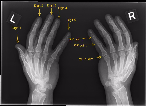
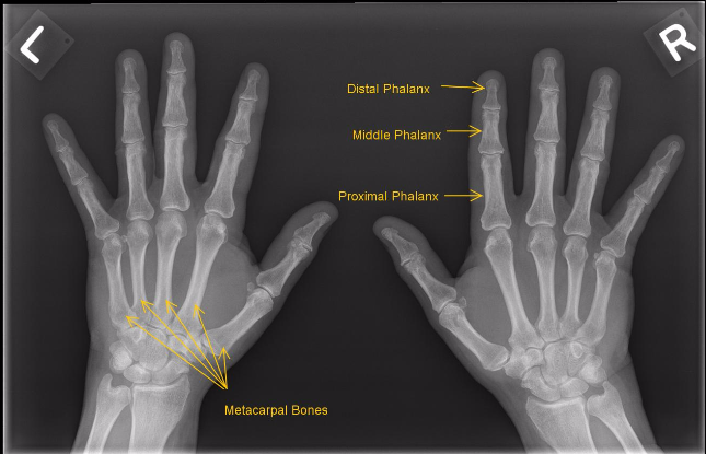
2. Wrist
The following is a normal, labelled, wrist x-ray:
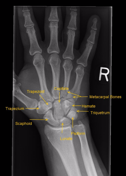
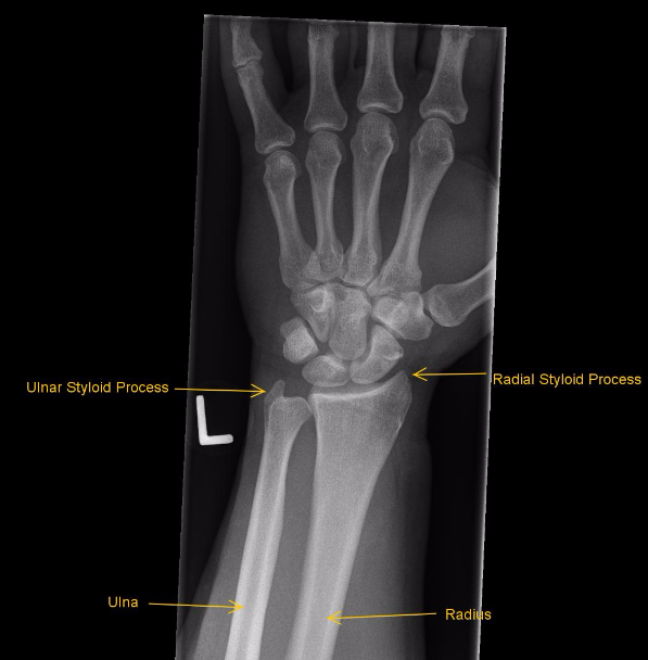
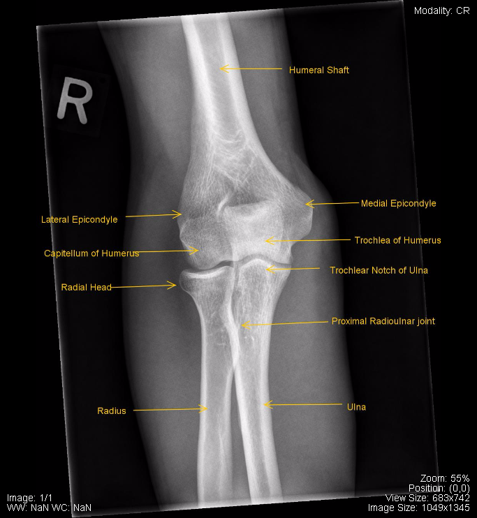
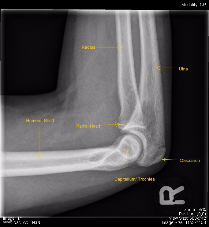
3. Elbow
The following is a normal, labelled, elbow x-ray:
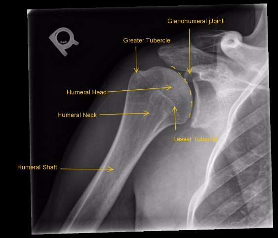
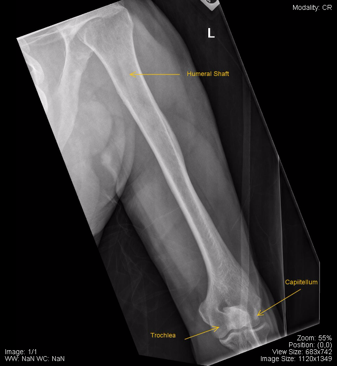
4. Shoulder
The following is a normal, labelled, shoulder x-ray:
5. Pelvis
The following is a normal, labelled, pelvis x-ray:
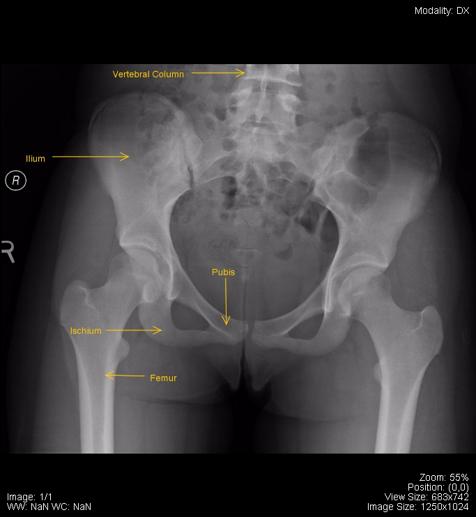
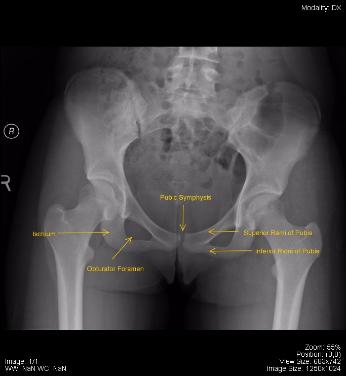
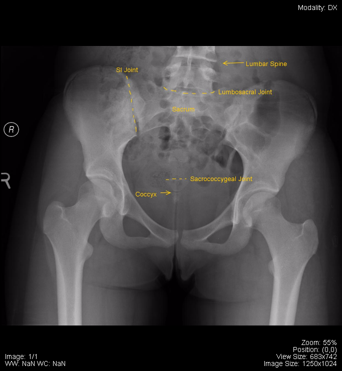
6. Knee
The following is a normal, labelled, knee x-ray:
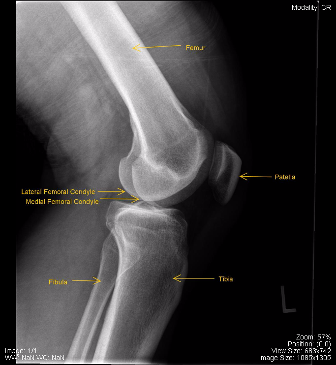
7. Ankle
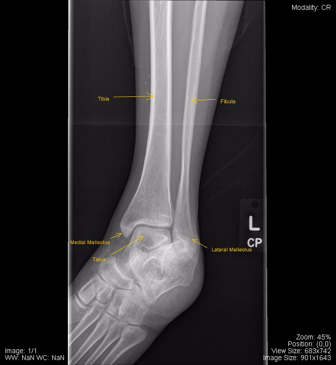
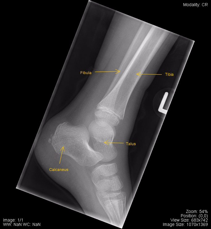
The following is a normal, labelled, ankle x-ray:
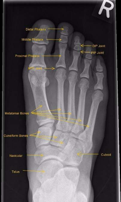
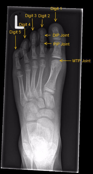
8. Foot
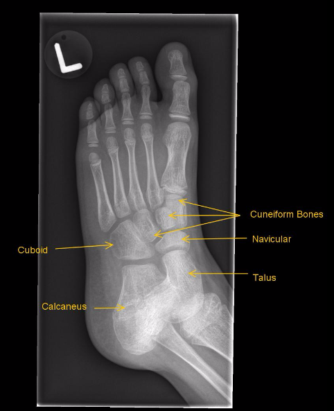
The following is a normal, labelled, foot x-ray:
Attributions
All figures in “Chapter 17: Musculoskeletal” by Dr. Brent Burbridge MD, FRCPC, University Medical Imaging Consultants, College of Medicine, University of Saskatchewan are used under a CC-BY-NC-SA 4.0 license.
