115
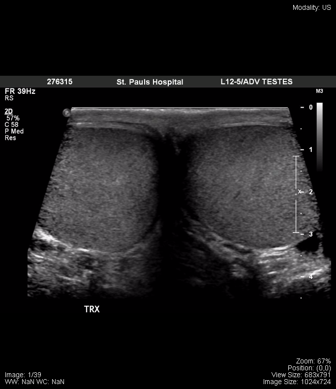
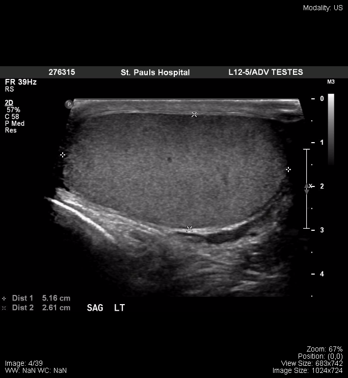
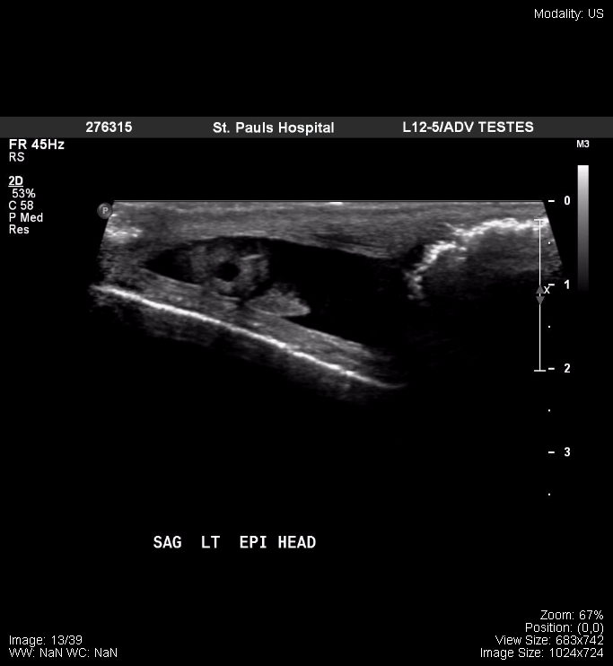
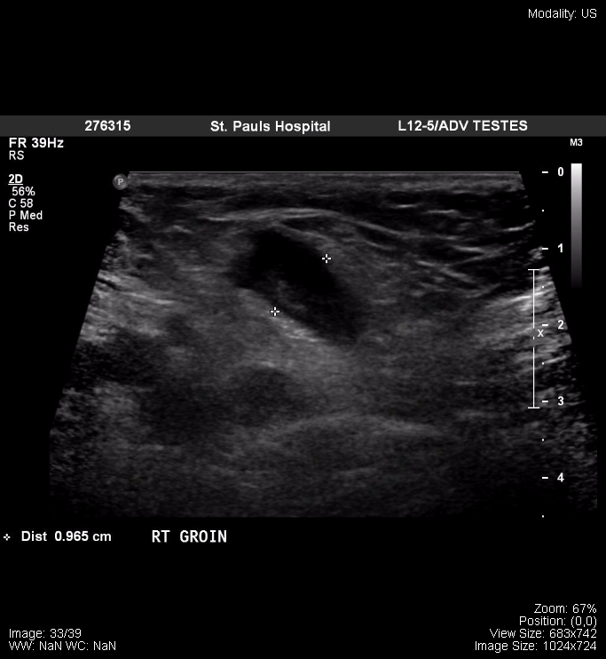
The following is a normal ultrasound of the testicles:
The following are from a normal female pelvic ultrasound:
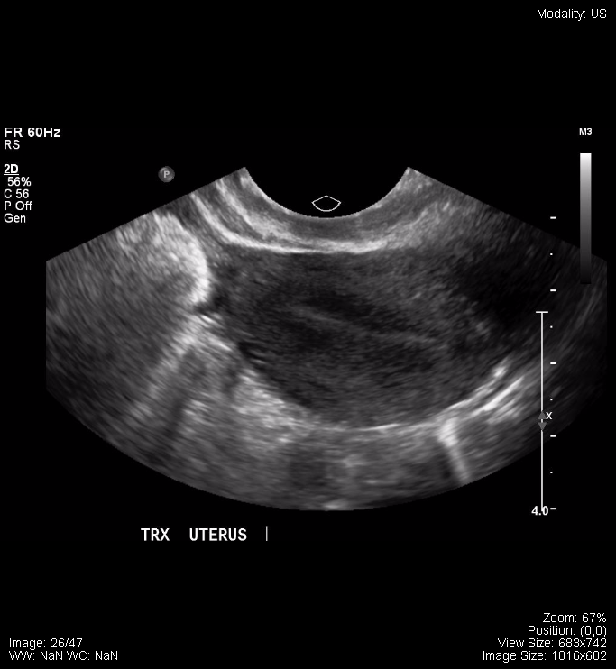
Transverse Uterus |
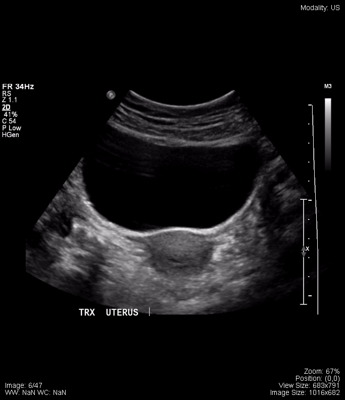
Transverse Uterus 2 |
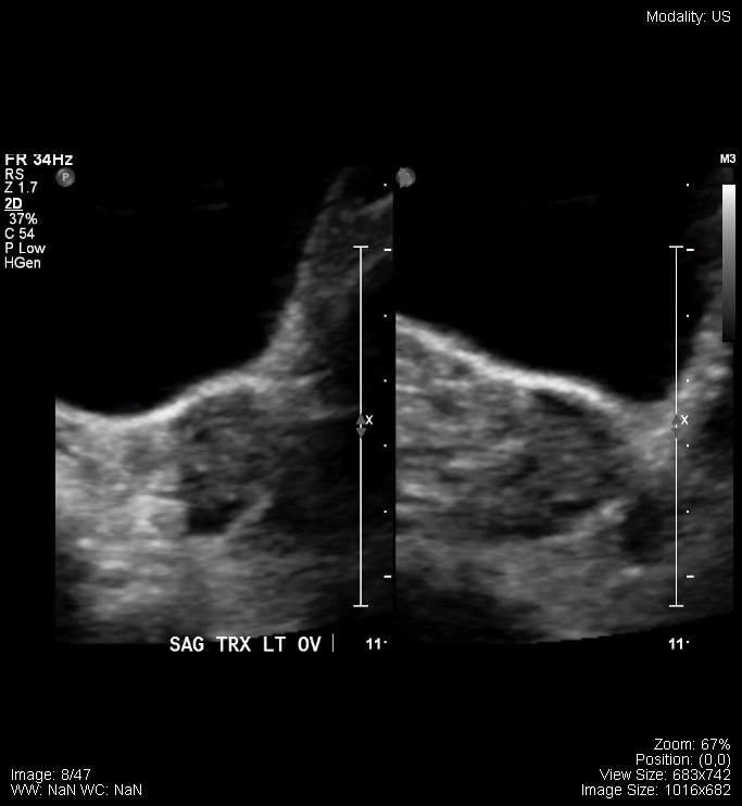
Sagittal Ovary |
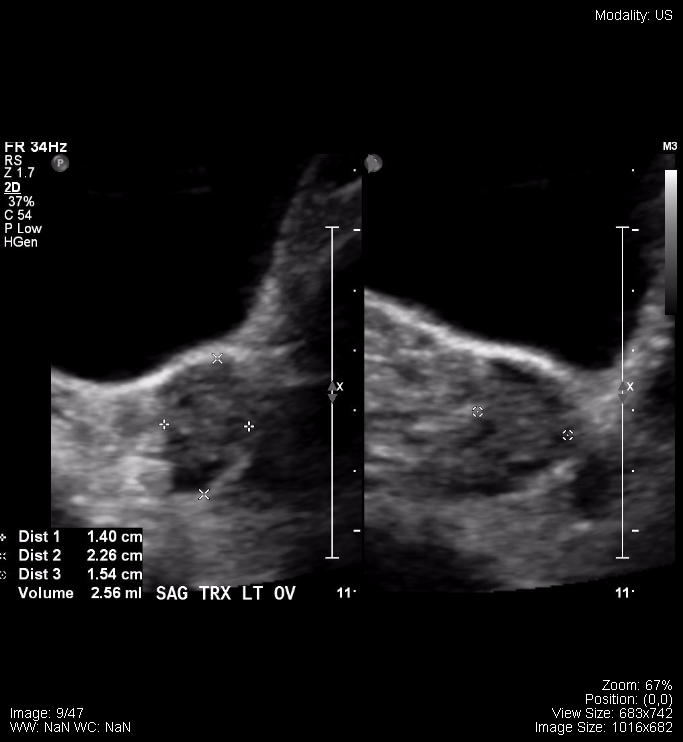
Sagittal Ovary, with measurements |
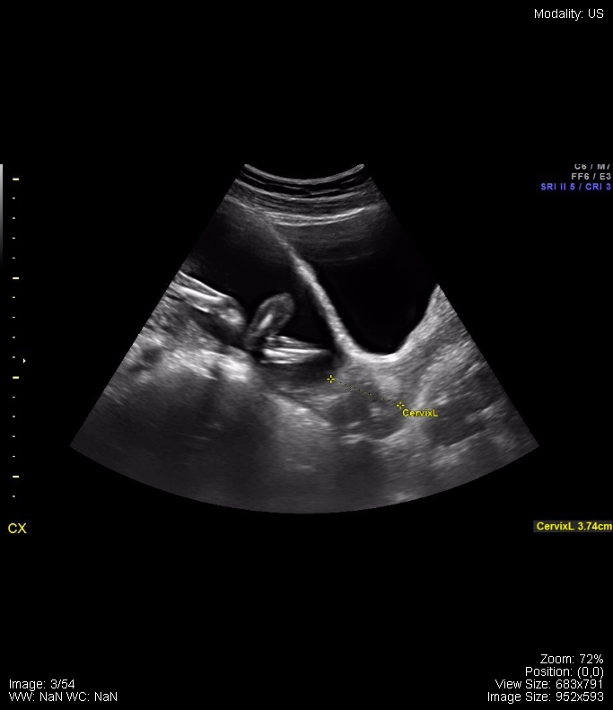
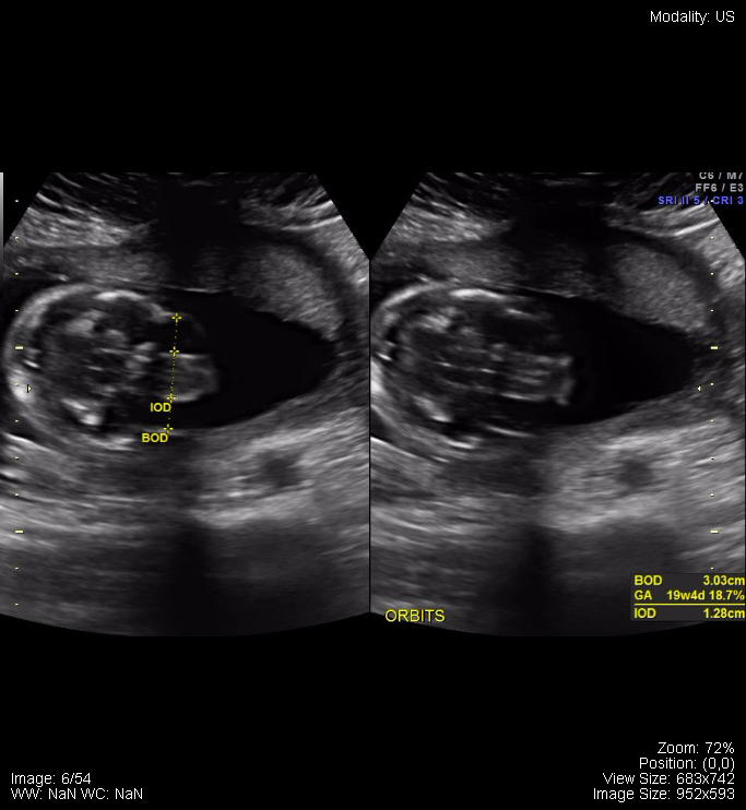
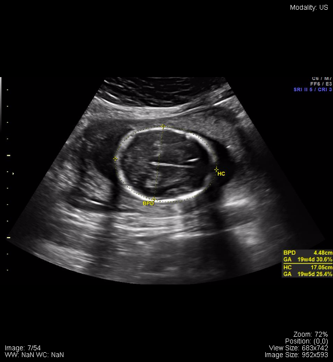
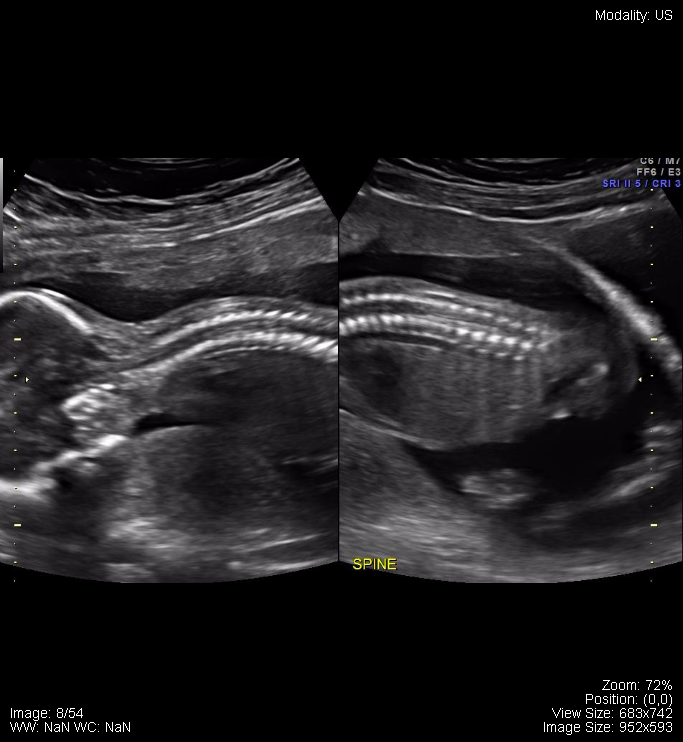
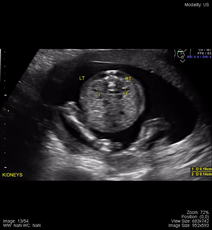
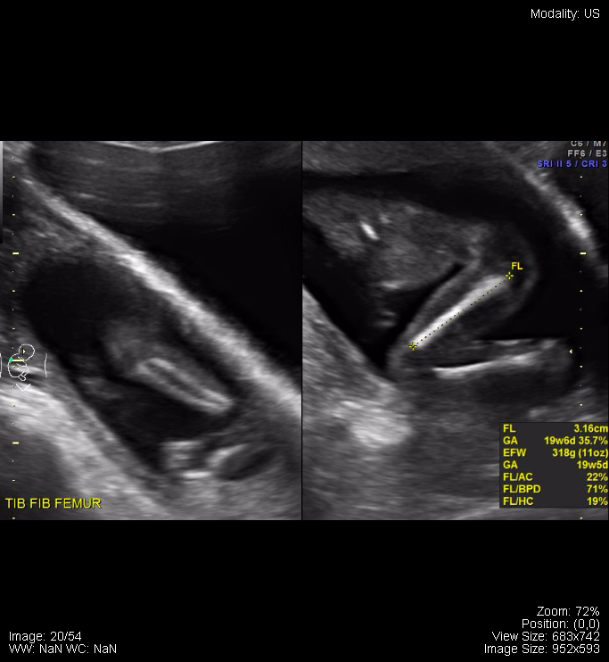
The following are samples from a normal pregnancy ultrasound:
Attributions
All figures in “Chapter 17: Pelvis” by Dr. Brent Burbridge MD, FRCPC, University Medical Imaging Consultants, College of Medicine, University of Saskatchewan is used under a CC-BY-NC-SA 4.0 license.
