13 Clubfoot
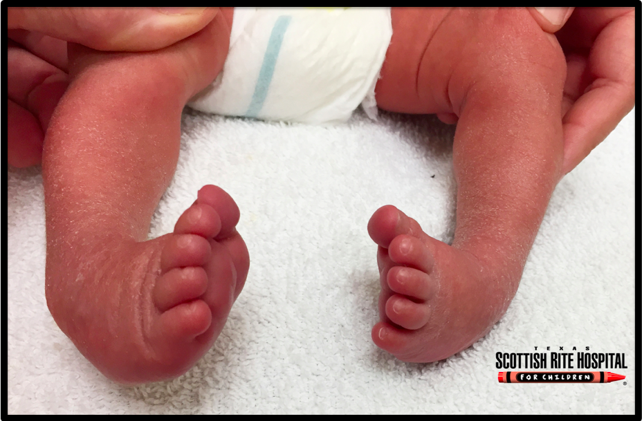
Talipes equinovarus, commonly known as “clubfoot,” is a congenital deformity of the foot (Figure 1). The condition is characterized by plantar flexion (equinus), inversion (varus), and an exaggerated arch (cavus) that may involve one or both feet. Taken together, these deformities cause the foot to resemble a club, hence the name. Clubfoot is often idiopathic and seen as an isolated birth defect, but it can also be caused by an underlying congenital disorder in approximately 20% of cases.
Structure and function
The foot can be divided into three regions: the hindfoot, the midfoot, and the forefoot.
The hindfoot consists of the talus and the calcaneus. These bones are joined at the subtalar joint to allow for inversion and eversion. The talus also articulates with the tibia and fibula at the ankle, to allow for dorsiflexion and plantarflexion.
The midfoot consists of the navicular, the cuboid, and the three cuneiform bones. The midfoot forms the arch of the foot and serves as a shock absorber.
The forefoot consists of the metatarsals and phalanges.
Clubfoot deformity primarily affects the hindfoot and midfoot. The pathological changes seen include an abnormally small calcaneus, talus, and navicular – and contracted ligaments between the hindfoot and midfoot. There is, accordingly, a plantarflexion deformity of the ankle (talocrural) joint, medial subluxation of the talonavicular and calcaneocubid joints and inversion and adduction of the calcaneus, navicular, and cuboid.
The deformity can extend distally to the forefoot where there can be plantar flexion (“equinus”) and adduction (“varus”) of the metatarsals and proximally to the calf, with atrophy, fibrosis and shortening of the muscle-tendon units of the posteromedial leg muscles seen.
There are many theories about the etiology of clubfoot but the definitive cause is still unknown. In the past, experts believed that the deformity was caused by the foot being stuck in the wrong position in the womb; today it is known that clubfoot is associated with multiple genetic abnormalities that influence the muscle contractile complex and bone development. For example, Gurnett et al (PMID: 18950742) found that abnormalities of the PITX gene, responsible for early limb development, has been associated with familial clubfoot.
Patient presentation
Clubfoot is a congenital deformity that is immediately apparent at birth. Some parents might know as early as the second trimester if the clubfoot is diagnosed via fetal ultrasound.
The affected foot is characteristically adducted (“varus”), plantarflexed (“equinus”), and possesses an exaggerated arch (“cavus”). Depending on the severity, the foot may be rigid or flexible. Half of all cases will involve both feet. In unilateral clubfoot, the involved foot, calf, and leg will be smaller and shorter than the unaffected side. Even after correction of the deformity, the foot, calf, and leg may have some residual problems including atrophied calf muscles and a smaller foot that loads more on the outside.
Despite its dramatic deformities, clubfoot is not painful, per se. However, if children begin to walk prior to successful correction of the deformity, they will bear weight on the dorsolateral aspect of the foot. This abnormal gait can cause focal loading on a small area and can be painful.
Objective evidence
Clubfoot is detectable via prenatal ultrasound in the second trimester. Early detection is important because it can prompt discussion with parents about treatment options and early screening for underlying neuromuscular diseases.
X-rays are not particularly useful in clubfoot evaluation because the neonatal bones are immature and poorly ossified. Radiographs are more useful for measuring progress in clubfoot treatment and long-term follow-up.
Epidemiology
Clubfoot occurs in 1 in 1000 births and affects males twice as frequently as females. Approximately 50% of all cases are bilateral and 25% have a positive family history of clubfoot. In the US, incidence ranges across ethnic groups from 0.4 in 1000 in the Chinese population to 7 in 1000 in the Polynesian population.
Most cases are idiopathic but about 20% are due to a genetic or chromosomal abnormality. The most common of these are disorders of the nervous system including myelomeningocele and arthrogryposis. These cases tend to be stiffer and more resistant to standard treatment than idiopathic cases.
Differential diagnosis
Metatarsus adductus is a congenital foot deformity that is superficially similar to clubfoot. Metatarsus adductus is characterized by the forefoot (metatarsus) pointing inward (adductus) with normal positing and mobility of the hindfoot, forming a “C” shape. The incidence of metatarsus adductus is approximately the same as clubfoot. Metatarsus adductus is distinguished from clubfoot by an examination of the hindfoot, which has normal mobility in metatarsus adductus but cannot be appropriately dorsiflexed or everted in the case of clubfoot.
Other congenital foot deformities include talipes calcaneovalgus (dorsiflexed and abducted foot) and vertical talus (rigid foot deformity similar to talipes calcaneovalgus). These are structurally and visually distinct from clubfoot.
Certain conditions, including spina bifida, arthrogryposis, and amniotic band syndrome can cause clubfoot. The evaluator should therefore pay close attention to the spine and motor function of the extremities.
Positional clubfoot is similar to clubfoot in that the foot is in equinus and varus. It is caused by a restrictive uterine environment that forces the baby’s feet into an abnormal position. However, it is different from classic clubfoot in that the foot and bony anatomy are completely normal.
Red flags
Although clubfoot is most often an idiopathic birth defect, some cases are secondary to underlying neuromuscular conditions such as spina bifida. Thus, the presence of clubfoot should be deemed a red flag, prompting a close diagnostic evaluation to exclude these conditions.
Treatment options and outcomes
Treatment options for clubfoot include serial casting, bracing, physical therapy, and surgery.
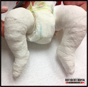
The standard of care for uncomplicated clubfoot deformities is the Ponseti Method, which involves repeated manipulation and casting (Figure 2) to guide the growth of the foot toward normal alignment. The child’s foot is manually stretched toward the correct position and a cast is then applied to maintain the correction. This process is repeated weekly over the course of 4-6 weeks.
Toward the end of the serial casting phase of treatment, most children will require a minor operation (percutaneous Achilles tendon tenotomy) to lengthen the Achilles tendon and release the foot from plantarflexion. Rarely, patients may also require a transfer of the tibialis anterior from its normal insertion on the first metatarsal to a new insertion on the third. This transfer will reduce supination of the foot with dorsiflexion. If the Ponseti Method of casting, bracing, and tenotomy is applied correctly, it will be successful in more than 95% of cases.
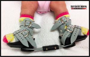
After casting, the child must wear a foot abduction brace (Figure 3) at night for a few years to maintain the correction. Without bracing, the deformity will likely recur because the muscles of the foot will pull it back into an abnormal position.
Recurrence (despite bracing) occurs in approximately 10% of patients, and most will respond to a repeated course of manipulation; a few will require additional surgery to prevent further relapse.
Although the Ponseti Method is highly effective in idiopathic clubfeet, some patients (especially those who present late or whose deformities are secondary to neuromuscular disease) will not respond to non-operative treatment and will require surgical intervention.
Surgery is normally performed at 9-12 months of age. The goal of surgery is to correct all the deformities in one operation. The surgical procedure varies from patient to patient but generally involves releasing all joint capsule contractures, lengthening any shortened muscle-tendon units, and realigning the bones of the foot.
Compared to the results of non-surgical methods, the long-term outcomes from surgery is associated with more pain, stiffness, deformity, and muscle weakness. However, it must be noted that surgery is reserved for tougher cases so this is not a true head-to-head comparison.
Even with optimal treatment, the corrected clubfoot will be functionally normal but structurally dissimilar from the unaffected foot. The affected foot is often smaller (requiring a different shoe size) and less mobile than the other foot. Additionally, the calf muscles in the affected leg may also be smaller. In some cases of clubfoot, the affected leg will stop developing before the other leg, leading to a significant difference in limb length. In these cases, it will be necessary to surgically lengthen the affected leg.
Risk factors and prevention
Honien et al (PMID: 11032161) found that family history of clubfoot is a major risk factor for developing clubfoot (OR = 6.52). Smoking exposure in utero is also associated with increased risk of clubfoot (OR = 1.34). This risk is dramatically increased in babies that already have a past family history of clubfoot (OR = 20.30). As a result, mothers with a positive family history of clubfoot should be strongly urged not to smoke while pregnant to minimize the risk of their child having clubfoot.
Clubfoot can be identified in the second trimester using fetal ultrasound, which can help prepare parents for the treatment course and provide an opportunity for counseling about genetic testing for associated conditions.
Miscellany
Figure-skater Kristi Yamaguchi, soccer star Mia Hamm, and NFL quarterback Troy Aikman were all born with clubfeet but still attained great success as professional athletes.
Clubfoot is uniformly treated at a very early age in the US, but in low-income countries it is not uncommon to see older children with neglected clubfoot (a deformity that was never treated), residual clubfoot (a deformity that was incompletely treated and retains aspects of the deformity years later), and recurrent clubfoot (a deformity that was completely treated but reverted back due to insufficient bracing). These forms of clubfoot can severely decrease quality of life by subjecting patients to physical disability and alienation.
Horses walk on their toes; hence a deformity of plantar flexion that places the toes in lone contact with the ground is called “equinus.”
Key terms
Clubfoot, Talipes equinovarus, Ponseti Method
Skills
Recognize the gross manifestations of clubfoot and differentiate these from other congenital foot deformities. Explain treatment options for clubfoot, including the Ponseti Method of serial casting, bracing, and tenotomy. Describe the possible outcomes of clubfoot treatment, including restoration of normal form and function, residual deformity, and recurrent deformity.
