12 Diabetic Foot Disorders
Diabetes is a medical condition characterized by many complications, including neuropathy and peripheral artery disease. Loss of sensation in neuropathy can lead to ulceration of the skin and accelerated degeneration of the joint (a condition known as Charcot arthropathy); and poor vascularity can impede healing.
Pathology and Pathophysiology
Neuropathy is the initiating event in the development of most diabetic foot ulcers and infections. However joint contractures, excess localized pressure and vascular disease also play a role in their formation. The development of peripheral neuropathy in individuals with diabetes is attributed to a complex interaction of glycosylated hemoglobin with arterioles of both central and peripheral nerves, leading to conduction defects in sensory, motor, and autonomic function.
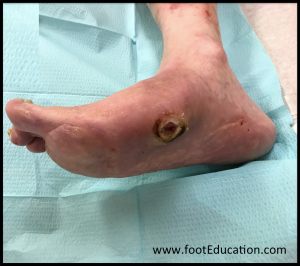
Prolonged exposure to even low pressure over a bony prominence (as may be seen with ill-fitting shoes) can cause skin breakdown. In patients without neuropathy this pressure will cause pain, and the person will shift his or her weight to limit the pressure. With a loss of such protective sensation, loading continues to the point that the skin breaks down and an ulcer forms (Figure 1). Without adequate off-loading of the area, ulcers can worsen leading to a local infection, progression to osteomyelitis, and eventual amputation in some patients.

Charcot arthropathy is characterized by joint fragmentation and subluxation caused by continued repetitive stress to the foot and ankle in patients with peripheral neuropathy. The condition is named after Jean Marie Charcot (1825-1893) who described the collapse of the bones of the foot in patients who had loss of sensation in the feet from tertiary syphilis. The three commonly affected locations in the foot and ankle are the tarsal-metatarsal joints (midfoot), the transverse tarsal joint (hindfoot just in front of the ankle), and the ankle joint.
According to the neurotraumatic theory, Charcot arthropathy occurs as a result of cumulative mechanical trauma. This is usually unrecognized microtrauma due to neuropathy, but can also occur from major trauma such as bumping the foot or twisting an ankle. The neurovascular theory holds that an autonomic peripheral neuropathy creates increased blow flow that leads to increased bone resorption and ligamentous weakening, leading to joint destruction. In a simplified version of this theory, increased blood flow “washes out” structural calcium from the bone, leading to localized osteopenia and structurally inferior bone that ultimately collapses. (A more detailed version posits that proinflammatory cytokines such as IL-1 and TNF alpha activate the RANK (Receptor Activator of Nuclear Factor κ B) ligand pathway, causing up-regulation of NF-κB (nuclear factor kappa-light-chain-enhancer of activated B cells) which leads to increased osteoclastogenesis and osteolysis.) Most likely, a combination of both theories contributes to this condition.
Patient presentation
Diabetic Foot Ulceration
Patients with diabetic foot ulcers present commonly with swelling, discharge or a foul-smelling odor from the affected foot. The ulcer can be a source of pain but many times, the foot will be painless due to the loss of sensation to the area. Ulcerations are often found incidentally by the patient or primary caregiver.
Physical examination should be focused on the location of the ulcer as well as the depth. The size of the ulcer, perfusion, loss of sensation, signs of inflammation, the presence of gangrenous tissue, and signs of infection such as exudate or odor should also be noted. Loss of sensation can be documented by lack of ability to detect a 5.07 Semmes-Weinstein monofilament
Typically, ulcers will occur over a prominent area such as the metatarsal heads, although any area of the foot that is subject to a concentrated, repetitive force is at risk for developing a diabetic ulceration.
Diabetic Foot Charcot Arthropathy
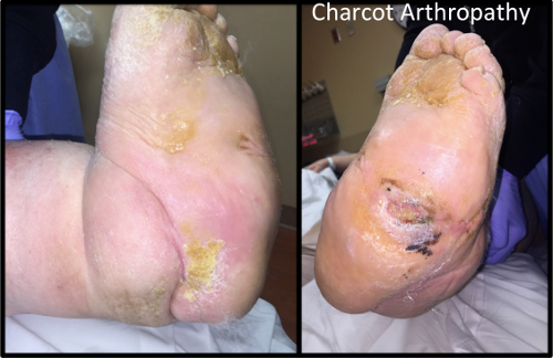
Patients with Charcot arthropathy of the foot often present with unilateral swelling, redness, and increased skin temperature around the midfoot or ankle (Figure 2). Most patients present in their fifth or sixth decade and have peripheral neuropathy. Many are obese and have had diabetes for many years.
Charcot arthropathy can be painful. However classically, the patient presents with a swollen, warm, and erythematous painless foot. Charcot arthropathy is commonly mistaken for an infection (e.g., osteomyelitis) especially when there is an ulcer present. One strategy for differentiating an acute Charcot foot from infection is to elevate the foot several minutes. In Charcot, the erythema will resolve, while in infection it does not.

Objective Evidence
Diabetic foot ulcers should be characterized objectively by location and severity. There are multiple classification systems to grade ulcers:
The Wagner system rates ulcers on a 0 to 6 scale, from “0: skin intact” to “6: ulcer with extensive foot gangrene.”
The Brodsky system considers both the extent to the ulcer (superficial, deep and exposed bone) as well as the extent of ischemia.
The vascular status of the foot should also be assessed objectively, noting the skin color and temperature, the quality of capillary refill and presence or absence of palpable pulses. The ankle-brachial index (ABI), namely the ratio of the blood pressure in the ankle relative to that in the arm can be assessed in the clinic, though calcification of the vessels may produce a falsely normal result. Values of 1.0-1.4 is a normal ABI result. Whereas, an ABI value of <0.5 represents severe arteriole disease.
Imaging Studies
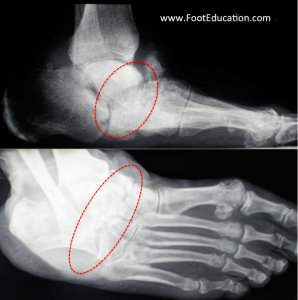
In patients presenting with an ulcer, standard weight-bearing anteroposterior, lateral, and oblique radiographs of the affected foot should be obtained. Weight-bearing x-rays should be performed whenever possible. These films can detect deformities of the foot in general, as well as bone changes such as periosteal reaction, bone fragmentation, joint subluxations, lucencies or osteolysis (Figure 3).
MRI may be helpful to determine the extent of bony and soft-tissue disruption, but MRI cannot differentiate between Charcot arthropathy and osteomyelitis with high specificity. Occasionally, a bone scan is indicated, although very often the results of the bone scan will not change the management and may not reveal any information that cannot be obtained from a detailed physical examination.

In cases of suspected infection, aerobic and anaerobic cultures of the deep wound should be obtained to help direct antibiotic therapy. Osteomyelitis is present in approximately 70% of ulcers that extend down to bone.
Laboratory tests such as white blood cell counts, sedimentation rate, and C-reactive protein levels may be used to establish the diagnosis of osteomyelitis, though a bone biopsy is the most specific method.
CT scans can be helpful to look at a more detailed picture of the collapse, but is not often necessary unless surgery is planned.
Epidemiology
Diabetic foot disorders are a common subset of pathologies seen in patients with diabetes mellitus. The prevalence of diabetes in the US is approximately 10% of the adult population, and in turn, approximately 10% of those patients will develop a lower extremity ulcer during the course of their disease and about 1% will develop Charcot arthropathy.
The rate of lower extremity amputations is at least 50% higher in men versus women. Mexican (Hispanic) Americans, Native Americans, and African Americans each have at least a 1.5- to 2-fold greater risk for diabetes related amputations than age-matched diabetic Caucasians.
Differential diagnosis
When a patient presents with a foot ulcer, it is essential to determine its cause. Thus, it is critical to evaluate foot ankle alignment and deformity, shoewear, assess for previous ulcerations, medical comorbidities, level of diabetic control and monitoring, and tobacco and intravenous drug use.
The presence of sensory neuropathy should be assessed. This can be done using a Semmes-Weinstein 5.07 monofilament to exert a consistent force (If the wire bows into a C shape when pressed against the skin for 1 second, 10g of force is applied). Sensation observed using the monofilament indicates protective sensation is maintained.
There are many potential causes for peripheral neuropathy besides diabetes. These can include: alcoholism, vitamin B1 and B12 deficiencies, and heavy metal poisoning. A focused history and appropriate laboratory studies can rule out other causes besides diabetes.
In the absence of diabetes, foot ulcers can be caused by atherosclerosis involving the lower extremities, vascular lesions, and even severe Raynaud’s phenomenon (vasospastic attacks in digits). A squamous cell carcinoma may also be responsible.
Charcot arthropathy may have a similar presentation as gout, cellulitis, osteomyelitis, and septic arthritis. Diabetic neuropathy is the most common cause of Charcot arthropathy, but other less common causes include spina bifida, cerebral palsy, meningomyelocele, syringomyelia, leprosy, alcohol abuse, and advanced syphilis.
Red flags
In all patients with diabetes, breaks in the skin are a red flag, as these can be a portal of entry for bacteria and subsequent infection. Any sign of increased pressure on focal areas of the foot (such as erythema or skin changes), is also considered a red flag and should be treated expeditiously.
Treatment options and Outcomes
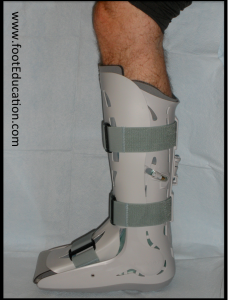
The primary goal for treatment of diabetic ulcers is to get the wound to close and prevent future ulcerations. Off-loading of the affected area is an important first step in treatment of ulcers, commonly done using a total contact cast or removable diabetic walker boot (Figure 4). These allow healing by redistributing plantar pressure, decreasing shear stresses, and reducing swelling.

Total contact casting has been shown to significantly increase the healing rate of neuropathic plantar foot ulcers at 12 weeks compared to removable cast walkers and half-shoes so it is the preferred method of treatment. It can take anywhere from 6 weeks to a year for an ulcer to heal depending on the size, depth, and duration of the ulcer. A patient should not return to unmodified shoes until the ulcer has completely healed. Unfortunately, the highest risk factor for a second ulcer is having an initial diabetic foot ulcer even after successful healing. As a result, life long careful evaluation and protection is needed in these patients.
Treatment of the ulceration may involve surgical debridement of the callus or necrotic tissue with scalpels and curved scissors. Usually, this can be done in the clinic setting, although sometimes it may need to be done in the operating room if the infection involves the bone and there is some need to remove part of the infected bone. Keeping the wound moist without excess fluids can accelerate re-epithelialization of the wound. Various topical agents and dressings may expedite healing. Negative pressure wound therapy, hyperbaric oxygen therapy, ultrasonic therapy, and electric stimulation are all being tested for treatment of diabetic foot ulcers.
The vascular status of the affected extremity is critical in determining the healing potential of foot ulcers and the need for possible surgical intervention. More than 60% of diabetic foot ulcers have decreased arterial blood flow due to concurrent peripheral vascular disease. Formal vascular studies are often needed to determine status of the posterior tibial and dorsalis pedis arteries and the need for surgical or endovascular procedures to perfuse the foot.
Equinus contractures cause plantarflexion and thus increased forefoot pressures. Therefore a patient with an equinus contracture may benefit from a tendon release. A percutaneous Achilles tendon lengthening prior to total contact casting markedly decreases the rate of recurrence of plantar ulcers.
Diabetic foot ulcers if not treated (or if they do not respond to treatment) can lead to gangrene, abscesses, and osteomyelitis. Amputation of the lower extremity may be needed. An amputation is a marker for severe diabetic disease, as the 5-year mortality rate following an amputation in diabetic patients is approximately 66%.
Charcot arthropathy can be difficult to treat. The overall goal of Charcot arthropathy treatment is to maintain a braceable and plantigrade foot to allow ambulation and prevent ulcerations over bony prominences. The best approach is early detection and prevention. Early treatment consists of a period of non-weight-bearing or limited weight-bearing in either a total contact cast or a diabetic removable boot. Swelling and redness will usually resolve or improve with elevation and immobilization. Later in the process, bone consolidation will occur and the added stability can allow the patient to improve his mobility and weight-bearing. Rolling knee walkers can help keep the weight off the bad foot while allowing patients to be mobile. The treatment of Charcot arthropathy can take up to 6-12 months or more for the involved joints to stabilize. Even after consolidation of the Charcot process, gross bony deformity may be present that could put the patient at risk for developing an ulcer over those areas.
Some studies suggest that inhibitors of osteoclasts (as would be used to treat osteoporosis) may be helpful in treating Charcot arthropathy, by limiting osteolysis, though good clinical results have not yet been attained. Electrical bone growth stimulation to promote rapid healing of fractures has been suggested as a supplement to the treatment of acute Charcot arthropathy. Similar to bisphosphonate therapy, there is no conclusive data for its efficacy.
Surgery may be recommended as a treatment if a severe deformity has occurred that results in repeated ulcerations, non-plantigrade foot, or a foot or ankle that has become unstable and cannot be corrected through immobilization and off-loading. Surgery ranges from exostectomy (removal of prominent bone) to reconstruction of the foot including fusion of the unstable joints after the deformity has been corrected. The goal of surgery is a foot and ankle that is stable for weight-bearing, plantigrade, and can accommodate a diabetic shoe or brace. There are many risks for patients with Charcot arthropathy undergoing surgery such as infection, wound complications, non-union, delayed union, and hardware failure.
Risk factors and prevention
Because diabetic foot disorders are caused by diabetes, all factors that increase the risk of the underlying condition increase the risk of resultant foot problems. Among patients with diabetes, cigarette smoking and poor glycemic control is associated with more diabetic neuropathy, peripheral artery disease, and in turn more foot disease. In addition, inappropriate footwear and poor toenail grooming increases the risk for ulcerations.
Previous ulcerations or amputations are associated with a higher risk of developing another ulcer.
A tight calf muscle (equinus contracture) causes the patient to place more weight on the forefoot and increases the risk of ulcers there.
Miscellany
There are reports of high complication rates associated with simple ankle fractures in the population of patients with diabetes, especially those with peripheral neuropathy. Many patients who present with Charcot arthropathy of the ankle initiated their disease process with an ankle fracture. Many experts recommend augmented internal fixation with prolonged non-weight-bearing to treat diabetic patients who have suffered an ankle fractures.
Key terms
Diabetic foot ulcer, Charcot arthropathy, neurotraumatic theory, neurovascular theory, neuropathy, infection, amputation
Skills
Thorough physical exam of feet in diabetic population. Classify ulcers to treat accordingly. Distinguish infection from Charcot arthropathy. Provide basic patient education information to diabetic patients.
