14 Epicondylitis of the Elbow
Epicondylitis of the elbow is a misnomer because it is neither primarily a disease of the epicondyle, nor is it exclusively inflammatory (as the suffix “itis” would suggest).
Instead, epicondylitis is a condition of degenerative tendinopathy within the wrist extensor tendon (as in lateral epicondylitis) or flexor tendon (as in medial epicondylitis), tendons which originate at the elbow.
The common term “tennis elbow” refers to lateral epicondylitis which affects the origin of the wrist extensor muscles, while the term “golfer’s elbow” refers to medial epicondylitis which affects the origin of the flexor/pronator muscles.
Epicondylitis is an overuse injury in which the rate of tendon damage exceeds the rate of tendon repair. This disequilibrium causes pain at the elbow and, ultimately, leads to impaired function – primarily in tasks that involve a power grip and require a stable wrist joint.
Structure and function
Epicondylitis is an overuse injury of the wrist extensor and flexor tendons including the extensor carpi radialis brevis, flexor carpi radialis, and pronator teres.
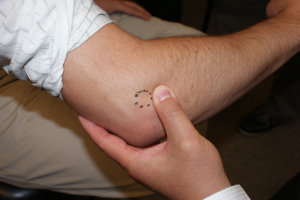
These muscles originate from the humerus (Figure 1) and cross the elbow joint, but their main action is at the wrist. They act at the wrist via concentric contraction (that is, shortening and changing the position of the joint), but they are also important in stabilizing the hand, wrist, and fingers, either isometrically (not moving at all) or via an eccentric contraction (actually lengthening while offering a resisting force). It is these eccentric contractions, defined as muscle contraction in one direction while another force elongates the muscle in the opposite direction, that are likely to cause microscopic tears within the tendon itself and its origin.
Lateral epicondylitis is commonly called “tennis elbow” because it is a common injury sustained by in tennis players hitting an off-center single arm backhand. When hitting the ball, the player’s wrist extensors are active, to hold the wrist in position. When the tennis ball hits the racket, it applies a force on the wrist in the direction of flexion. This force pulls on the contracting extensor muscles and can create microscopic tears in the fibers that originate from the lateral epicondyle.
On the lateral side the primary focus of damage is within the extensor carpi radialis brevis; on the medial side it is within the flexor carpi radialis and pronator teres origin.
The histology of epicondylitis has been described as “angiofibroblastic hyperplasia”: namely, the presence of fibroblasts and vascular tissue, along with degenerative and torn tendon fibers. (Note that pathological specimens are obtained at the time of surgery, and surgery is, of course, reserved for only severe cases. Thus, as far as we know, “angiofibroblastic hyperplasia” may be present only in end-stage disease.)
Patient presentation
Patients with epicondylitis present with a chief complaint of pain in the elbow on the affected side, especially with grasping.
Patients with epicondylitis often volunteer a history of repetitive forearm use, including sports involving rackets or bats, but also occupational activities such as working with tools.
In early phases of the condition, patients experience a minor ache after extensive activity. As the disease progresses, the signs and symptoms of epicondylitis are not only more severe, but may also be present without recent activity.
The presence of focal epicondylar pain and tenderness that is worse with provocation (stress on the wrist) is key to the physical diagnosis of epicondylitis.
Patients with epicondylitis complain of pain localized to the elbow; however, the best physical examination maneuver to provoke pain involves manipulation of the wrist, as the affected muscles are primarily used to stabilize the wrist. This is because while the lateral structures (especially the extensor carpi radialis brevis) are active wrist extensors, they also stabilize the wrist by resisting wrist flexion (with so-called eccentric or isometric contraction).
The importance of a stabilizing force against wrist flexion is seen dramatically in the mechanics of a tennis backhand stroke. This action requires that the wrist be held, fixed, in a neutral position despite the impulse of the ball, which, unopposed, would flex the wrist. A similar stabilizing force is employed on a routine basis in everyday grasping. Recall that the muscles of finger flexion originate on the volar surface of the forearm and cross the wrist on their journey to the phalanges. Unless these muscles are opposed by a countervailing wrist extension force, their firing will flex the wrist as well as the fingers. Put simply, in order to flex the fingers without flexing the wrist, the wrist extensor muscles must act. Repetitive grasping with the fingers therefore requires repetitive use of the wrist extensors, even if no wrist movement is produced.
Lateral epicondylitis can be caused by routine grasping tasks because they require the use of wrist extensors.
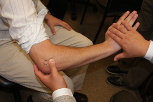
Symptoms of epicondylitis can be provoked on physical exam by asking the patient to hold his or her hand in maximal wrist extension, with the examiner acting against the patient to flex the wrist (Figure 2). Pain at the origin of the extensor muscles in the vicinity of the epicondyle is diagnostic. Note that the point of maximal tenderness is not at the epicondyle but is, rather, slightly distal, within the tendon itself.
objective Evidence
Epicondylitis is, for the most part, a clinical diagnosis: tenderness, especially provocative tenderness, in a patient with an apt history, makes the diagnosis.
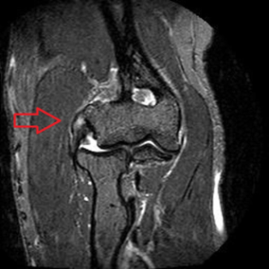
An MRI (Figure 3) may show abnormal signal within the tendon at its origin, typically signifying chronic granulation tissue at the injured site. This test may also be used to exclude other conditions.
Similarly, an EMG may be helpful to exclude compression neuropathy. This is not 100% reliable, however. Plain radiographs may help exclude arthritis or loose bodies.
Epidemiology
The exact incidence of epicondylitis is not known, as there are mild forms of the condition that do not present for medical attention.
In general, it is safe to assume that epicondylitis is very common and should be high on the differential diagnosis of elbow pain.
Clinical experience suggests that medial epicondylitis is far less common than lateral epicondylitis. Moreover, the medial side of the elbow has other structures that may be the source of pain, e.g. the medial collateral ligament and the ulnar nerve. Medial epicondylitis therefore perhaps deserves a less prominent place on the “default” list of causes of medial elbow pain.
The incidence of epicondylitis is highest in the fourth and fifth decades of life. It can be present in both older and younger patients, but like most tendinopathies, epicondylitis is mostly prevalent in middle age.
Differential diagnosis
Cubital tunnel syndrome, i.e., a compression neuropathy of the ulnar nerve at the elbow, is commonly seen in association with medial epicondylitis, but also may be seen on its own.
Entrapment of the radial nerve in the arcade of Frohse may mimic lateral epicondylitis (this can be diagnosed by EMG, clinical examination for radial tunnel syndrome, or diagnostic injection).
Cervical radiculopathy can also present as elbow pain: C5 or C6 nerve roots for lateral elbow pain; C8 or T1 nerve roots for medial elbow pain.
In addition, primary pathology within the joints, including arthritis, loose bodies and synovitis, may mimic the symptoms.
Treatment options and outcomes
As epicondylitis is an overuse injury, it is best treated initially by a period of relative rest. Avoiding provocative activities, assuming they can be identified and omitted, may be helpful.
So-called “counterforce bracing” may also be helpful. This bracing involves a strap worn distal to the elbow that helps redirect force away from the origin (please see “Miscellany”, below).
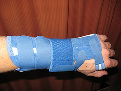
Splinting the wrist may also be helpful for this elbow pain (Figure 4). Note that the finger flexor tendons of course cross the wrist as well. As such, active finger flexion will flex the wrist unless there is a so-called isometric contraction of the wrist extensors. That is, wrist extension is needed to oppose the flexion force. By stabilizing the wrist with a brace, the wrist extensor muscles can rest even while grasping.
Physical therapy, especially modalities such as ultrasound and tendon glides, may be helpful.
Cortisone injections are also an accepted mode of treatment. Cortisone’s precise mechanism of action is not known with certainty, however, it is relatively effective at minimizing pain in the short term.
Surgical treatment of epicondylitis is reserved for severe cases. Lateral epicondylitis can be treated by excision of the chronic granulation tissue of the extensor carpi radialis brevis and subsequent repair with appropriate immobilization while the tendon heals.
In general, patients with epicondylitis do well – especially if they interpret the pain as a signal to stop doing what is causing the pain. Pain, to the extent that it enforces a period of rest, promotes recuperation.
Controlled studies documenting the natural history of treated and untreated epicondylitis, however, have not been performed.
Risk factors
Epicondylitis is an overuse injury that often occurs in the setting of aging. There is not much that can be done to prevent aging, of course. Patients who are entering the fourth and fifth decade of life, however, may need to modulate their activities accordingly.
It is also worth noting that poor form and technique – and the use of poorly maintained or adjusted equipment – may increase the risk of epicondylitis. For example, playing tennis with a racket poorly matched to one’s skill and size may help inflict damage.
Miscellany
The concept of counterforce bracing (Figures 5 and 6) is similar to the capo used in a guitar, which effectively changes the “origin point” of the guitar string.
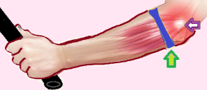

Just as a dog can have lice and fleas, a patient can have two simultaneous conditions, e.g. lateral and medial epicondylitis. This combination of tennis and golfer’s elbow has been described by Nirschl as “country club elbow.”
Key terms
epicondylitis, angiofibroblastic hyperplasia
Skills
Perform physical examination to diagnose epicondylitis.
