4 Scapulothoracic Disorders
The scapulothoracic joint is the articulation between the scapula and the thorax. It is not a true joint, but rather the broad contact between the inner surface of the scapula and the rib cage. The scapula is able to slide relative to the rib cage to allow for elevation and depression, along with protraction/retraction, rotation and shoulder abduction. When the arm moves relative to the body, approximately two-thirds of this motion is at the glenohumeral joint and one-third at the scapulothoracic joint. Three relatively common conditions are seen at the scapulothoracic joint. In scapular winging, the serratus anterior or trapezius fail to stabilize the scapula. (The muscle weakness is in turn caused by dysfunction of the long thoracic or spinal accessory nerves, respectively.) Snapping scapula syndrome is characterized by a grating sensation, often from overuse and scapulothoracic bursitis. With so-called scapulothoracic dyskinesis, abnormal scapula motion disrupts the normal glenohumeral joint mechanics and leads to shoulder pain. It is also possible to have a traumatic disruption of the scapulothoracic joint with high energy trauma. This is known as a scapulothoracic dissociation.
Structure and Function
The scapulothoracic joint is not a true synovial articulation, but rather is the broad contact between the scapula and the thorax. The only ligamentous connection of the scapula to the body is via the clavicle: the medial clavicle is attached to the sternum, whereas its lateral end is attached to the scapula at the acromion.
There are 17 muscles that connect the scapula to the arm and the torso. Scapula-to-arm muscles include the four muscles of the rotator cuff, the biceps, the triceps, the coracobrachialis, the deltoid and part of the latissimus dorsi. Scapula-to-torso muscles include the rhomboids, the serratus anterior, and the trapezius. The serratus anterior muscle originates from the ribs and inserts on the medial scapular border; the trapezius originates from the vertebrae and inserts along the scapular spine. Dysfunction of the scapula-to-torso muscles can produce abnormal scapulothoracic motion and thereby disrupt normal glenohumeral joint mechanics. This scapular abnormality can thus be the cause of shoulder pain.
The scapula is internally rotated about 30° in the coronal plane and is slightly tipped anteriorly in the sagittal plane.
The scapula is able to move in multiple directions. These motions include elevation (as in shrugging the shoulders) and depression, protraction (moving the scapula laterally and anteriorly along the chest wall) and retraction (moving the scapula medially), and rotation.

Motion of the scapula affects the position of the glenoid fossa, and in turn influences shoulder function. If the scapula does not move properly, the glenoid fossa will not be oriented for optimal contact with the humeral head. Also, about one-third of the arc of shoulder motion is at the scapulothoracic joint. As such, full range of motion of the shoulder requires normal scapular motion.
Motion of the scapula when motion is not desired can also lead to dysfunction; a lack of scapulothoracic stability can impede arm function as well.
The scapulothoracic bursae (plural) facilitate the gliding of the scapula on the chest wall.
Scapulothoracic bursitis is caused by poor scapular mechanics or a structural lesion (such as an osteochondroma of the scapula). Chronic inflammation leads to fibrosis, which can produce a snapping sensation. Snapping can also be the result of masses such as an osteochondroma or rib cage abnormalities (as may be seen with scoliosis).
Patient Presentation
A patient with a scapulothoracic disorder may present with shoulder pain, especially with overhead activities, or with focal scapular complaints such as audible or palpable crepitus.
As always, a thorough physical examination is needed, starting with posture. Scoliosis or kyphosis that may contribute to bony incongruity should be noted. Active and passive glenohumeral motion should be evaluated, assessing scapular motion and the presence of crepitus, if any. Scapulothoracic crepitus is present in about of one-third of individuals without symptoms. Thus, this finding must be correlated to complaints.
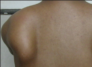
Asking the patient to perform a “wall pushup” can reveal medial winging. The etiology of medial scapular winging is dysfunction of the serratus anterior or the long thoracic nerve that supplies it. There is weakness in protraction of the scapula, and thus the rhomboids and trapezius work unopposed.

Lateral scapular winging is caused by dysfunction of the trapezius or the spinal accessory nerve that supplies it. In that setting, the serratus anterior works unopposed. This is usually an iatrogenic injury during neck surgery. The scapular flip sign can detect a spinal accessory nerve palsy. This sign is present when the medial scapular border “flips” or lifts from the thoracic wall during resisted shoulder external rotation with the arm at the side.
“Pseudo-winging” of the scapula can be seen without neuromuscular disease. One such instance would be when a tumor (e.g. osteochondroma) pushes the scapula off the posterior aspect of the chest wall. Psuedo-winging of the scapula can also be seen when patients learn to avoid painful positions by moving their scapula abnormally.
Objective Evidence
Plain x-rays will help detect osseous abnormalities. Snapping may be due to a mass. If a mass is seen on x-ray, then CT scans can be used for further definition. MRI may identify bursitis and soft-tissue masses.
Plain x-rays can also find abnormalities that upset normal scapular motion, including AC arthrosis, glenohumeral arthritis, thoracic kyphosis or scoliosis, and clavicular shortening (often due to fracture non-union or malunion).
Diagnostic injections of local anesthetic can identify or confirm symptomatic bursitis.
An EMG may confirm the presence of a nerve injury that is responsible for winging.
Epidemiology
Scapulothoracic disorders are thought to be rare, but may be under-diagnosed. That is, shoulder pain may be incorrectly attributed to primary disorders of the glenohumeral joint, when in fact scapulothoracic dysfunction is the true cause.
Scapulothoracic dyskinesis is usually found in athletes.
Differential Diagnosis
If snapping is present, the differential diagnosis is whether a mass is present or not. Imaging usually resolves this.
Scapular dyskinesis is often a diagnosis considered for a patient with shoulder pain that is not responding to treatment. Scapular dyskinesis is rarely the diagnosis considered first. Rather, it is a diagnosis made after all other diagnoses have been considered and rejected – a so-called “diagnosis of exclusion”. It should be suspected in any patient with non-specific shoulder pain especially if the medial scapula is prominent at rest.
Red Flags
Winging is usually the product of nerve dysfunction, thus its presence should prompt a close examination to exclude any other neurological finding.
Two other points, not quite “red flags” but related to the general topic of vigilance, are worthy of notice. First, it is easy to miss a scapulothoracic diagnosis if the patient is insufficiently disrobed (or if the scapula is not palpated in lieu of direct visualization. As such, a shoulder examination with a fully clothed patient is a red flag for a possibly missed diagnosis. Second, the lung is near the scapula. Thus, if one is performing a bursal injection, it is critical to stay parallel to the undersurface of the scapula to avoid giving the patient a pneumothorax.
Treatment Options and Outcomes
Non-surgical treatment could be utilized as the first line of treatment for patients with snapping scapula syndrome. Often, a non-surgical approach will alleviate patient’s symptoms, especially patients with a problem with soft tissues. One to two cortisone injections can reduce symptoms associated with bursitis.
If there is a symptomatic mass, removal of the mass is indicated.
Arthroscopic bursectomy can be employed for patients who have a confirmed diagnosis (such as a positive response to a diagnostic injection and bursitis on MRI) who fail non-operative treatment.
The indications for surgery for those patients without an identified bursa or mass are less well defined. Resection of the superomedial or inferomedial angle of the scapula should be reserved for patients who localize their pain to precisely those areas and report adequate (albeit temporary) relief with an injection.
The mainstay of scapulothoracic dyskinesis is physical therapy, though if this condition is caused by a primary abnormality elsewhere, such as a clavicular or AC joint abnormality, physical therapy is unlikely to produce more than frustration. The underlying abnormality must be addressed.
For scapular conditions in general, physical therapy is focused on relative rest, increasing core muscle strength and balance, and improving posture.
Winging is usually monitored, though in some cases exploration of the nerve with neurolysis and possible repair is indicated. Winging that does not improve at all may require a muscle transfer or scapulothoracic fusion.
Snapping usually resolves over time. Snapping due to masses respond very well to resection. Because there can be a significant psychological overlay, surgery for snapping scapula in the absence of a mass lesion produces unpredictable and often disappointing results.
Scapulothoracic dyskinesis due to overuse also responds well to rest and rehabilitation. The outcomes in patients with scapulothoracic dyskinesis due to underlying abnormalities are governed by the response to treatment of those conditions.
Medial winging usually resolves over time as well, as it is caused by chronic compression. Lateral winging has a worse prognosis, as it usually caused by overt trauma to the nerves.
Risk Factors and Prevention
Scapulothoracic dysfunction can be exacerbated by overuse. Recreational activities such as baseball pitching or occupations with repetitive overhead actions (e.g. carpentry) are also risk factors for this condition.
Miscellany
Scapulothoracic dyskinesis can be thought of as “SICK” scapula, which can remind the examiner of three associated findings: Scapular malposition, Inferior medial border prominence, Coracoid pain and malposition, leading to dysKinesis of scapular movement.
Key Terms
Snapping scapula, scapular winging, scapulothoracic dyskinesis
Skills
Recognize scapular winging on examination.
