Levels of Organization
8 Higher-Order Structures
Introduction
At this point, we have learned that all matter is made of atoms and that atoms combine into molecules through chemical bonding. In living systems such as human beings, smaller molecules are assembled into huge macromolecules by machinery inside our cells. We’ve also discussed metabolism, the anabolic and catabolic reactions that allow life. The energy to power these reactions is derived from cellular respiration, a complex chain of reactions that allows cells to “recharge their batteries.” This involves extracting chemical bond energy from fuel molecules such as glucose and using it to reassemble ATP from ADP and inorganic phosphate. You’ve had a chance to investigate cells, the smallest unit of organization that has the emergent property we call life. Eukaryotic cells contain compartments that allow for much greater complexity, setting the stage for higher-order structures and multicellular life. You’ve already learned quite a bit!
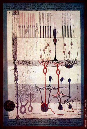
We now turn our attention to higher levels of organization: tissues, organs, organ systems, organisms, and populations. Keep in mind that all of these levels are built by combining elements from the level below and that each level has new and distinct properties. Figure 8.1 shows a variety of nervous cells (neurons) found in the light-sensing part of our eyes, called the retina. Nervous tissue is one of the four fundamental tissue types. Not shown are the cells of other tissues that provide physical and nutritional support to the neurons.
Tissues
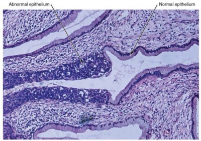
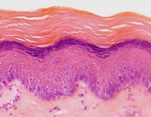
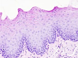
The body contains at least 200 distinct cell types. These cells contain essentially the same internal structures, yet they vary enormously in shape and function. The different types of cells are not randomly distributed throughout the body; rather they occur in organized layers, a level of organization referred to as tissue. The micrograph to the right shows the high degree of organization among different types of cells in the tissue of the female cervix. You can also see how that organization breaks down when rogue cancer cells divide without restraint.
The variety in cell shape reflects the many different roles that cells fulfill in your body. The human body starts as a single cell at fertilization. As this fertilized egg divides, it gives rise to trillions of cells, each built from the same blueprint but organized into tissues and becoming irreversibly committed to a developmental pathway.
Learning Objectives
- Define tissue.
- Describe the structure and basic function of the four major tissue types.
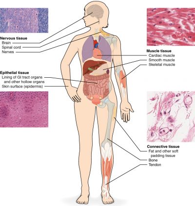
The tissues of multicellular, complex animals are four primary types: epithelial, connective, muscular, and nervous. A tissue is defined as a group of similar cells carrying out related functions. These tissues combine to form organs—like the skin or kidney—that have specific, specialized functions within the body. Organs are organized into organ systems to perform functions. Examples include the cardiovascular system, which consists of the heart, blood, and blood vessels, and the digestive system, which includes several organs, including the stomach, intestines, liver, and pancreas. Organ systems come together to create an entire organism.
There are a number of instances when we use the same term (such as muscle or bone) to refer to both an organ and a type of tissue. As an example, consider a muscle like the extensor digitorum. The contractile cells of skeletal muscle are bundled together to make muscle fiber tissue. In turn, endomysium cells form enclosing tissue that wraps around bundles of muscle fibers, like a tortilla around the filling of a burrito. Several of these structures are in turn wrapped by another tissue, perimysium. Finally, bundles of these are surrounded by a sheath of yet another tissue, epimysium, which covers the outside of the whole muscle. Yet more tissue is necessary for the muscle to function in the body. Connective tissue comprising ligaments attaches the muscle to the skeleton, and nerve tissue conducts impulses from the nervous system to signal the muscle to contract.
The body is made of dozens of different tissues, but broadly speaking there are four types of tissues.
Muscular tissue, which is in turn divided into skeletal, smooth, and cardiac muscle, is contractile and elastic. It allows movement of the body. It also allows necessary contractions of various organs such as the heart and respiratory and digestive system organs. Nervous tissue comprises the body’s wiring system. It conducts signals between the nervous system and various organs. Connective tissue holds the body together. It is found in most organs, anchoring them to the skeleton and other organs. Types of connective tissue include fibrous, fatty, loose, and cartilage. Connective tissue also includes bone, blood, and lymph. Epithelial tissue is the body’s protection against the outside environment. Skin tissue helps to maintain homeostasis. It helps monitor and control temperature and resists abrasion, foreign bodies, and damaging chemicals. Internally, epithelial tissue lines most internal cavities, secreting hormones and lubricating fluids or absorbing nutrients.
Embryonic Origin of Tissues
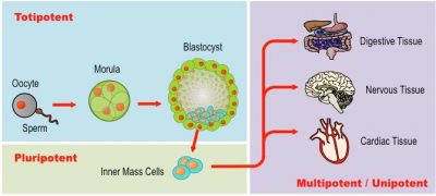
Tissues form during development. The zygote, or fertilized egg, is a single cell formed by the fusion of an egg and sperm. After fertilization the zygote divides rapidly, generating many cells to form the embryo. The first embryonic cells generated have the ability to differentiate into any type of cell in the body and, as such, are called totipotent, meaning each has the capacity to divide, differentiate, and develop into a new organism.
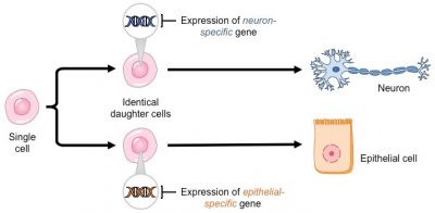
Differentiation is the process during development whereby newly formed cells become more specialized and distinct from one another as they mature. All cells of an organism share an identical genome, meaning that each cell contains the entire set of genetic instructions for that organism. If all cells in your body carry the same information, then what leads to the variation we observe in cell structure and function? The activation of different instructions (genes) within a given cell by chemical signals causes the cell to differentiate. When a cell differentiates and becomes specialized, it loses its capacity to form alternative cell types.
(Credit: BioNinja, CC BY NC SA)
Stem cells are unspecialized cells that have two key qualities: self-renewal and potency. Self-renewal refers to the ability to divide and replicate continuously. Potency refers to the capacity to differentiate into specialized cell types. There are four main types of stem cells present at various stages of human development. As you read above, a totipotent cell can form any cell type, as well as extra-embryonic (placental) tissue. Only the first few cells produced after fertilization are totipotent. Pluripotent stem cells can form any cell type (e.g., embryonic stem cells). Multipotent stem cells can differentiate into a number of closely related cell types (e.g., stem cells in adults that can give rise to any type of blood cell). Unipotent stem cells can not differentiate but are capable of self-renewal (e.g., progenitor cells in the epidermis of skin).
As cell proliferation progresses, three major cell lineages are established within the embryo. Each of these lineages of embryonic cells forms the distinct germ layers from which all the tissues and organs of the human body eventually form. Each germ layer is identified by its relative position: ectoderm (ecto- = “outer”), mesoderm (meso- = “middle”), and endoderm (endo- = “inner”). The figure below shows the types of tissues and organs associated with each of the three germ layers. Note that epithelial tissue originates in all three layers, whereas nervous tissue derives primarily from the ectoderm and muscle tissue from the mesoderm.
Stem cells in the embryo differentiate into various cell types. The necessary genes in the cells turn on or off, resulting in the production of proteins that characterize a cell’s structure and function. Early in embryonic growth, the cells migrate to the appropriate location in the body. Once there, they proliferate so that the tissue can perform its needed function.
Learn By Doing 8.1
The process by which a less specialized cell matures into a more specialized cell is called ________.
- differentiation
- maturation
- modification
- specialization
The zygote is described as totipotent because it ultimately gives rise to all the cells in your body including the highly specialized cells of your nervous system. Describe this transition, discussing the steps and processes that lead to these specialized cells.
Epithelial Tissues
Epithelial tissue also referred to as epithelium, refers to the sheets of cells that cover exterior surfaces of the body, line internal cavities, and passageways, and form certain glands. Epithelial tissues may occur as a single layer or multiple layers of cells. The shapes of cells present and the number of layers of cells are used to classify the types of epithelia. Epithelia composed of a single layer of cells are called simple epithelia; epithelial tissue composed of multiple layers is called stratified epithelia. Epithelia are always described with two terms, one that refers to the number of cell layers (simple or stratified) and one that refers to the shape of the cells (squamous, cuboidal, or columnar). There are two types of epithelia that appear stratified but are composed of a single layer of cells: pseudostratified epithelium and transitional epithelium.
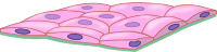
Squamous Epithelia
 |
 |
Squamous epithelial cells are generally round and flat and have a small, centrally located nucleus. The cell outline is slightly irregular, and cells fit together to form a covering or lining. When the cells are arranged in a single layer (simple epithelia), they facilitate diffusion in tissues, such as the areas of gas exchange in the lungs and the exchange of nutrients and waste at blood capillaries. In the figure to the left, the image on the top left illustrates a layer of squamous cells with their plasma membranes joined together to form an epithelium. The image on the right is an example of simple squamous epithelium in the cardiovascular system. Blood vessels are lined with a single layer of epithelial cells termed endothelium (indicated with arrows). Note the red and white blood cells on the lower right in both images.
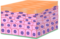
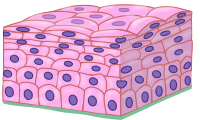
Squamous epithelial cells also exist in layers called strata. This type of epithelial tissue, termed stratified squamous epithelium, is found in areas of the body that require significant protection from abrasion. For example, stratified squamous epithelium occurs in the skin and in tissues lining the mouth, esophagus, and vagina. These epithelia may contain layers of dead cells full of the protein keratin, as in skin, or they may not, as in the vagina and esophagus.
Cuboidal Epithelia
Cuboidal epithelial cells are cube-shaped with a single central nucleus. They are most commonly found in a single layer representing simple epithelia in glandular tissues throughout the body where they prepare and secrete glandular material. They are also found in the walls of tubules and in the ducts of the kidney and liver.
    Atlas of Plant and Animal Histology, University of Vigo, CC BY NC SA) |
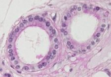
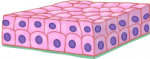
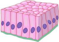
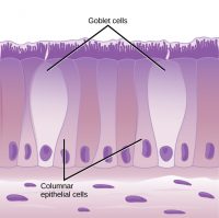
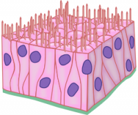
Columnar epithelial cells are taller than they are wide: they resemble a stack of columns in an epithelial layer and are most commonly found in a single-layer arrangement. The nuclei of columnar epithelial cells in the digestive tract appear to be lined up at the base of the cells, as illustrated on the right in the table above. These cells absorb material from the lumen of the digestive tract and prepare it for entry into the body through the circulatory and lymphatic systems.
  |
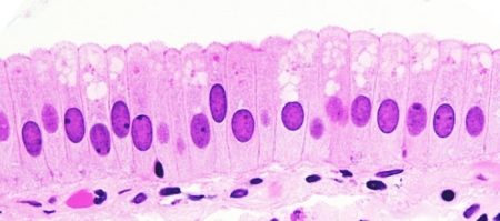 |
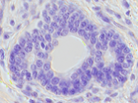
Columnar epithelial cells lining the respiratory tract appear to be stratified. However, each cell is attached to the base membrane of the tissue and, therefore, they are simple tissues. The nuclei are arranged at different levels in the layer of cells, making it appear as though there is more than one layer, as seen in the image to the right. This is called pseudostratified columnar epithelia (pseudo means false). This is a true simple epithelium since all the cells directly contact the connective tissue layer below, called the basal lamina (base layer).
 |
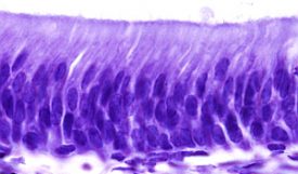 |
This cellular covering has cilia at the apical(free) surface of the cells. The cilia enhance the movement of mucous and trapped particles out of the respiratory tract, helping to protect the system from invasive microorganisms and harmful material that has been breathed into the body. Goblet cells are interspersed in some tissues (such as the lining of the trachea). The goblet cells contain mucous that traps irritants, which in the case of the trachea keep these irritants from getting into the lungs.
Transitional or uroepithelial cells appear only in the urinary system, primarily in the bladder and ureter. These cells are arranged in a stratified layer, but they can appear to pile up on top of each other in a relaxed, empty bladder. As the urinary bladder fills, the epithelial layer unfolds and expands to hold the volume of urine introduced into it. As the bladder fills, it expands, and the lining becomes thinner. In other words, the tissue transitions from thick to thin.
|
|
(Credit: Atlas of Plant and Animal Histology, University of Vigo, CC BY NC SA) |
(Credit: Atlas of Plant and Animal Histology, University of Vigo, CC BY NC SA) |
The table below summarizes the different types of epithelial tissues.
| Cell shape | Description | Location |
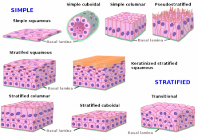 (Credit: Atlas of Plant and Animal Histology, University of Vigo, CC BY NC SA) Epithelium is described with two adjectives. The first describes the number of cell layers. Simple refers to a single layer of cells, while stratified refers to more than one layer of cells. |
| squamous | flat, irregular round shape | simple: lung alveoli, capillaries
stratified: skin, mouth, vagina |
|
| cuboidal | cube-shaped, central nucleus | glands, renal tubules | |
| columnar | tall, narrow, nucleus toward base; tall, narrow nucleus along cell | simple: digestive tract
pseudostratified: respiratory tract |
|
| transitional | round, simple but appear stratified because cells are crowded together | urinary bladder |
Connective Tissues
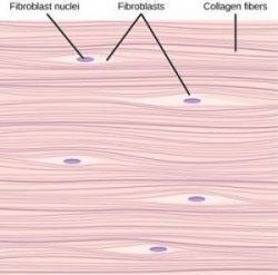
Connective tissues are made up of a matrix consisting of living cells and a non-living substance called the ground substance. The ground substance is made of an organic substance (usually protein) and an inorganic substance (usually a mineral or water). The combination of ground substance and proteins is referred to as the extracellular matrix. The principal cell of connective tissues is the fibroblast. This cell makes the fibers found in nearly all of the connective tissues. Fibroblasts are motile, able to carry out cell division and can synthesize whichever connective tissue is needed. Certain white blood cells (macrophages and lymphocytes) can be found in some of the tissues. Some tissues have specialized cells that are not found in others. The matrix in connective tissues gives the tissue its density. Connective tissue with a high concentration of cells or fibers has a proportionally less dense matrix.
The organic portion or protein fibers found in connective tissues are either collagen, elastic, or reticular fibers. Collagen fibers provide strength to the tissue, preventing it from being torn or separated from the surrounding tissues. Elastic fibers are made of the protein elastin; this fiber can stretch to one and one-half of its length and return to its original size and shape. Elastic fibers provide flexibility to the tissues. Reticular fibers are the third type of protein fiber found in connective tissues. This fiber consists of thin strands of collagen that form a network of fibers to support the tissue and other organs to which it is connected. The various types of connective tissues, the types of cells and fibers they are made of, and sample locations of the tissues are summarized in the table below.
| Tissue | Cells | Fibers | Location |
|---|---|---|---|
| loose/areolar | fibroblasts, macrophages, some lymphocytes, some neutrophils | few: collagen, elastic, reticular | around blood vessels; anchors epithelia |
| dense, fibrous connective tissue | fibroblasts | mostly collagen | irregular: skin regular: tendons, ligaments |
| cartilage | chondrocytes, chondroblasts | hyaline: few collagen fibrocartilage: large amount of collagen | shark skeleton, fetal bones, human ears, intervertebral discs |
| bone | osteoblasts, osteocytes, osteoclasts | some: collagen, elastic | vertebrate skeletons |
| adipose | adipocytes | few | adipose (fat) |
| blood | red blood cells, white blood cells | albumins, globulins, fibrinogen | blood vessels |
| lymph | lymphocytes | antibodies | lymphatic vessels |
Loose Connective Tissue
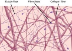
Loose connective tissue, also called areolar connective tissue, has a sampling of all of the components of connective tissue. As illustrated on the left, loose connective tissue has some fibroblasts; macrophages are present as well. Collagen fibers are relatively wide and stain a light pink, while elastic fibers are thin and stain dark blue to black. The space between the formed elements of the tissue is filled with the matrix. The material in the connective tissue gives it a loose consistency similar to a cotton ball that has been pulled apart. Loose connective tissue is found around every blood vessel and helps to keep the vessel in place. The tissue is also found around and between most body organs. In summary, areolar tissue is tough yet flexible and is part of many membranes.

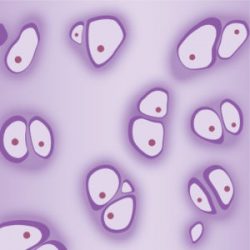
Adipose tissue, or fat tissue, is considered connective tissue even though it does not have fibroblasts or a real matrix and only has a few fibers. Adipose tissue is made up of cells called adipocytes that collect and store fat in the form of triglycerides for energy metabolism. Adipose tissues additionally serve as insulation to help maintain body temperatures, allowing animals to be endothermic, and they function as cushioning against damage to body organs. Under a microscope, adipose tissue cells appear empty due to the extraction of fat during the processing of the material for viewing, as seen on the right. The thin lines in the image are the cell membranes, and the nuclei are the small, black dots at the edges of the cells.
Fibrous Connective Tissue
Fibrous connective tissues contain large amounts of collagen fibers and few cells or matrix material. The fibers can be arranged irregularly or regularly with the strands lined up in parallel. Irregular fibrous connective tissues are found in areas of the body where stress occurs from all directions, such as the dermis of the skin. Regular fibrous connective tissue, shown in the middle below, is found in tendons (which connect muscles to bones) and ligaments (which connect bones to bones).
Cartilage
Cartilage is a connective tissue with a large amount of the matrix and variable amounts of fibers. The cells, called chondrocytes, make the matrix and fibers of the tissue. Chondrocytes are found in spaces within the tissue called lacunae.
A cartilage with few collagen and elastic fibers is hyaline cartilage, illustrated to the right. The lacunae are randomly scattered throughout the tissue, and the matrix takes on a milky with routine histological stains. Sharks have cartilaginous skeletons, as does nearly the entire human skeleton during a specific pre-birth developmental stage. A remnant of this cartilage persists in the outer portion of the human nose. Hyaline cartilage is also found at the ends of long bones, reducing friction and cushioning the articulations of these bones. Hyaline cartilage found in movable joints such as the knee and shoulder becomes damaged as a result of age or trauma. Damaged hyaline cartilage is replaced by fibrocartilage, and this results in the joints becoming “stiff.”
Elastic cartilage has a large number of elastic fibers, giving it tremendous flexibility. The ears of most vertebrate animals contain this cartilage as do portions of the larynx or voice box. Fibrocartilage contains a large number of collagen fibers, giving the tissue tremendous strength. Fibrocartilage comprises the intervertebral discs in vertebrate animals.
Bone
Bone, or osseous tissue, is a connective tissue that has a large amount of two different types of matrix material. The organic matrix is similar to the matrix material found in other connective tissues, including some amount of collagen and elastic fibers. This gives strength and flexibility to the tissue. The inorganic matrix consists of mineral salts—mostly calcium salts—that give the tissue hardness. Without adequate organic material in the matrix, the tissue breaks; without adequate inorganic material in the matrix, the tissue bends.
There are three types of cells in bone: osteoblasts, osteocytes, and osteoclasts. Osteoblasts are active in making bone for growth and remodeling. Osteoblasts deposit bone material into the matrix, and after the matrix surrounds them, they continue to live but in a reduced metabolic state as osteocytes. Osteocytes are found in the lacunae of the bone. Osteoclasts are active in breaking down the bone for bone remodeling, and they provide access to calcium stored in tissues. Osteoclasts are usually found on the surface of the tissue.
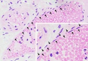
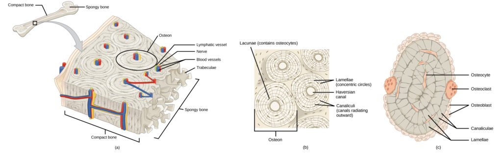
Bone can be divided into two types: compact and spongy. Compact bone is found in the shaft (or diaphysis) of a long bone and the surface of the flat bones, while spongy bone is found in the end (or epiphysis) of a long bone. Compact bone is organized into subunits called osteons, as illustrated below. A blood vessel and a nerve are found in the center of the structure within the Central (or Haversian) Canal, with radiating circles of lacunae around it known as lamellae. The wavy lines seen between the lacunae are microchannels called canaliculi; they connect the lacunae to aid diffusion between the cells. Spongy bone is made of tiny plates called trabeculae. These plates serve as struts to give the spongy bone strength. Over time, these plates can break causing the bone to become less resilient. Bone tissue forms the internal skeleton of vertebrate animals, providing structure to the animal and points of attachment for tendons.
Blood
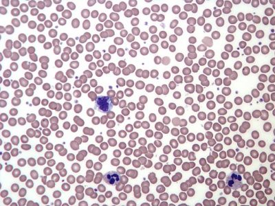
Blood is considered a connective tissue because it has a matrix, though it is fluid rather than fibrous or solid. The living cell types are red blood cells (RBC), also called erythrocytes, and white blood cells (WBC), also called leukocytes. The matrix is commonly called plasma.
The cell found in greatest abundance in blood is the erythrocyte. Erythrocytes are counted in millions in a blood sample: the average number of red blood cells in primates is 4.7 to 5.5 million cells per microliter. Mammalian erythrocytes lose their nuclei and mitochondria when they are released from the bone marrow where they are made. The principal job of an erythrocyte is to carry and deliver oxygen to the tissues.
Leukocytes are white blood cells (WBC) found in the peripheral blood. Leukocytes are counted in the thousands in the blood per microliter. There are many types, each with a specific role to play in guarding us again pathogens and cancer. We will discuss the different types of white blood cells and their functions in greater detail later in the course.
The slightly granular material among the cells is a cytoplasmic fragment of a cell in the bone marrow. This is called a platelet or thrombocyte. Platelets participate in the stages leading up to blood coagulation to stop bleeding through damaged blood vessels. Blood has a number of functions, but primarily it transports material through the body to bring nutrients to cells and remove waste material from them.
Muscular Tissues
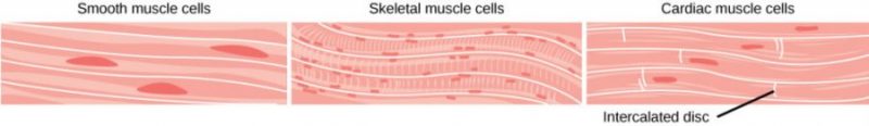
There are three types of muscle in animal bodies: smooth, skeletal, and cardiac. They differ by the presence or absence of striations or bands, the number and location of nuclei, whether they are voluntarily or involuntarily controlled, and their location within the body. The table and image below summarize these differences.
| Characteristic | smooth muscle | skeletal muscle | cardiac muscle |
| Striations | no stripes | striped | striped |
| Nuclei | single, centrally located | many, at periphery | single, in center |
| Control | involuntary | voluntary | involuntary |
| Location | visceral organs | skeletal muscles | heart |
 |
|||
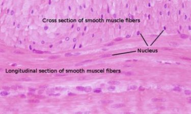
Smooth muscle does not have striations in its cells. It has a single, centrally located nucleus, as shown in the figure to the right. Constriction of smooth muscle occurs under involuntary, autonomic nervous control and in response to local conditions in the tissues. Smooth muscle tissue is also called non-striated as it lacks the banded appearance of skeletal and cardiac muscle. The walls of blood vessels, the tubes of the digestive system, and the tubes of the reproductive systems are composed of mostly smooth muscle.
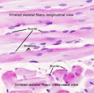
Skeletal muscle has striations across its cells caused by the arrangement of the contractile proteins actin and myosin. These muscle cells are relatively long and have multiple nuclei along the edge of the cell. Skeletal muscle is under voluntary, somatic nervous system control and is found in the muscles that move bones. The figure to the right illustrates the histology of skeletal muscle.
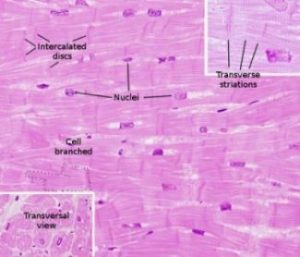
Cardiac muscle, shown to the left, is found only in the heart. Like skeletal muscle, it has cross striations in its cells, but cardiac muscle has a single, centrally located nucleus. Cardiac muscle is not under voluntary control but can be influenced by the autonomic nervous system to speed up or slow down. An added feature to cardiac muscle cells appears as a line that extends along the end of the cell where it abuts the next cardiac cell in the row. This line is called an intercalated disc: it assists in passing electrical signals efficiently from one cell to the next and maintains a strong physical connection between neighboring cardiac cells.
Nervous Tissues
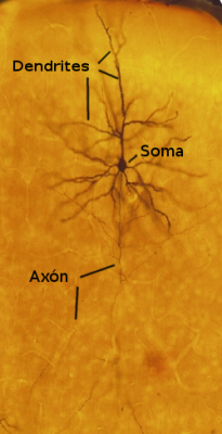
Nervous tissues are made of cells specialized to receive and transmit electrical impulses from specific areas of the body and to send them to specific locations in the body. The main cell of the nervous system is the neuron. The large structure with a central nucleus is the neuron’s cell body (soma). Projections from the cell body are either dendrites specialized in receiving input or a single axon specialized in transmitting impulses.
Glial cells are generally considered to be the support cells of the nervous system. Astrocytes regulate the chemical environment of the nerve cell, and oligodendrocytes insulate the axon, so the electrical nerve impulse is transferred more efficiently. Other glial cells that are not shown to support the nutritional and waste requirements of the neuron. Some of the glial cells are phagocytic and remove debris or damaged cells from the tissue.
The table below summarizes the characteristics of the four main tissue types.
| Type of Tissue | General Features | Additional Characteristics |
| Epithelial | Organized in sheets. Provides covering: Lines body surface, hollow organs, cavities, and tubes. Provides tissues for secretory glands. | Cells are polarized with a ‘surface’ and ‘basement’ side. The surface side may have special characteristics such as cilia or microvilli. The membrane functions also often differ. |
| Connective | Characterized by a varied and extensive matrix. Cells are usually scattered irregularly in this matrix. Cell shape is irregular to round. | Found throughout body, forms the support and structure for organs and the body itself. |
| Nervous | Cells can generate electrical signals. Highly branched. | Found throughout body with concentrations in the brain, the spinal cord and the enteric nervous system of the gut. |
| Muscular | Cells can generate electrical signals, which result in contraction. ‘Voluntary’ muscle makes up skeletal muscles; Cardiac muscle powers the heart; smooth muscle surrounds hollow organs and tubes. | Intestinal Smooth muscle and Cardiac muscle both have cell-to-cell communication via gap junctions. |
Learn By Doing 8.2
What types of tissues are in your hand?
- epithelial
- connective
- muscle
- nervous
- all of the above
Our bodies are made of 4 basic tissue types: epithelial, connective, muscular, and nervous. For each of the following parts of the hand, decide what type of tissue it is made of.
- The skin that covers the hand
- The bones in the fingers
- The abductor pollicis brevis muscle
- Cartilage in the joints
- The tissue that conveys the sense of touch
- Blood in the blood vessels
- The nerves that control the muscles
How does connective tissue provide structure and support?
- its cells are strong
- it contains structural fibers and a dense extracellular matrix
- it doesn’t
- it is responsible for force generation
In observing epithelial cells under a microscope, the cells are arranged in a single layer and look tall and narrow, and the nucleus is located close to the bottom of the cell. The specimen is what type of epithelial tissue?
- columnar
- stratified
- squamous
- transitional
Connective tissue is made of which three essential components?
- cells, ground substance, and carbohydrate fibers
- cells, ground substance, and protein fibers
- collagen, ground substance, and protein fibers
- matrix, ground substance, and fluid
Ligaments connect bones together and withstand a lot of stress. What type of connective tissue should you expect ligaments to contain?
- areolar tissue
- adipose tissue
- dense regular connective tissue
- dense irregular connective tissue
Striations, cylindrical cells, and multiple nuclei are observed in ________.
- skeletal muscle only
- cardiac muscle only
- smooth muscle only
- skeletal and cardiac muscles
The cells responsible for the transmission of the nerve impulse are ________.
- neurons
- oligodendrocytes
- astrocytes
- microglia
Which features of neuron morphology (morph = form) make them suitable for the transmission of nerve impulses?
Tissue Membranes
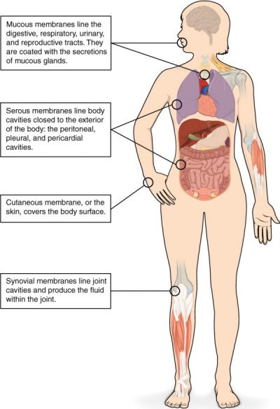
A tissue membrane is a thin layer or sheet of cells that covers the outside of the body (for example, skin), the organs (for example, pericardium), internal passageways that lead to the exterior of the body (for example, abdominal mucosa), and the lining of the moveable joint cavities. There are two basic types of tissue membranes: connective tissue and epithelial membranes.
Connective Tissue Membranes
The connective tissue membrane is formed solely from connective tissue. These membranes encapsulate organs, such as the kidneys, and line our movable joints. A synovial membrane is a type of connective tissue membrane that lines the cavity of a freely movable joint. For example, synovial membranes surround the shoulder, elbow, and knee joints. Fibroblasts in the inner layer of the synovial membrane release a molecule called hyaluronan into the joint cavity. The hyaluronan effectively traps available water to form the synovial fluid, a natural lubricant that enables the bones of a joint to move freely against one another without much friction. This synovial fluid readily exchanges water and nutrients with blood, as do all body fluids.
Epithelial Membranes
The epithelial membrane is composed of epithelium attached to a layer of connective tissue, for example, your skin. The mucous membrane is also a composite of connective and epithelial tissues. Sometimes called mucosae, these epithelial membranes line body cavities, and hollow passageways that open to the external environment and include the digestive, respiratory, excretory, and reproductive tracts. Mucous, produced by the epithelial exocrine glands, covers the epithelial layer. The underlying connective tissue called the lamina propria (literally “own layer”) helps support the fragile epithelial layer.
A serous membrane is an epithelial membrane composed of mesodermally derived epithelium called the mesothelium that is supported by connective tissue. These membranes line the cavities that do not open to the outside and cover the organs within those cavities. They are essentially membranous bags, with mesothelium lining the inside and connective tissue on the outside. Serous fluid secreted by the cells of the thin squamous mesothelium lubricates the membrane and reduces abrasion and friction between organs. Serous membranes are identified according to location. Three serous membranes line the thoracic cavity; the two pleura that cover the lungs and the pericardium that covers the heart. A fourth, the peritoneum, is the serous membrane in the abdominal cavity that covers abdominal organs and forms double sheets of mesenteries that suspend many of the digestive organs.
The skin is an epithelial membrane also called the cutaneous membrane. It is a stratified squamous epithelial membrane resting on top of connective tissue. The apical (top) surface of this membrane is exposed to the external environment and is covered with dead cells that help protect the body from desiccation and pathogens.
Organs
Learning Objectives
- Define organ.
- Locate the organ level within the larger hierarchy of human physiology.
The organ level of organization in the body may be the most familiar to us from our everyday experiences. Many of the common ailments we hear about—an upset stomach, a broken bone, lung disease, skin cancer—are named for the organs they affect.
An organ is made up of tissues that work together to perform a specific function for the body as a whole. Groups of organs that perform related functions are organized into organ systems, which perform more general functions. We will spend much of the rest of this course exploring the structure and function of organs and organ systems. The table below describes the structures and functions of some common organs.
| Organ | Primary function(s) | Tissues it contains | Organ system(s) it is a part of |
| brain | control of body systems and behavior; cognition | nervous, connective, epithelial | nervous system; endocrine system |
| skin | protection; support and containment; temperature and fluid regulation | epithelial, nervous, connective, muscular | integumentary system |
| stomach | chemical and mechanical digestion of food | epithelial, connective, muscular, nervous | digestive system |
| sternum (breastbone) | support; protection; blood cell production | epithelial, connective, nervous | skeletal system; immune system; cardiovascular system |
| kidney | waste removal; fluid regulation | epithelial, connective, nervous | urinary system |
So, an organ is a functional unit of the body. A system, such as the cardiovascular system, is a number of structures; some of them are organs that carry out one or several functions. However, as usually happens in biology, it is difficult to define the precise role of an organ or system in the many tasks that an animal must accomplish. So that how the organs are distributed in tasks depends on the author or the focus of a particular study (physiology or anatomy, general or particular functions). Thus, an organ may take part in different systems at the same time, doing more than one function or sharing functions with other organs.
Organ Systems, The Whole Body, and Populations
Learning Objective
Discuss how the organ systems work together in the whole body and how the body interacts with the environment to influence physiology.
Organ systems
Organ systems are made up of organs that work together to perform a specific function for the body as a whole. The table below describes the organ systems and their primary organs and physiological functions.
| Organ system | Key organ(s) | Primary function(s) |
| integumentary | skin | support; protection; regulation of fluid levels and temperature |
| skeletal | bones, cartilage | support; protection; movement; blood cell production |
| muscular | muscles, tendons | support; movement |
| urinary | kidneys, bladder, urethra | waste removal; regulation of fluid levels |
| digestive | tongue, esophagus, stomach, small intestine, large intestine, gallbladder, rectum | digestion of food; waste removal |
| respiratory | trachea, lungs | gas exchange; regulation of temperature |
| cardiovascular | heart, blood vessels | transport of materials through the body; regulation of temperature |
| nervous | brain, spinal cord | control of behavior and body systems; cognition |
| endocrine | glands | control of body systems and development |
| immune | thymus, tonsils, spleen | defense against infection |
| lymphatic | lymph nodes, lymphatic vessels | immunity; regulating fluid balance |
| reproductive | penis, testes, prostate (males); uterus, ovaries, vagina (females) | reproduction |
The Whole Body
The organ systems of the body all work together to maintain proper physiological functions. Many times in the arena of anatomy and physiology, including in this course, we closely examine the molecules, cells, tissues, and organs of the body to learn their forms and functions. However, it is important to consider that every molecule works as part of the entire system. Endocrine disorders such as diabetes affect glucose levels in the body. Altered blood glucose levels can affect many organ systems. For example, the immune system may not heal as well, the urinary system may experience kidney damage, and the cardiovascular system can experience vascular damage, even to the point of causing blindness. In the body, everything is interconnected.
Populations
Beyond the body, populations and environment can impact physiology and health. Some diseases and disorders are common to certain populations, most likely because of genetic connections. Also, environmental conditions can impact health. Particulates in the air can impact respiratory function. We are also affected by food, exercise, sun exposure, and other environmental conditions.
Did I Get This? 8.1
If you eat a hamburger, you are mainly eating ground-up beef muscle. What levels of organization are certainly represented in this ground-up muscle?
- organ, organ system, and organism
- organelle, cell, tissue
- tissue, organ, organ system
Summary
The human body contains more than 200 types of cells that can all be classified into four types of tissues: epithelial, connective, muscle, and nervous. Epithelial tissues act as coverings controlling the movement of materials across the surface. Connective tissue integrates the various parts of the body and provides support and protection to organs. Muscle tissue allows the body to move. Nervous tissues propagate information.
The study of the shape and arrangement of cells in tissue is called histology. All cells and tissues in the body derive from three germ layers in the embryo: the ectoderm, mesoderm, and endoderm.
Different types of tissues form membranes that enclose organs, provide a friction-free interaction between organs, and keep organs together. Synovial membranes are connective tissue membranes that protect and line the joints. Epithelial membranes are formed from epithelial tissue attached to a layer of connective tissue. There are three types of epithelial membranes: mucous, which contain glands; serous, which secrete fluid; and cutaneous which makes up the skin.
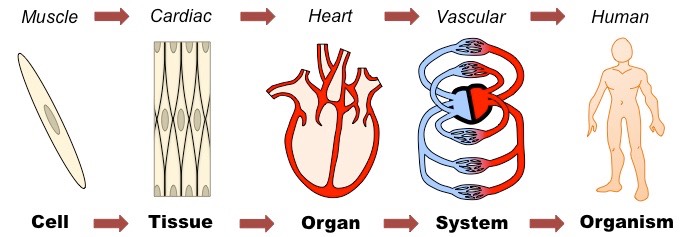
In multicellular organisms:
- Cells may be grouped together to form tissues
- Organs are then formed from the functional grouping of multiple tissues
- Organs that interact may form organ systems capable of carrying out specific body functions
- Organ systems collectively carry out the life functions of the complete organism.
*
“Learn By Doing” and “Did I Get This?” Feedback
Learn By Doing 8.1
The process by which a less specialized cell matures into a more specialized cell is called ________.
- differentiation
- maturation
- modification
- specialization
The zygote is described as totipotent because it ultimately gives rise to all the cells in your body including the highly specialized cells of your nervous system. Describe this transition, discussing the steps and processes that lead to these specialized cells.
The zygote divides into many cells. As these cells become specialized, they lose their ability to differentiate into all tissues. At first they form the three primary germ layers. Following the cells of the ectodermal germ layer, they too become more restricted in what they can form. Ultimately, some of these ectodermal cells become further restricted and differentiate in to nerve cells.
Learn By Doing 8.2
What types of tissues are in your hand?
- epithelial
- connective
- muscle
- nervous
- all of the above
Our bodies are made of 4 basic tissue types: epithelial, connective, muscular, and nervous. For each of the following parts of the hand, decide what type of tissue it is made of.
- The skin that covers the hand: epithelial and connective
- The bones in the fingers: connective
- The abductor pollicis brevis muscle: muscular
- Cartilage in the joints: connective
- The tissue that conveys the sense of touch: nervous
- Blood in the blood vessels: connective
- The nerves that control the muscles: nervous
How does connective tissue provide structure and support?
- its cells are strong
Incorrect. Some cells are stronger than others, but not strong enough to provide support to a whole organism. - it contains structural fibers and a dense extracellular matrix
Correct. - it doesn’t
Incorrect. Structure and support are hallmarks of connective tissue; blood is a connective tissue that doesn’t seem to be very stiff, but the large fluid content of the body is very mechanically relevant. - it is responsible for force generation
Incorrect. Muscle is the type of tissue that generates force.
In observing epithelial cells under a microscope, the cells are arranged in a single layer and look tall and narrow, and the nucleus is located close to the bottom of the cell. The specimen is what type of epithelial tissue?
- columnar
- stratified
- squamous
- transitional
Connective tissue is made of which three essential components?
- cells, ground substance, and carbohydrate fibers
- cells, ground substance, and protein fibers
- collagen, ground substance, and protein fibers
- matrix, ground substance, and fluid
Ligaments connect bones together and withstand a lot of stress. What type of connective tissue should you expect ligaments to contain?
- areolar tissue
- adipose tissue
- dense regular connective tissue
- dense irregular connective tissue
Striations, cylindrical cells, and multiple nuclei are observed in ________.
- skeletal muscle only
- cardiac muscle only
- smooth muscle only
- skeletal and cardiac muscles
The cells responsible for the transmission of the nerve impulse are ________.
- neurons
- oligodendrocytes
- astrocytes
- microglia
Which features of neuron morphology (morph = form) make them suitable for the transmission of nerve impulses?
Neurons are well suited for the transmission of nerve impulses because short extensions called dendrites, receive impulses from other neurons, while a long extension, called the axon, carries electrical impulses away from the cell and this allows communication with other neurons.
Did I Get This? 8.1
If you eat a hamburger, you are mainly eating ground-up beef muscle. What levels of organization are certainly represented in this ground-up muscle?
- organ, organ system, and organism
No: When you eat hamburger, you are eating ground-up muscle tissue, so you might be eating an entire muscle organ. However, you certainly are not eating an entire cow (the organism). - organelle, cell, tissue
Yes: Hamburger is made from ground-up muscle tissue. The tissue contains cells, which contain organelles. - tissue, organ, organ system
No: Your hamburger definitely contains muscle tissue, and it might contain an entire muscle organ. However, it definitely does not contain an entire organ system, which consists of all the muscle organs in the cow’s body. That would be a big hamburger.
