Review and Synthesis: Putting It All Together
20 Review of Body Systems
Review of Body Systems
Now that you have reviewed ways to describe location and orientation, you will learn about the organ systems that are necessary for the vital functions of life. You will also get a chance to practice using body planes and directional orientations to explain the anatomical integration and relative location of structures within organ systems. The next section will systematically describe the organ systems of the body, as well as the major anatomical structures and functions.
An organ system is an integrated collection of organs in the body that work together to perform a vital function. This course will organize the organ systems of the body based on the vital functions defined earlier.
Example: Skeletal System
As an example of how the components of an organ system work together, let’s look at the skeletal system. The most obvious components of this system are the bones, which form a rigid framework for the body. The bones contribute to our ability to stand upright and move around, but they cannot do it alone. Ligaments and cartilage are also parts of the skeletal system. Ligaments connect the bones to each other. Cartilage cushions the spaces between the bones, allowing for smooth movement. And the bones could not move at all without the skeletal muscles and tendons that connect muscles to bones (parts of the muscular system). The bones provide the muscles with something to pull against.
If one component of an organ system is damaged or malfunctions, the function of the organ system will be affected. Think about a broken bone. If the femur breaks, it will be much harder to maintain an upright posture or to walk or run. These might also be more difficult if the cartilage of the femur is destroyed by arthritis or a ligament in the knee is injured while playing a sport. If any component of the skeletal system is damaged—bone, ligament, or cartilage—the knee will not function properly.
The sections that follow will briefly describe the organ systems that perform the vital functions of life. You will learn how they contribute to homeostasis and how imbalances in homeostasis lead to various disease states.
The major organ systems of the body are shown in Table 20.1 below.
| Function | Organ System |
| Protection from the Environment | Integumentary System
Immune System |
| Exchange with the Environment | Digestive System
Respiratory System |
| Fluid Transport within the Body | Cardiovascular and Lymphatic Systems
Urinary System |
| Structure, Support, and Movement | Skeletal System
Muscular System |
| Control and Regulation | Nervous System
Endocrine System |
Exchange with the Environment
Learning Objectives
- Describe the digestive system, listing the major organs and structures, describing the major functions, and using anatomical planes and directional terms to identify organs and their relationships to each other.
- Describe the respiratory system, listing the major organs and structures, describing the major functions, and using anatomical planes and directional terms to identify organs and their relationships to each other.
Every cell needs to take in nutrients and get rid of waste products. This is accomplished via processes that exchange molecules across the cell membrane. For a single-celled organism, this is accomplished through direct exchange with the environment. It gets more complicated with larger organisms where cells are not in direct contact with the “outside” environment. In such organisms, including humans, there are specialized organ systems that serve as interfaces with the environment, moving things into and out of the body to keep it functioning. The two major organ systems that serve to exchange nutrients and waste with the environment are the digestive system (food and feces) and respiratory system (oxygen and carbon dioxide).
Humans take in food, water, and oxygen. During digestion and metabolism, food is broken down into simpler substances (including some waste products), and some of those simpler substances—for example, simple sugars—can be metabolized in a way that they release energy, especially when oxygen is present. The products of these processes include solid waste, liquid waste, and carbon dioxide. These waste materials must be eliminated from the body to prevent illness. The urinary system also helps with the removal of wastes being transported in the body fluids, but the “exchange” is all internal, so it will be explored more in next the vital function category.
Digestive System
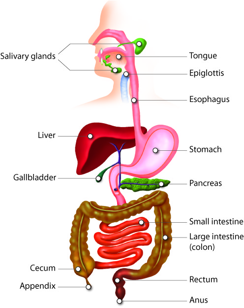
Also called the gastrointestinal system, the digestive system breaks down eaten material into nutrient molecules, absorbs water and ions, and eliminates undigested residue.
The digestive system is a continuous tube (the digestive tract or alimentary canal). Areas along this tube are specialized to perform different functions related to getting the nutrients from your food to the cells that need them. Accessory organs add secretions into different areas along the tube.
Your cells cannot use the pizza you had for lunch in pizza form. It needs to be broken down into molecules that are small enough to be absorbed. As the pizza travels along the digestive tract, each organ along the way breaks it down further. Muscles in the walls of the digestive tract keep things moving along, and glands in the tissues secrete digestive juices—mostly enzymes and acids—that break up the larger substances in the pizza into smaller molecules. Food is physically broken into smaller pieces in a process termed mechanical digestion. These pieces are then chemically broken down into smaller units in a process termed chemical digestion. Proteins are broken down into amino acids. Carbohydrates are broken down into simple sugars. Fats are broken down into molecules like fatty acids and cholesterol. It is important that the large particles be broken into their smallest units so they can be absorbed from the digestive tract into the bloodstream. Therefore, the main functions of the digestive system are to ingest, break down, and absorb the nutrients from our food. It also eliminates the wastes (anything not absorbed) as feces.
The specialized organs of the digestive tract extend in a roughly superior to inferior direction from the mouth (where food goes in) to the anus (where waste comes out) in the following order:
- Mouth
- Pharynx
- Esophagus
- Stomach
- Small intestine (including the duodenum, jejunum, and ileum)
- Large Intestine (including the cecum, colon, and rectum )
- Anus
Accessory organs in the digestive system are connected to the digestive tract and secrete additional digestive juices.
- The salivary glands produce saliva containing (among others) amylase, an enzyme that breaks down carbohydrates.
- The pancreas secretes a variety of enzymes that break down fats, carbohydrates, and proteins, as well as bicarbonate ions that neutralize stomach acids. It is important to note that this function corresponds to the exocrine portion of the pancreas.
- The liver produces bile, which aids in fat digestion and absorption.
- The gall bladder stores and concentrates bile and secretes it into the small intestine.
The stomach is a sort of muscular sac that can expand to hold a large meal. Glands in the walls of the stomach secrete enzymes and acids that break down food. Muscles in the walls of the stomach churn the food and digestive juices together. Although the stomach can receive a large amount of food at a time, it releases its contents gradually into the small intestine so that the intestine can better perform its function.
Digestion continues in the small intestine, with additional digestive juices produced by the pancreas, liver, and the walls of the small intestine itself. The walls of the small intestine have numerous tiny folds, which increase its surface area, allowing for efficient absorption of nutrients into the circulatory system, which in turn takes the nutrients to all the cells of the body.
Excess water is reabsorbed in the large intestine, and the undigested portion of your pizza leaves the body. Resident microbes of the large intestine (gut microbiota) can digest substances that our cells cannot.
Respiratory System
When humans breathe, air enters and exits via the respiratory system. This allows the body to obtain oxygen, which is needed for metabolic processes and to eliminate carbon dioxide, which is a metabolic waste product and can affect the body’s pH homeostasis.
Like the digestive system, the respiratory system can be thought of as a tube, or rather, as a branching series of tubes that get smaller and smaller as they branch off. Unlike the digestive system, which moves solids and liquids in a single direction, the respiratory system moves gases in both directions when we inhale and exhale.
When we inhale, air passes through the nose or mouth into the pharynx, larynx, trachea, and lungs, and into smaller and smaller airways termed bronchi and then bronchioles until it reaches the air sacs or alveoli. Only a single cell thick, the walls of the alveoli allow the oxygen in the air to diffuse into the blood, and the cardiovascular system carries it to each cell in the body.
Carbon dioxide, a waste product of cell metabolism, also diffuses through the alveolar walls, but in the opposite direction, from the blood to the airways. Carbon dioxide is then exhaled through the airways to the external environment.
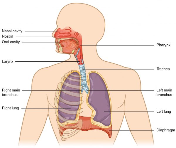
The organs of the respiratory system are arranged in a roughly superior to inferior direction and include:
- Nose
- Mouth
- Pharynx
- Larynx
- Trachea
- Lungs
Within the lungs, the respiratory system can be further divided into:
- Bronchi (singular bronchus)
- Bronchiole tree
- Alveoli (singular alveolus)
Note that the pharynx (the part of the throat just behind the mouth) is listed as a part of both the digestive and respiratory systems. Food and beverages pass through the pharynx on the way through the digestive tract. Air passes through the pharynx on its way to and from the lungs. Food and water are prevented from entering the airway when we swallow by a structure called the epiglottis. It is not uncommon for organs to be part of more than one organ system. The pancreas, for example, has both digestive and endocrine functions, and the kidneys play a role in both the urinary and endocrine systems.
The respiratory system is superior to the abdomen and internal to the ribs.
Did I Get This? 21.1
Which of the following is not a major function of the digestive system?
- Secrete hormones to control homeostasis
- Chew food to break it down into smaller pieces
- Absorb nutrients into the blood
- Release enzymes to chemically break down food into smaller units
Hint: The digestive system has several important functions which all relate to getting the nutrients from what we eat available to our body cells.
After food goes in the mouth, it travels in what direction?
- Superior
- Inferior
- Distal
- Lateral
Hint: Which direction is from the mouth toward the stomach?
With acid reflux, acid from the stomach goes into the esophagus. Which direction is that?
- Superior
- Inferior
- Distal
- Lateral
Hint: This direction is from the stomach toward the mouth.
Correct. With acid reflux, acid travels in the superior direction, along the sagittal plane, into the esophagus.
The respiratory system functions to…
- Exchange oxygen and carbon dioxide with the environment
- Transport oxygen and carbon dioxide in the blood
Hint: The respiratory system uses pulmonary ventilation or breathing to begin this process.
When you inhale, your rib cage moves away from the coronal and transverse planes in which directions?
- anterior and superior
- inferior and medial
- lateral and deep
- posterior and intermediate
Hint: Take a breath and watch where your ribs go. The ribcage goes out and up.
Fluid Transport Within the Body
Learning Objectives
- Describe the cardiovascular system: list the major organs and structures, describe the major functions and use anatomical planes and directional terms to identify organs and their relationships to each other.
- Describe the urinary system: list the major organs and structures, describe the major functions and use anatomical planes and directional terms to identify organs and their relationships to each other.
Our body constantly transports molecules from one place to another to carry out the required metabolic functions for daily living. As we take in nutrients and oxygen, these molecules must be delivered to the cells, tissues, and organs that need them for metabolic processes. Furthermore, our body must have a system for disposing of wastes generated by metabolism and foreign particles, which may potentially disturb optimal body function. The cardiovascular and lymphatic systems coordinate a diverse set of transport functions.
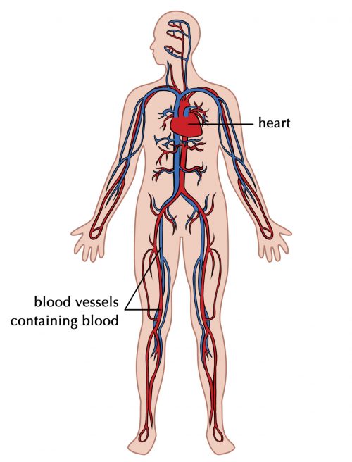
The cardiovascular system consists of the blood, heart, and blood vessels. The heart supplies the force to circulate blood throughout the body via the blood vessels. Blood is the main fluid used to deliver nutrients to the body’s tissues and to receive wastes that will ultimately be removed from the body by the urinary system. The respiratory and cardiovascular systems work together to take oxygen in from the external environment and deliver it, via blood, to cells. In a similar fashion, these systems also use the blood to remove carbon dioxide from cells and release it into the external environment. The red blood cells or erythrocytes are responsible for the transport of respiratory gasses. The lymphatic system consists of lymphatic vessels, which are near blood vessels and several organs, such as lymph nodes, the thymus gland, and the spleen. It returns excess tissue fluid to the cardiovascular system while initiating immune responses to ensure that the delivered blood is not compromised, responding to foreign invasion in multiple ways. The lymphatic system is an integrated part of the immune system to cleanse body fluids of dead cells, toxins, and pathogens. Together, the transport systems of the body coordinate the efforts of nutrient delivery and removal of wastes and other harmful substances. In addition to transporting the fluid, fluid/electrolyte balance must be maintained by integrated functions of these organ systems: urinary, lymphatic, and cardiovascular.
Cardiovascular System
The cardiovascular system transports, from one part of the body to another: nutrients, oxygen, ions, proteins, hormones, and other signaling molecules, as well as waste products, including carbon dioxide. This system also helps to maintain homeostasis of fluid volume, pH, and temperature.
The primary components of the cardiovascular system are blood, the heart, and the vessels of the circulatory system, which work together to transport nutrients, wastes, and gases to every cell in the body.
The blood that is circulated throughout the body contains two main components:
- Plasma contains water, electrolytes, glucose, proteins (including enzymes, hormones, and blood clotting factors), and metabolic wastes
- Formed elements or blood cells
There are three types of formed elements:
- Red blood cells transport oxygen and carbon dioxide.
- White blood cells fight infection by attacking foreign cells and clearing old or diseased cells.
- Platelets are important for hemostasis (not to be confused with homeostasis); hemostasis is our ability to stop bleeding after vascular injury (injury to blood vessels).
The blood functions to transport molecules and blood cells and contributes to the maintenance of pH balance. Blood cells are formed in the red bone marrow.
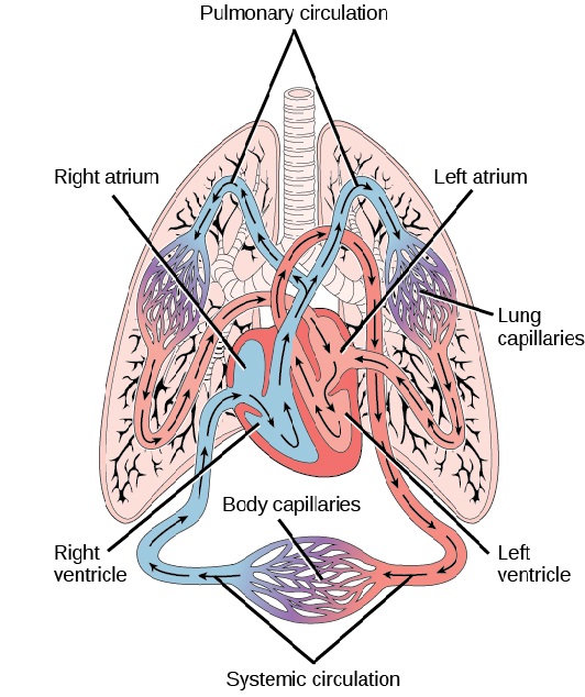
The heart is divided into four chambers. The two lower chambers, called ventricles, force blood out into the arteries. The two upper chambers of the heart, called atria, receive blood returning from the veins. The heart contracts as a unit; both atria (named right and left) contract together to move blood into the ventricles, and then both ventricles contract at the same time to move blood out of the heart into the pulmonary artery and the aorta.
The cardiovascular system is divided into two functional subsystems.
- The systemic circuit transports blood and its components to the body.
- The pulmonary circuit transports blood and its components between the heart and the lungs.
Arteries of the systemic circuit (also known as the systemic circulatory circuit) carry oxygenated blood from your heart to provide oxygen and nutrients dissolved in the blood to every cell in your body. When blood leaves the left ventricle, it first enters the aorta, the largest artery in the human body. Arteries gradually branch into smaller and more numerous arterioles, which then supply blood to the smallest vessels, termed capillaries. It is estimated that your body contains approximately 60,000 miles of capillaries, which is enough to encircle earth three times! Capillaries allow the exchange of oxygen, nutrients, and waste between the blood and tissue cells. After waste has been picked up, blood is moved through vessels of increasing size venules into the larger veins. Veins return oxygen-poor blood back to the heart, where the blood is passed to the pulmonary circuit to the lungs to pick up oxygen.
The pulmonary artery (part of the pulmonary circuit) carries oxygen-poor blood from the right ventricle of the heart to the lungs for oxygenation and removal of carbon dioxide. The pulmonary veins carry oxygenated blood from the lungs to the left side of the heart.
Without this system that pumps the heart to squeeze blood out and without the network of vessels to distribute the pumped blood, the cells of your body would not have an adequate supply of nutrients and oxygen.
The heart lies medial to the lungs, anterior to the spinal cord, posterior to the sternum, and superior to the diaphragm. The heart is divided into four chambers. The two lower chambers, called ventricles, force blood out into the arteries. The two upper chambers of the heart, called atria, receive blood returning from the veins.
Urinary System
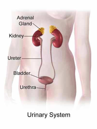
The urinary system filters blood and adjusts the composition of blood/interstitial fluid by removing excess water, salt, acid, and metabolic waste from the body as urine. This allows the urinary system to control body fluid volume, blood pressure, pH, and electrolyte balance. It is a critical system for maintaining homeostasis.
We have seen how the digestive and respiratory systems remove some wastes from the body—undigested food leaves the digestive tract through the anus, and carbon dioxide leaves through the lungs and airways. The urinary system (or excretory system) filters blood to remove excess water, electrolytes, and other metabolic wastes and reabsorbs water, electrolytes, and other molecules as needed to maintain homeostasis in the body fluid. The resulting excess and wastes are excreted as urine. In this way, the urinary system also works with the respiratory system to maintain the pH balance in the body.
The organs of the urinary system include:
- Kidneys
- Ureters
- Urinary Bladder
- Urethra
The kidneys, the main organs of the urinary system, are located against the posterior wall of the abdomen. They serve as a filtration and reabsorption system, where soluble substances are filtered, and then those that the body needs to keep are reabsorbed. Those that are not reabsorbed (or not reabsorbed fully), such as our metabolic waste products, end up in the urine. Because our bodies are constantly producing waste, the kidneys continuously work to prevent the buildup of waste products and toxins, filtering about 180 liters of fluid a day. Because the average person has about three liters of plasma (the fluid fraction of blood), this means that our plasma is filtered about 60 times a day!
One of the most important waste products removed from the blood is urea, the main end product of protein metabolism. Other waste products and some toxins are also removed from the blood by the kidneys.
In addition, ions and water are also filtered by the kidneys, but a large fraction of these are reabsorbed to keep the fluid and electrolyte concentration of the blood and other body fluids within an optimal range for proper cell function. The kidneys also play an important role in the regulation of pH by managing the amount of acid in the urine.
As previously described, the kidneys filter the blood to form urine. Urine leaves the kidneys and flows through the ureters to the urinary bladder, where it is stored until it passes out of the body through the urethra. On average, two liters of urine are produced per day, but this can vary greatly depending on fluid intake, fluid loss through perspiration, and other factors.
The right and left kidneys are located against the posterior wall of the abdominal cavity. The kidneys’ location is also described as retroperitoneal because they are behind the peritoneal cavity that encloses the intestines.
Did I Get This? 21.2
Which component of blood is most responsible for transporting oxygen?
- Platelets
- Plasma
- red blood cells
- white blood cells
When oxygenated, these cells take on a red color.
Which structure is most responsible for generating the “blood pressure” that is measured in the doctor’s office?
- Arteries
- Blood
- Heart
- Veins
Hints: This structure performs the most work, which generates pressure. When your blood pressure is measured, it should be measured at the same level as this structure. This is the structure that contracts to supply the force to move blood through the systemic circuit.
What direction best describes the location of the heart relative to the shoulders?
- medial to the shoulders
- superficial from the shoulders
- distal from the shoulders
- lateral from the shoulders
Hint: The heart is slightly off-center (to the left) in the body but is still in between the shoulders.
Where is the heart relative to the digestive organs?
- Superior
- distal
- anterior
- superficial
Hint: The heart is in the chest, and most of the digestive organs are in the middle and lower abdomen.
A key organ in the urinary system, this organ filters the blood and reabsorbs the water, electrolytes, and ions needed to maintain optimal body fluid levels.
- Kidney
- Ureter
- Urethra
- Urinary Bladder
Hint: This organ involves sifting through soluble substances in the blood to separate waste products and toxins from the water, ions, and electrolytes that are reusable fluids needed by the body.
Where are the kidneys compared to the spinal column?
- Superior
- inferior
- lateral
- proximal
Hint: The two kidneys are equally distant from the spinal column. The spinal column is bisected by the midsagittal plane.
Urine travels in what direction from the kidney through the ureter to the bladder?
- superior
- inferior
- proximal
- deep
Hint: Typically, gravity aids in moving urine into the bladder.
Protection from Pathogens
Learning Objectives
- Describe the integumentary system: list the major organs and structures, describe the major functions and use anatomical planes and directional terms to identify organs and their relationships to each other.
- Describe the lymphatic system: list the major organs and structures, describe the major functions and use anatomical planes and directional terms to identify organs and their relationships to each other.
Previous sections contained an overview of all of the inputs and outputs necessary for life as well as a mechanism to deliver them within the body. This section will introduce the structures the human body needs to compartmentalize and protect these vital components and the structures that enable the body to move to obtain nutrients and avoid predators. Organ systems are required to coordinate these many efforts.
Integumentary System
We often don’t think of the skin as a complex organ, but it is. The skin is the primary organ in the integumentary system, which also includes hair, nails, and certain glands. The integumentary system helps to provide support and structure for the body, but it also plays several other important roles:
- It is the first line of defense against foreign organisms and the external environment.
- It helps to regulate body temperature.
- It senses changes in the environment (pain, pressure, touch).
- It supports the removal of wastes (as sweat).
- It aids in the production of vitamin D.
The integumentary system is one of the most active parts of our body, even though we are not as aware of its activity as we are of the heart, lungs, or stomach. The integumentary system encapsulates and protects the body. The skin is actually the largest organ in the body because of its large surface area. In some ways, the skin can be thought of as an immune system organ since it protects the body from foreign organisms. In other ways, the skin can be thought of as a sensory organ because it contains many nerves that are related to the sense of touch. The skin also integrates with muscles and allows for movements such as facial expressions.
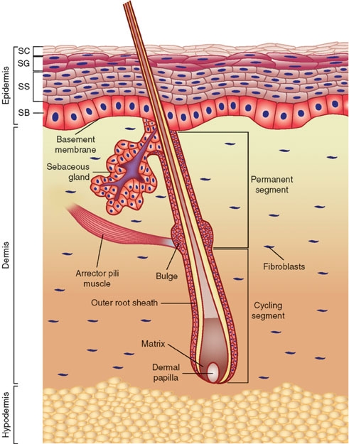
If we take a closer look at the skin, we can see that there are many layers. Within the skin, there are hair follicles from which hair grows. Also, there are sweat glands that produce sweat for thermal regulation of the body and sebaceous glands that secrete oil to waterproof and moisturize the skin. Nails are also included in the integumentary system, as are horns, feathers, claws, and hooves…but hopefully, you don’t have any of those.
The major structures within the integumentary system are:
- skin
- hair, nails
- sebaceous glands
- sweat glands
Skin, the largest organ of the body, is the primary organ of the integumentary system. Skin is composed of three main layers, each of which has specific functions related to its structure. The three main layers of the skin are:
- the epidermis, which acts as a seamless, waterproof barrier to the external environment and prevents excessive water loss from the body (the root “epi-” means “above”).
- the dermis, which provides the tensile strength and elasticity of the skin, contains nerves, sensory receptors, and contains blood vessels that aid in regulating body temperature.
- the hypodermis, which attaches skin to other structures below it and acts as an insulator and shock absorber (the root “hypo-” means “below”); the hypodermis is also known as the subcutaneous layer.
Hair, another component of the integumentary system, is found in nearly all regions of the skin except on the palms, soles of the feet, and some parts of the genitals. Hair grows from hair follicles that are part of the epidermis, even though they extend down and the dermis extends up around them. Hair helps regulate body temperature and protect the surface of the body, including eyelashes that protect the eyes.
Nails are located on the end of each distal phalanx (each finger and each toe). They protect the phalanges from trauma and provide mechanical support for manipulating objects. Nails grow from epidermal cells in the nail beds.
Glandular structures are also part of the epidermis and are present in different regions of the skin. They secrete substances that are important for many physiological functions. There are two main types of glands:
- sebaceous glands, whose secretions maintain the softness and hydrophobicity (water repellency) of the hair and skin.
- sudoriferous glands, whose secretions moisten the skin during pain, fear, sexual arousal, and emotional upset, or who secrete sweat to regulate body temperature.
The skin covers the outside surface of the body; thus, it is superficial to many skeletal muscles. Special structures such as hair, nails, and glands are part of the integumentary system.
Lymphatic and Immune Systems
Our body is in constant exchange with the environment through breathing, eating, and other activities. Therefore, it is important to screen the body and its components regularly to identify foreign invaders that might enter during these activities (or in any other manner). Further, it is important to rapidly and effectively remove these invaders before they can cause significant harm. Our body has specialized transport systems to carry out these functions. The cardiovascular and lymphatic systems work together to transport excess fluids (blood and lymph fluid, respectively) away from body tissues. Once fluid enters the lymphatic system, it is termed lymph. Lymph travels through lymph vessels and passes through many lymph nodes, which filter and clean the lymph. The immune system also produces and matures immune cells, which protect the body from invasion by agents that cause disease. One additional function of the lymphatic system is to transport absorbed fat from the digestive system to the body cells.
The immune system coordinates the activities required to respond to disease and infection. This response can provide two types of immunity:
- Specific immunity, in which specialized cells (such as T and B cells) recognize specific foreign molecules called antigens within the body and respond to them.
- Nonspecific immunity, in which the body uses several general methods such as physical barriers (that is, the skin and mucous membranes), fever, inflammation, specific action by immune cells, and enzyme activity to protect itself against general harmful agents.
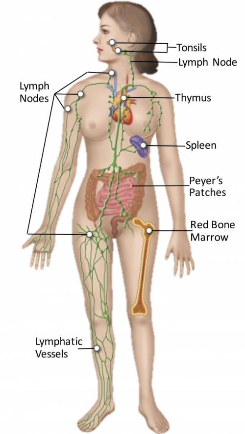
The major organs of the lymphatic and immune systems (described below) can be classified based on their role in lymphocyte (a type of white blood cell) maturation. The maturation of lymphocytes takes place within the red bone marrow and the thymus gland, which are primary lymphoid organs. Antigens become trapped within secondary lymphoid organs such as the lymph nodes, spleen, and tonsils. These organs are sites that contain lymphocytes for the destruction of invading pathogens.
| Organ | Description |
| Tonsils and Adenoids | Adenoids are one of three sets of tonsils. They trap pathogens that enter through the mouth and nose. Also, the tonsils monitor the external environment that the mouth and nose are exposed to and can react with an appropriate immune response for certain pathogens. |
| Thymus | A lobular (of or pertaining to a lobe) structure which contains many immature, inactive lymphocytes. As the lymphocytes mature, they leave the thymus to attack infected cells in lymphatic tissues throughout the body. |
| Spleen | The largest of the lymphatic organs, it houses lymphocytes for potential immune response. Also, the resident phagocytes within the spleen perform the most basic function of removing cell debris from the blood. |
| Lymph Nodes | These house lymphocytes and macrophages, which destroy foreign material contained in the lymph fluid. |
| Lymph Vessels | These transport lymphatic fluid throughout the lymphatic system. |
| Red Bone Marrow | All of our blood cells are generated from red bone marrow stem cells. These stem cells differentiate into red blood cells, platelets, and several cells that play roles in immunity. These “immune cells” include lymphocytes, which carry out specific immunity, and neutrophils and macrophages (macrophages start as monocytes and mature into macrophages in the tissues), which are nonspecific phagocytic cells. |
While many people know that we are protected from foreign micro-organisms by an immune system, few people realize how the immune system is able to patrol the entire body. White blood cells of the immune system are produced in the red bone marrow and travel through the blood. They can leave blood capillaries to travel through tissues. White blood cells are then able to remove dead or damaged cells and “foreign” organisms they encounter and recognize specific foreign organisms again if necessary. Additionally, lymph collects from tissues and circulates through lymph vessels, making “rest stops” in discrete points throughout the body called lymph nodes or lymph organs. In these nodes and organs, including the spleen, tonsils, and other tissue clusters, there are large collections of white blood immune cells. Lymph slowly travels through these organs, ensuring that the lymphocytes have plenty of time to react to these foreign organisms in the lymph before returning it to the blood.
The lymph nodes are located in several regions along the path of lymphatic vessels in our body. The thymus is located within the upper chest, lies posterior to the upper portion of the sternum, and extends from the root of the neck onto the pericardium. The spleen is found in the upper left abdominal cavity; it lies superior, posterior, and lateral to the stomach. The tonsils are masses of lymphatic tissue within the nasopharyngeal (nose and mouth) region.
Did I Get This? 21.3
How does the integumentary system contribute to immunity?
- It produces immune cells.
- It makes us waterproof.
- It feels when microorganisms are trying to enter.
- It provides a physical barrier to most microorganisms.
Hint: Open wounds often become infected.
Which of the following is not a function of the integumentary system?
- Regulates body temperature
- Protection from pathogens
- Excretes wastes
- Production of vitamin A
What is the role of red bone marrow in immune function?
- trap antigens for immune response
- produces immune cells from stem cells
- monitors the lymph fluid interacting with invaders
- It has no function related to immunity
Hint: People taking anti-cancer drugs often have greatly decreased bone marrow activity, leading to a poorly functioning immune system.
What lymphatic organ is inferior to the thymus?
- Spleen
- Lymph nodes
- Tonsils
- The thymus is the most inferior lymph organ.
Hint: The organ you are asked to identify is the largest organ in the lymphatic and immune systems.
Support and Movement
Learning Objectives
- Describe the skeletal system: list the major organs and structures, describe the major functions and use anatomical planes and directional terms to identify organs and their relationships to each other.
- Describe the muscular system: list the major organs and structures, describe the major functions and use anatomical planes and directional terms to identify organs and their relationships to each other.
Maintenance of a consistent physical structure is important for the body to function efficiently. Support and structure are among the primary functions of the integumentary system, the skeletal system, and the muscular system. These organ systems also allow the body to move in response to internal and external stimuli.
Skeletal System
Although we often think of bones as the only organs of the skeletal system, cartilage, ligaments, and tendons are equally important organs. These structures of the skeletal system work together to:
- provide leverage and resist forces from muscles and gravity.
- keep joints together.
- allow flexibility within joints.
- maintain a safe range of motion.
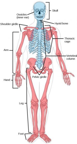
Bones are found throughout the body, from the skull in the head to the 26 bones in the foot. Bones allow us to maintain our stature, they protect softer internal organs, and they let us move around. Bones are interconnected by articulations, another word for joints. In an articulation, where bone meets bone, there is a layer of softer cartilage. Articulations are then stabilized by ligaments, which help keep the bones aligned properly. Bones are connected to the muscular system by tendons, which allow the body to move.
The major structures within the skeletal system are:
- bones
- cartilage
- ligaments
- tendons
The skeletal system consists of bones, ligaments, tendons, and cartilage. Bone is the primary organ of the skeletal system. Although there are different types of bones in our body, the basic components of all bone tissue are the same:
- Osteoblasts, osteoclasts, and osteocytes are specialized cells that are responsible for bone formation, regulation, and repair.
- Collagens and other proteins give bone flexibility.
- Inorganic calcium and phosphate minerals give bone its hardness.
- Red bone marrow produces all blood cells in a process termed hematopoiesis (or hemopoiesis).
These basic components give bone tissue its load-bearing, protective qualities. The living cells in bone allow it to sense and respond to stress. The inorganic matrix of bone gives the bone rigidity and also acts as a storage depot for calcium and phosphorus in the body.
The other parts of the skeletal system facilitate the movement of the bones. Cartilage is a firm, flexible, and smooth connective tissue found at the ends of bones. Cartilage is present in joints to protect the bone and to evenly distribute forces to the underlying bone. Ligaments are band-like elastic structures that surround joints to hold them together. Ligaments connect one bone to another bone and allow movement in very specific directions. Tendons are band-like structures similar to ligaments. However, tendons are more rigid and connect bones to muscles. Tendons play a role in integrating the force generation of the muscle with the rigid bone, which helps actuate large-scale motion.
The numerous organs and structures of the skeletal system allow it to serve an important role in the support and protection of our body. Bones are very strong yet flexible, which makes them perfect for supporting our weight and allowing movement. The connective tissues, such as cartilage, ligaments, and tendons, aid in protecting our joints and providing stability. The red bone marrow inside the bone is vital for hematopoiesis, or the production of all blood cells. Bones are also a reservoir for calcium. If your diet is deficient in calcium, a hormone will mobilize calcium from the bones to the blood, and your bones will be weaker.
Bones are found throughout the body. Regions capable of more intricate movements, such as the hands and feet, have more articulations and, therefore more bones. Each articulation has cartilage and is stabilized by ligaments.
Muscular System
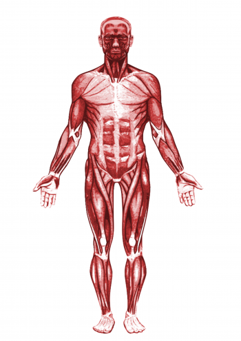
The muscular (musculoskeletal) system generates force for movement of bones around articulations, facial expression, breathing, posture, and assists with temperature regulation. The muscular system only contains skeletal muscle, although the body also has smooth and cardiac muscle tissue, which are important in other body systems. There are over 650 skeletal muscles in the human body!
The skeletal muscle converts signals from the nervous system into movement via muscle contractions. Muscles, like the biceps and triceps, are the organs of the skeletal muscular system. The main functions of skeletal muscles include:
- responding to neural information (conscious control)
- applying forces to the bones to cause movement
- producing heat to warm the body
- changing the size of the thoracic cavity for breathing
- applying forces for conscious control of openings to the outside of the body (sphincters)
The muscular system contains muscle tissues and interconnects with both the nervous system and skeletal system. Nerves control the muscles and allow us to consciously direct movements. Some muscles, such as the muscles that control the pupil of your eye, cannot be controlled consciously but react to nerve stimuli. The skeletal system provides a stiff support for muscles to pull on. Muscles generate force to lift as well as to balance us. The energy produced by contracting muscles (such as when shivering) in the muscle system helps keep us warm. There are many muscle fiber types throughout the body that vary based on function. Parallel muscles form along the long bones, pennate and convergent muscle fibers attach to tendons and circular muscles assist with closing our eyes or puckering our lips.
The major structures within the muscular system are:
- skeletal muscles
- tendons
Skeletal muscles are voluntary muscles that attach to, and contract to move the bones. Skeletal muscles often work in pairs. When one muscle is contracting, the other is relaxing. For example, to bend your arm at the elbow, your biceps muscle contracts, and your triceps muscle relaxes. To straighten your arm, the biceps relaxes, and the triceps contracts. The diaphragm is skeletal muscle that contracts and relaxes for inhalation and exhalation. Hiccups are a spasm in your diaphragm muscle.
Skeletal muscles are made of long cylinder shaped cells called muscle fibers, which have many nuclei within each cell. Therefore, we say that skeletal muscle is multi-nucleate. The functional unit within a skeletal muscle fiber, called a sarcomere (note that “sarc” means flesh), contains filaments of the proteins actin and myosin. Myosin is a thicker protein (appears darker) than actin and the two proteins create a pattern so the muscle appears striped or striated.
A muscle contraction occurs when the myosin filaments pull on the actin to shorten the sarcomere. This results in shortening of the muscle fiber and ultimately the entire muscle shortens or contracts to pull on the bone.
An electrical signal from the nervous system is necessary to cause a skeletal muscle contraction. The area where the nerve meets the muscle to stimulate it is termed neuromuscular junction. When a nerve signal reaches the neuromuscular junction, the muscle fiber is stimulated and the muscle contracts.
Tendons are grouped in both the skeletal system and the muscular system since they connect the two systems (connect muscle to bone). Tendons play a role in transmitting force from the muscles to the bones to permit movement.
Like bones, skeletal muscles are found throughout the body. Skeletal muscles are found under the skin of the integumentary system and attached to and surrounding the bones of the skeletal system.
Other Muscle Types
Although only skeletal muscle is part of the muscular system, there are three types of muscle tissue. Smooth muscle and cardiac muscle are similar to skeletal muscle, but perform specialized functions in the body. Most of these functions are involuntary and do not include the skeletal system.
Smooth muscles control involuntary functions of the body, such as arterial contractions to move blood and peristaltic contractions in the digestive system to move food. Smooth muscles lack striations thus, are termed smooth due to their appearance. They are composed of muscle fibers with a single nucleus in each cell and are uninucleate. Smooth muscles do not have any attachment to the skeletal system. Smooth muscle has the ability to produce its own contractions involuntarily. However, as with skeletal muscle, electrical signals from the nervous system can modulate the activities of smooth muscle. The organization of smooth muscle on a cellular level is irregular and unorganized. Therefore, smooth muscle does not contain sarcomeres.
Cardiac muscle contains similarities to both skeletal and smooth muscle. Like skeletal muscle, cardiac muscle is composed of organized muscle fibers and sarcomeres, and is striated. However, cardiac muscle does not attach to the skeletal system and is under involuntary control, and is uninucleate. Cardiac muscle is not long and cylinder shaped like skeletal muscle but is more branched.
Did I Get This? #4
Where is the skin in relation to the muscles?
- superior
- inferior
- deep
- superficial
Hint: The skin is the most exterior layer of the body.
What is the tissue which connects muscles to bones?
- Cartilage
- Tendon
- Osteocyte
- Ligaments
- Bones
Hint: This structure is firmer than a ligament.
What is the most numerous structures of the skeletal system?
- Ligaments
- Cartilage
- Tendons
- Bones
- Osteocytes
Hint: You think of these structures when you hear the term skeletal system.
Knees and ankles are articulations in the legs. From the knee, the ankle is in which direction?
- distal
- proximal
- anterior
- inferior
Hint: The ankle is further from the axis of the body than the knee.
The skull bones are also articulated; you will learn more about the articulations of the skull in a later section of the course. What direction is the skull from the rest of the skeleton?
- superior
- inferior
- medial
- lateral
Hint: The skull is at the top of the body.
Which of the following are structures of the muscular system?
- tendons
- skeletal muscle
- cardiac muscle
- ligaments
- both tendons and skeletal muscle
- tendons, skeletal muscle, and ligaments
Hint: The muscular system is associated with movement of bones.
If you were given a paralytic drug that blocks skeletal muscle function, your biggest concern would be:
- Your heart would no longer beat.
- All motility of your digestive tract would stop.
- You would not be able to breathe.
- You would not be able to move.
Hint: Paralytic drugs are often administered during general surgery in order to keep the patient from moving, but the patient must be put on a ventilator.
Skeletal muscle functions to
- move food through the digestive tract
- produce heat
- move bones
- all of the above are functions of the muscular system
- both production of heat and movement of bones are functions of the muscular system
Hint: The muscular system works with the skeletal system to accomplish its main goal but has several functions.
Where are the muscles in relation to the bones and skin?
- intermediate
- superficial
- deep
- inferior
Hint: Muscles cover the bones and skin covers the muscles; therefore, muscles are between the skin and bones.
Control and Regulation
Learning Objectives
- Describe the nervous system: list the major organs and structures, describe the major functions, and use anatomical planes and directional terms to identify organs and their relationships to each other.
- Describe the endocrine system: list the major organs and structures, describe the major functions, and use anatomical planes and directional terms to identify organs and their relationships to each other.
The nervous and endocrine systems work together to control many of the body’s other organ systems through electrical signals and chemical messengers. The systems integrate with each other, so that control systems within the nervous system regulate many activities of the endocrine system and hormones of the endocrine system can affect some of the functions of the nervous system. The chemical messengers produced by each individual system are responsible for many homeostatic functions, through feedback mechanisms. The activities of the two systems coordinate many of the body’s metabolic activities.
Nervous System
The basic functional units of the nervous system that transmit messages are cells called neurons. Signals travel through a neuron as electrical impulses. Neurons release chemical substances, known as neurotransmitters, to transmit information to other neurons, to muscles, or to glands. The chemical messages of the nervous system are transmitted over short distances, and their effects are short-lived. The nervous system allows for control and coordination of skeletal muscular movements that may be consciously predetermined, or may happen automatically, such as reflexes. Other parts of the nervous system control and coordinate subconscious body activities, including heart rate, gland secretions and smooth muscle movement in the digestive system. Some activities, such as breathing, can be controlled both subconsciously and consciously. The nervous system typically works quickly. It also allows us to integrate and store information, such as when you are learning.
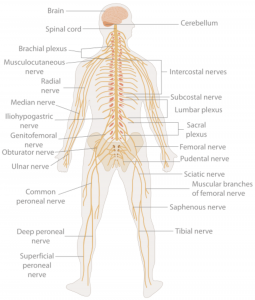
The nervous system transmits signals to different parts of the body to coordinate function. Electrochemical signals are processed in the brain and sent down the spinal cord, which runs the length of the back. From the spinal cord, peripheral nerves send signals out to the extremities. Return signals come in through sensory nerves and either return to the spinal cord for processing or back to the brain. The spinal cord processes reflexes and repeated patterns.
The nervous system is often divided into two functional parts:
- The central nervous system, which processes incoming information and initiates a response.
- The peripheral nervous system, which brings sensory information to, or carries motor output from, the central nervous system to initiate a reaction.
The major structures within the central nervous system are:
- brain
- spinal cord
The brain has several lobes, each of which carries out specific functions and processes information associated with specific parts of the body. The spinal cord is located within the vertebral column and processes some reflexes but primarily transmits information to and from the brain along neurons. Specialized membranes called meninges cover the brain and the spinal cord to protect them. Additionally, a special fluid, called cerebrospinal fluid, chemically and mechanically protects the brain and spinal cord.
The major structures within the peripheral nervous system are:
- cranial nerves
- spinal nerves
The peripheral nervous system is composed of nerves outside the brain and spinal cord. Nerves are bundles of extensions from neurons that extend through the body in the peripheral nervous system. These nerves are categorized into the following functional groups:
- sensory nerves, which carry sensory input to the brain or spinal cord from the environment
- motor nerves, which carry motor impulses from the brain or spinal cord to muscles or glands
- mixed nerves, which have a combination of sensory and motor neurons in one nerve.
The peripheral nervous system can be subdivided into two subdivisions: the somatic and autonomic divisions. The somatic nervous system includes sensory neurons that send sensory information from sensory receptors of the skeletal muscle, skin and special senses (including smell, taste, sight, hearing and equilibrium) to the central nervous system and motor neurons that control skeletal muscle.
The autonomic nervous system monitors and regulates changes in the body’s internal environment. These changes are not under voluntary control. Body processes controlled by the autonomic nervous system include the contractions of the stomach and other digestive organs, the heart rate, and contractions of blood vessels to control blood pressure and flow though the body.
The autonomic nervous system is further divided into the sympathetic and parasympathetic divisions. The sympathetic nervous system controls functions that speed up the heart and increase energy usage during emergencies or times of stress. On the other hand, the parasympathetic nervous system controls functions that have the opposite effect—they reduce heart rate and decrease overall energy usage when the body is returning to normal after an emergency or during normal functioning.
The brain is protected inside the skull. The spinal cord runs from the brain down through the bones of the spinal column. From the brain and spinal cord, nerves run throughout the body, including to the limbs.
Endocrine System
The endocrine system is an equally important method of sending messages within the body for control and coordination of multiple body systems. The functional unit of the endocrine system is a gland, or a group of cells that secrete chemicals called hormones. Hormones circulate throughout the body within the bloodstream and act as long-term messengers. In comparison with neurotransmitters, hormones act over long distances for a longer time.
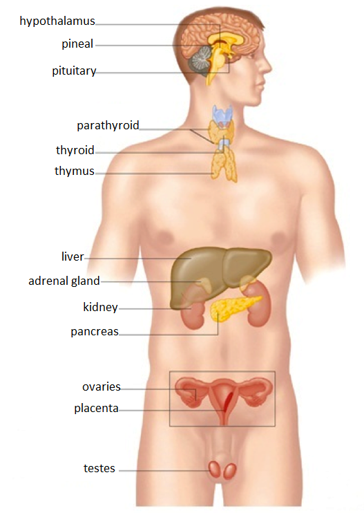
The major organs of the endocrine system are:
- hypothalamus
- pituitary gland
- pineal gland
- thyroid and parathyroid glands
- thymus (note that this gland is also part of the lymphatic system)
- adrenal glands
- pancreas (note that this gland is also part of the digestive system)
- gonads (that is, ovaries or testes—note that these glands are also part of the reproductive system)
As a specialized part of the brain, the hypothalamus is an endocrine gland that produces hormones that regulate many basic functions such as hunger, thirst and sleep through control of the pituitary gland. The hypothalamus receives sensory input from receptors and perceptual information from the brain, such as changes in emotional state, temperature, and lighting.
The pituitary gland is sometimes called the master gland, because it controls the release of hormones from many other endocrine glands.
The pineal gland secretes the hormone melatonin, which is important for transmitting information about environmental lighting and inducing sleep.
The thyroid gland and parathyroid glands are located together in the neck. The thyroid glands secrete hormones that regulate metabolism and calcium levels. The parathyroid gland also secretes hormones that regulate calcium levels.
The thymus gland secrets the hormones thymosin and thymopoietin that stimulate the production of special lymphocytes (white blood cells) called T-cells, which play an important role in the immune system by attacking foreign or abnormal cells.
The adrenal glands produce steroid hormones that regulate metabolic functions during stress, kidney function, and sexual function. The adrenal glands also secrete epinephrine (adrenaline) when stimulated by the autonomic nervous system.
The pancreas secretes insulin, to lower blood sugar levels, and glucagon to raise blood sugar levels. Therefore, the pancreas is an important endocrine organ for regulating the fuels available for energy production by cells.
The gonads, or sex organs (ovaries and testes) secrete sex hormones which control production of sperm and eggs as well as other secondary sex characteristics that are different for males and females. The secretion of sex hormones by the gonads is under the control of pituitary gland hormones.
The hypothalamus is found deep inside the brain and lies inferior to the thalamus. The pituitary gland is located at the base of the brain, inferior to the hypothalamus. The pineal gland is a small gland on the midline at the posterior of the brain. The thyroid and parathyroid glands lie inferior to the larynx, around the trachea. The parathyroid gland lies on the posterior surface of the thyroid gland. The adrenal glands are located superior to each kidney. The pancreas is located posterior to the stomach and is connected to the part of the small intestine called the duodenum. The ovaries are located in the pelvic cavity lateral to the uterus, while the testes are held external to the abdominopelvic cavity inside the scrotum.
Did I Get This? #5
The chemicals released by neurons which allow communication with other cells are termed what?
- neurotransmitters
- meninges
- hormones
Hint: These chemicals are released by neurons and critical for the functioning of the nervous system.
Which of the following consists of the brain and spinal cord?
- Peripheral Nervous System
- Mixed Nerves
- Sensory Nerves
- Somatic Nervous System
- Central Nervous System
Hint: The brain and the spinal cord form the “headquarters” of the nervous system.
What is the division of the nervous system functions to trigger the fight or flight response?
- autonomic
- somatic
- parasympathetic
Hint: Remember “auto” means automatic.
If a motor nerve is cut, which of the following would happen?
- The muscle controlled by that particular nerve will not contract.
- The bone the muscle is attached to will not move.
- Certain motor reflexes may no longer function.
- All of the above.
Hint: The nervous system controls many things.
What term(s) could be used to communicate the location of the brain?
- [A] cephalic
- [B] superior
- [C] inferior
- [D] caudal
- Both A and B
- Both C and D
Hint: Opposite of inferior.
How do functions of the endocrine system differ from functions of the nervous systems?
- Endocrine glands are found outside of the central nervous system, neurons are not.
- The molecules secreted by endocrine glands are typically active longer and travel further than those secreted by neurons.
- Endocrine glands secrete signaling molecules, neurons do not.
- The endocrine system controls functions of the body.
Hint: Both use chemical signaling, but the way the signaling occurs is different.
Which endocrine gland is most responsible for the release of estrogen and testosterone?
- gonads
- pituitary
- adrenal
- thymus
- pancreas
Hint: These organs control the formation of gametes and release sex hormones.
The nervous system releases chemicals termed __________, while the endocrine releases ___________.
- neurotransmitters, hormones
- hormones, neurotransmitters
Hint: The endocrine system releases these chemicals directly into the blood.
Which of the following glands would not be cut with a mid-sagittal body section?
- Adrenal gland
- Pineal gland
- Pancreas
- Hypothalamus
*
“Learn By Doing” and “Did I Get This?” Feedback
Did I Get This? #1
Which of the following is not a major function of the digestive system?
- Secrete hormones to control homeostasis
- Chew food to break it down into smaller pieces
- Absorb nutrients into blood
- Release enzymes to chemically break down food into smaller units
Secreting hormones is a function of the endocrine system, not the digestive system.
After food goes in the mouth it travels in what direction?
- Superior
- Inferior
- Distal
- Lateral
Food travels in the inferior direction, down the sagittal plane, to the stomach.
With acid reflux, acid from the stomach goes into the esophagus. Which direction is that?
- Superior
- Inferior
- Distal
- Lateral
With acid reflux, acid travels in the superior direction, along the sagittal plane, into the esophagus.
The respiratory system functions to…
- Exchange oxygen and carbon dioxide with the environment
- Transport oxygen and carbon dioxide in the blood
When we breathe, we move air and respiratory gases in and out of our lungs. This allows the exchange of carbon dioxide and oxygen.
When you inhale, your rib cage moves away from the coronal and transverse planes in which directions?
- anterior and superior
- inferior and medial
- lateral and deep
- posterior and intermediate
When you breathe air into your body, the rib cage moves away from the middle of the coronal plane towards the front, referred to as the anterior of the body, and through the transverse plane towards the upper, also called the superior, area of the body.
Did I Get This? #2
Which component of blood is most responsible for transporting oxygen?
- Platelets
- Plasma
- red blood cells
- white blood cells
Red blood cells transport oxygen and carbon dioxide, Red blood cells are also called erythrocytes (“eryth” means “red” and “cyte” means “cell”).
Which structure is most responsible for generating the “blood pressure” that is measured in the doctor’s office?
- Arteries
- Blood
- Heart
- Veins
The heart generates the pressure. Measuring blood pressure is a way to determine heart and vascular (artery and vein) function.
What direction best describes the location of the heart relative to the shoulders?
- medial to the shoulders
- superficial from the shoulders
- distal from the shoulders
- lateral from the shoulders
The heart is between the shoulders, slightly to the left of the center.
Where is the heart relative to the digestive organs?
- superior
- distal
- anterior
- superficial
The heart is superior to (above) the digestive organs.
A key organ in the urinary system, this organ filters the blood and reabsorbs the water, electrolytes, and ions needed to maintain optimal body fluid levels.
- Kidney
- Ureter
- Urethra
- Urinary Bladder
The kidneys filter soluble substances in the blood, discarding waste as urine and reabsorbing water, electrolytes, and ions needed to maintain body fluid levels within the optimal range (called homeostasis) for proper cell functioning.
Where are the kidneys compared to the spinal column?
- superior
- inferior
- lateral
- proximal
The kidneys are located laterally, on either side of the spinal column; the spinal column is located between the kidneys. The right kidney is usually slightly lower than the left. Its close proximity to the liver pushes the right kidney downward.
Urine travels in what direction from the kidney through the ureter to the bladder?
- superior
- inferior
- proximal
- deep
Urine travels inferiorly, down from the kidneys through the ureters into the bladder.
Did I Get This? #3
What is the role of red bone marrow in immune function?
- trap antigens for immune response
- produces immune cells from stem cells
- monitors the lymph fluid interacting with invaders
- It has no function related to immunity
Immune cells and other blood cells originate in red bone marrow; some mature in red bone marrow while others move to thymus to mature before moving to other structures in the body.
What lymphatic organ is inferior to the thymus?
- Spleen
- Lymph nodes
- Tonsils
- The thymus is the most inferior lymph organ.
The thymus is near the neck, and the spleen is in the abdomen. This means that the spleen is inferior to the thymus.
How does the integumentary system contribute to immunity?
- It produces immune cells.
- It makes us waterproof.
- It feels when microorganisms are trying to enter.
- It provides a physical barrier to most microorganisms.
Correct. The skin might actually be our most important immune organ because of this function.
Which of the following is not a function of the integumentary system?
- Regulates body temperature
- Protection from pathogens
- Exretes wastes
- Production of vitamin A
The skin makes vitamin D not vitamin A.
Did I Get This? #4
Where is the skin in relation to the muscles?
- superior
- inferior
- deep
- superficial
Correct. Skin covers the muscles and provides the external surface of the body.
What is the tissue which connects muscles to bones?
- Cartilage
- Tendon
- Osteocyte
- Ligaments
- Bones
Tendons are a type of connective tissue that binds bones to muscles.
What is the most numerous structures of the skeletal system?
- Ligaments
- Cartilage
- Tendons
- Bones
- Osteocytes
There are 206 bones in the human body.
Knees and ankles are articulations in the legs. From the knee, the ankle is in which direction?
- distal
- proximal
- anterior
- inferior
The ankle is further from the axis of theDistal means away from the trunk. The ankles and knees are on the limbs. The ankles are further away from the trunk of the body than the knees.
The skull bones are also articulated; you will learn more about the articulations of the skull in a later section of the course. What direction is the skull from the rest of the skeleton?
- superior
- inferior
- medial
- lateral
The skull is the top-most structure of the skeleton.
Which of the following are structures of the muscular system?
- tendons
- skeletal muscle
- cardiac muscle
- ligaments
- both tendons and skeletal muscle
- tendons, skeletal muscle, and ligaments
The muscular system is comprised of two main structures: tendons and skeletal muscle.
If you were given a paralytic drug that blocks skeletal muscle function, your biggest concern would be:
- Your heart would no longer beat.
- All motility of your digestive tract would stop.
- You would not be able to breathe.
- You would not be able to move.
The biggest concern is an inability to breathe, because the diaphragm, which allows you to breath, is a skeletal muscle.
The skeletal muscle functions to?
- move food through the digestive tract
- produce heat
- move bones
- all of the above are functions of the muscular system
- both production of heat and movement of bones are functions of the muscular system
In addition to these functions the muscular system helps with breathing.
Where are the muscles in relation to the bones and skin?
- intermediate
- superficial
- deep
- inferior
Muscles are between the bones and skin.
Did I Get This? #5
The chemicals released by neurons which allow communication with other cells are termed what?
- neurotransmitters
- meninges
- hormones
Correct. Neurotransmitters are released at a synapse which is a junction between two neurons or between a neuron and another cell.
Which of the following consists of the brain and spinal cord?
- Peripheral Nervous System
- Mixed Nerves
- Sensory Nerves
- Somatic Nervous System
- Central Nervous System
Correct. The Central Nervous System consists of the brain and spinal cord.
What is the division of the nervous system functions to trigger the fight or flight response?
- autonomic
- somatic
- parasympathetic
The sympathetic division of the autonomic nervous system causes the increased heart rate and blood pressure typical in the fight of flight response.
If a motor nerve is cut, which of the following would happen?
- The muscle controlled by that particular nerve will not contract.
- The bone the muscle is attached to will not move.
- Certain motor reflexes may no longer function.
- All of the above.
Motor nerves control all of these things.
What term(s) could be used to communicate the location of the brain?
- [A] cephalic
- [B] superior
- [C] inferior
- [D] caudal
- Both A and B
- Both C and D
The brain is in the head, so it is at the cephalic end of the body, and this is also in the superior part of the body.
How do functions of the endocrine system differ from functions of the nervous systems?
- Endocrine glands are found outside of the central nervous system, neurons are not.
- The molecules secreted by endocrine glands are typically active longer and travel further than those secreted by neurons.
- Endocrine glands secrete signaling molecules, neurons do not.
- The endocrine system controls functions of the body.
Hormones typically have longer-lasting effects than neurotransmitters and by definition hormones travel through the bloodstream to act at a distant site, whereas neurotransmitters act at a very short distance across synapses.
Which endocrine gland is most responsible for the release of estrogen and testosterone?
- gonads
- pituitary
- adrenal
- thymus
- pancreas
Estrogen and testosterone are necessary for egg and sperm development and come from the gonads, the ovaries in women and the testes in men
The nervous system releases chemicals termed __________, while the endocrine releases ___________.
- neurotransmitters, hormones
- hormones, neurotransmitters
Remember that neurotransmitters are faster acting than hormones but hormones often have a longer duration.
Which of the following glands would not be cut with a mid-sagittal body section?
- Adrenal gland
- Pineal gland
- Pancreas
- Hypothalamus
The sagittal plane divides the body vertically into the left and right sides. An adrenal gland exists on each side and would not be cut by mid-sagittal sectioning.
