Punch Biopsy

Step 1: Health history and physical exam

Assess the duration of time the lesion has been present, location, presence of infected tissue around biopsy site, allergies, bleeding disorders, and ensure the patient is aware that all biopsies result in a scar.

Assess for risk factors for poor wound healing:
• impaired nutritional status
• advanced age
• medications
• diabetes
• peripheral vascular disease
• autoimmune disease
• immunosuppression
• COPD
• CHF
• anemia
• smoking
• substance use

Assess lesions using the ABCDE rule to identify potentially malignant lesions.

Step 2: Position patient comfortably

Relaxed skin tension lines can be better visualized when the patient is positioned in an anatomically neutral position. Punch biopsies done with consideration of skin tension lines are more likely to achieve a cosmetically appealing scar.

Step 3: Cleanse the area with chlorhexidine or other approved skin cleanser

Wound cleansing reduces risk of sepsis to the wound site. Cleanse a wide area around the wound to prevent contamination of suture material and surgical instruments during the procedure. The area thus prepared should measure at least 3 inches wide around the edges of the wound or planned incision.

Step 4:  Anesthetize area

Inject the anesthetic agent at the base of the lesion or area to be biopsied to raise an intradermal welt or wheal. Alternatively, a field block technique can be used if an entire small lesion is to be removed. Using anesthetic with epinephrine will constrict blood vessels and decrease bleeding during the procedure. Avoid use of epinephrine in the digits, ears, and nose due to the risk of ischemia. See chapter titled “Local Anesthesia” for full procedure.

Step 5: Choose the appropriate size punch

Punch tools range in diameter from 2mm to 10mm. 3mm is the smallest size that is likely to give a specimen large enough for accurate diagnostic assessment. 4mm is the general size required to provide a good specimen for diagnosis.

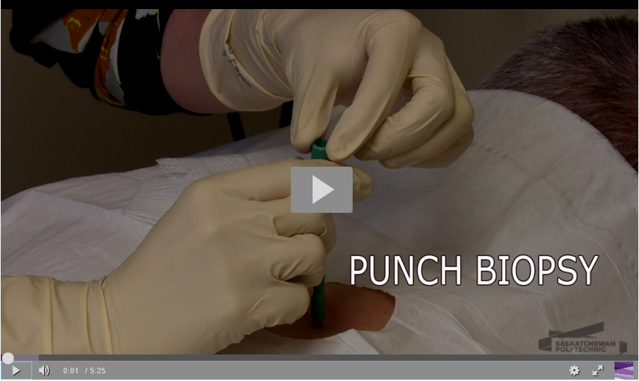
Step 6: Perform the punch

i) Use your first finger and thumb to stabilize the skin, stretching it slightly perpendicular to the skin tension lines

This will produce an oval rather than a circular wound, allowing the wound edges to approximate easily.

ii) Place the punch perpendicular to the skin and apply constant firm and downward pressure with a circular twisting motion

Turn the punch biopsy tool in a circular one direction motion. Do not to reverse the direction as this will help prevent shearing forces that separate the layers of the skin.

A “give” will be felt when the punch has reached the subcutaneous fat. Releasing pressure to soon will not result in a full-thickness cut. Avoid removing the punch to check the progress as this can also result in ragged wound edges or a shredded specimen.

iii) Remove the punch and place downward pressure to the wound edges

The specimen will remain in situ as it is still attached at the base. Applying downward pressure to the wound edges will pop up the core of the wound and allow you to grasp it for detachment. Gently grasp the core with forceps or a needle tip to fully elevate it and detach the core at its base using tissue scissors or a scalpel. Take care not to crush the tissue sample as this will affect laboratory analysis.

iv) Place the specimen in formalin for transfer to the lab

Step 7: Approximate wound edges as needed

Punches less than or equal to 3mm in diameter can heal by secondary intention. Punches greater than 3mm in diameter are best closed with one or two sutures to achieve hemostasis and produce a cosmetically acceptable scar. See chapter titled “Suturing of Uncomplicated Lacerations” for full procedure.

Step 8: Apply dressing to incision site

Apply an occlusive dressing to facilitate a moist environment and speed wound healing. Occlusive dressings can be made by using a topical antibiotic ointment or petroleum jelly on the wound and then covered with non-adherent gauze and tape, or a dressing such as Mepore.

Step 9:  Send specimen to the lab


https://videos.saskpolytech.ca/media/Punch+Biopsy/0_48to925i/108282082

License

Icon for the Creative Commons Attribution 4.0 International License

Advanced Procedural Skills Copyright © 2018 by Brittany Stephenson NP, BScN, MN is licensed under a Creative Commons Attribution 4.0 International License, except where otherwise noted.