Chapter Objectives
This chapter will enable you to identify:
- The various histological characteristics found in each layer of thin and thick skin
- The various histological characteristics of pigmented skin
- A lamellar corpuscle and a tactile corpuscle Before you begin to study the histological characteristics of the skin and its derivatives, it is important for you to review the anatomy of the various layers of the skin, as well as the functions of these layers.
Thick Skin
Thick skin (Figure 17-1), which lacks hair, is found on the palms of the hands and the soles of the feet. The most superficial layer of the epidermis is termed the stratum corneum. This is a rather thick layer composed of clear, dead, scale-like cells termed keratinocytes. They have a thickened cellular membrane and no nuclei, and they are closely interdigitated. The cytoplasm has been replaced by keratin proteins.
Deep to the stratum corneum is the stratum lucidum, a rather thin layer that has staining characteristics different from those of the stratum corneum. (In this photomicrograph, the stratum lucidum stains significantly darker than the more superficial stratum corneum.)
The stratum granulosum is found deep to the stratum lucidum and is composed of two to five rows of flattened, lightly staining cells, the longitudinal axes of which run parallel to the surface. Cells of this layer contain numerous cytoplasmic granules that usually stain intensely with hematoxylin.
The cells of the next two layers possess quite similar histological characteristics. Their nuclei are deeply chromatic, whereas the cytoplasm is deeply basophilic and contains numerous fine filaments that are not visible with the light microscope. The cells of the stratum spinosum, which are found in the layer immediately deep to the stratum granulosum, are polygonal in shape and are held together closely by desmosomes. During fixation, the cells of this layer often pull apart, thereby creating the “spines,” characteristic of this layer, that actually are fixation artifacts.
The deepest layer of the epidermis is the stratum basale. It is composed of a single layer of columnar or high cuboidal cells on a basal lamina (see description of basal lamina in section on Epithelial Sheets in Chapter 2). The shape of these cells varies from ovoid in the deepest layer to round in the cells closest to the stratum spinosum. These cells are somewhat smaller in overall size than those of the stratum spinosum.
The dermis of the skin is composed mostly of dense, irregular connective tissue (see section on Dense Irregular Connective Tissue [Dermis of the Skin] in Chapter 3) containing all three connective tissue fiber types. The dermis is subdivided into two layers: a deep reticular layer and a superficial papillary layer. The reticular layer is characterized by dense, collagenous fibers arranged into bundles that are often seen to unite to form secondary bundles that interlace frequently.
The papillary layer is composed of loose connective tissue (see section on Loose, Irregular Connective Tissue [Lamina Propria of Duodenum] in Chapter 3) with fine and closely arranged connective tissue fibers. Papillae are seen to indent the deepest layers of the epidermis. These papillae contain blood vessels (see section on Blood Vessels in Chapter 8) that nourish (but do not enter) the more superficial epidermal layers and peripheral nerves (see section on Peripheral Nerves in Chapter 7) that either terminate in the dermis or penetrate the basal lamina and enter the epidermis.
Thin Skin (Pigmented)
The terms thick and thin skin refer to the thickness of the epidermis. Thin skin lacks a stratum lucidum and therefore is composed of only four layers.
Thin skin (Figure 17-2) covers most of the body and usually possesses hair. However, thin skin that lacks hair may be found in the lips, terminal and lateral portions of the fingers and toes, and portions of the external genitalia. In thin skin, all of the layers are reduced in thickness, with the stratum corneum, stratum spinosum, and stratum basale being the only layers that are consistently represented in all parts of the body.
The color of the epidermis is due to a combination of the dermal blood supply, the thickness of the epidermis, and variable quantities of two pigments: carotene and melanin. Note the presence of pigment granules within the keratinocytes located in the deeper layers of the epidermis.
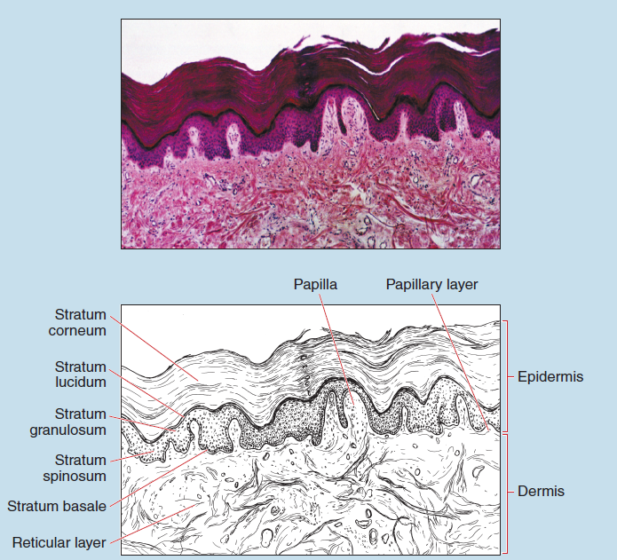
Figure 17-1 (25X): Thick skin (Caucasian).
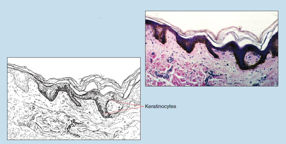
Figure 17-2 (25X): Thin skin (pigmented).
Scalp
Hairy scalp contains hair follicles and their associated sebaceous glands and arrector pili muscle (Figure 17-3).
Lamellar and Tactile Corpuscles
Lamellar Corpuscle
Lamellar (also termed pacinian) corpuscles (Figure 17-4) are found in association with joints, internal organs, periosteum of bones, the hypodermis, and the deeper layers of the skin— especially within the fingertips—and connective tissue in general. They are responsive to pressure and vibratory stimuli.
A lamellar corpuscle is composed of a myelinated axon surrounded by a capsular structure. As the neuron enters the corpuscle, its myelin covering remains intact for only one to three nodal segments. Then the myelin is replaced by flattened Schwann cells that will form the inner core of the capsule. Around this inner core are concentric lamellae composed of flattened cells similar to those found within the endoneurium of peripheral nerves. Delicate collagenous fibers, a few isolated capillaries, and fluid fill the space between these lamellae.
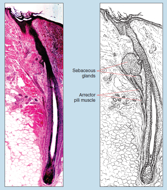
Figure 17-3 (25X): Scalp demonstrating a hair follicle.
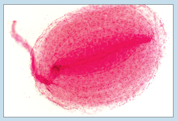
Figure 17-4 (70X): Lamellar corpuscle.
Tactile Corpuscle
Tactile corpuscles (also termed Meissner’s corpuscles) (Figures 17-5 and 17-6) are touch receptors found within hairless skin. They are especially numerous in the lips, hands (palmar surface and fingers), soles of the feet, and the undersurface of the toes. These structures are typically found just deep to the stratum basale within the dermal papillae and are orientated perpendicular to the surface of the skin.
Tactile corpuscles resemble a tapered cylinder. They are composed of one or two sensory nerves that penetrate the corpuscle and spiral within a cellular component of flattened Schwann cells, which then form several irregular lamellae around the sensory nerves.
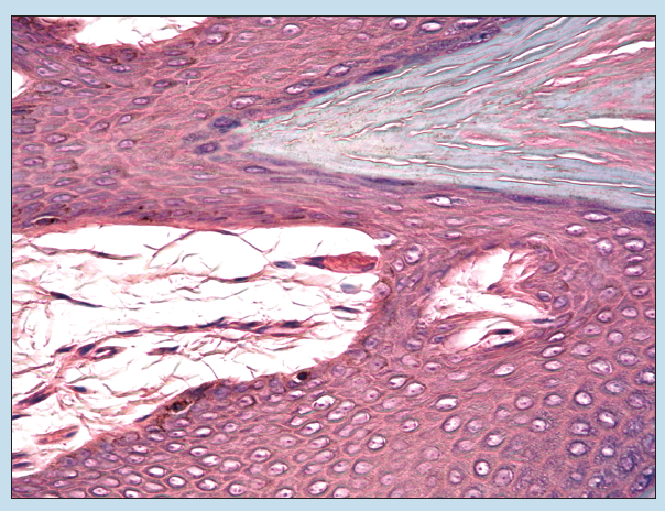
Figure 17-5 (70X): Tactile corpuscle.
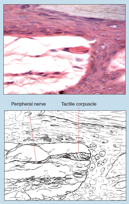
Figure 17-6 (140X): Tactile corpuscle.
