Chapter Objectives
This chapter will enable you to differentiate between and correctly identify:
-
- Elastic and muscular arteries
- Muscular arteries and arterioles
- Arteries and veins
- Veins and venules
- Venules, arterioles, and capillaries
- The various layers in the walls of arteries, veins, arterioles, and venules
- Anatomical and histological components of the heart
The cardiovascular system consists of the heart and blood vessels. The histology of cardiac muscle has been discussed previously (Chapter 6). However, to place cardiac muscle within the context of the cardiovascular system, it is discussed again in this chapter.
Heart
Cardiac Muscle
Cardiac muscle is limited to the heart. The second form of striated muscle, it exhibits a banding pattern that is quite similar to that seen in skeletal muscle. Cardiac muscle is commonly called a functional syncytium, in that excitation of one cardiac muscle cell quickly causes the excitation of all adjacent cells. This is due to the specialized junctions, termed intercalated discs, which are found between cardiac muscle cells.
Cardiac muscle, like skeletal muscle, is striated; as a result, these two tissues are often misidentified. In Chapter 6 (see section on Cardiac Muscle) you were asked to keep in mind the following histological differences between skeletal and cardiac muscle:
- Cardiac muscle cells are considerably smaller than skeletal muscle fibers. The exception to this rule is Purkinje fibers.
- Cardiac muscle cells branch frequently and therefore demonstrate a greater size variation when viewed in cross section.
- Cardiac muscle cells (except for Purkinje fibers) have a single, centrally located nucleus.
- Cardiac muscle cells possess specialized cell junctions called intercalated discs, which are visible in longitudinal sections of cardiac muscle.
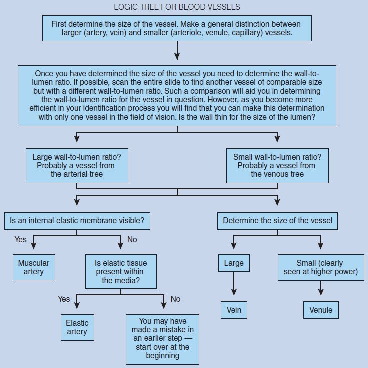
Figure 8-1 is a photomicrograph of cardiac muscle in longitudinal section. Note that cardiac muscle has only one centrally located nucleus per cell. Compare nucleus location and size variation demonstrated in this photomicrograph with that seen in comparable cross sections of skeletal muscle (see Figures 6-1 and 6-2).
Figure 8-2 clearly demonstrates the single, centrally located nucleus of cardiac muscle cells. Intercalated discs, seen between adjacent cardiac muscle cells, are specialized junctions that facilitate the spread of electrical impulses from one cardiac muscle cell to another. They also anchor adjacent cardiac muscle cells to one another, thereby allowing the spread of contractile forces from one cell to another within the heart.
Compare this figure to skeletal muscle in Figure 6-4. Note the following characteristics of cardiac muscle; they will help you to differentiate between skeletal and cardiac muscle when viewed in longitudinal section:
- Note how cardiac muscle cells branch.
- Note that the banding pattern of cardiac muscle is not as prominent as that of skeletal muscle.
- Note that the banding pattern between individual cardiac muscle cells is not “in register,” as was seen in skeletal muscle.
These three characteristics are all major histological features of cardiac muscle when viewed in longitudinal section.
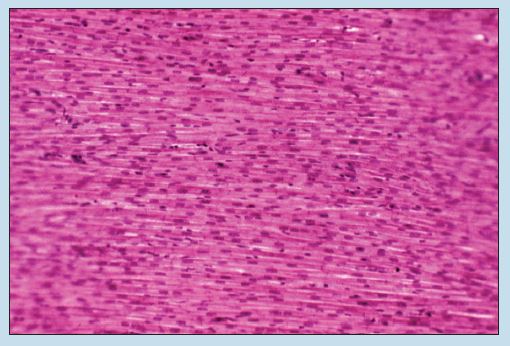
Figure 8-1 (100X): Cardiac muscle (longitudinal section).
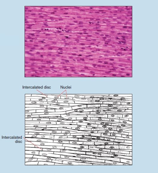
Figure 8-2 (200X): Cardiac muscle demonstrating intercalated discs (longitudinal section).
Figure 8-3 is a photomicrograph of cardiac muscle in cross section. This figure clearly demonstrates two major histological features of cardiac muscle when viewed in cross section:
- Note that cardiac muscle has only one, centrally located nucleus per cell.
- In addition, this tissue demonstrates an increased variation in cell size compared with skeletal muscle viewed in cross section (see Figure 6-1). This increased size variation is due to the branching of the cardiac myocytes.
Cardiac Muscle – Purkinje Fibers
Purkinje fibers are a part of the subendocardiac conducting network of the heart. Purkinje fibers are cardiac muscle cells specialized for high-velocity transmission of electrical impulses throughout the ventricle. Figure 8-4 demonstrates that Purkinje fibers:
- Usually occur in bundles
- Demonstrate a significantly increased muscle fiber diameter compared to other cardiac muscle cells
- Have an increased intracellular glycogen content, resulting in lighter staining properties
- Have a reduced number of myofibrils, and therefore a reduction in the banding pattern (when viewed in longitudinal section)
- Are multinucleate. The nuclei are more rounded and often found in groups of two or three within a single Purkinje fiber.
Figure 8-3 (200X): Cardiac muscle (cross section).
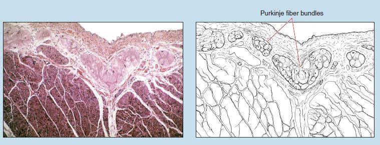
Figure 8-4 (50X): Cardiac muscle demonstrating Purkinje fibers.
Blood Vessels
All blood vessels within the cardiovascular system, with the exception of capillaries, follow the same histological organization. The walls of arteries, arterioles, veins, and venules all have three layers, or tunics.
- The innermost layer is the intima (or tunica intima), which consists of an endothelial tube of longitudinally arranged, simple, squamous epithelial cells termed endothelial cells.
- A sheet of elastic tissue, termed the internal elastic membrane (internal elastic lamina), forms the boundary between the intima and the second layer of the vessel, the media (or tunica media).
- The thin, squamous endothelial cells are separated from the internal elastic membrane by a layer of loose connective tissue termed the subendothelial connective tissue. The subendothelial connective tissue contains a few fibrocytes, occasional smooth muscle cells, and thin collagen fibers.
- The media is composed of multiple concentric layers of circularly arranged, smooth muscle cells.
- An external elastic membrane (external elastic lamina) serves as the boundary between the media and the outermost layer of the vessel, the adventitia (or tunica adventitia).
- In larger vessels you may find small blood vessels, termed vasa vasorum, within the media. The vasa vasorum serve to nourish the vessel.
- The adventitia consists of fibrocytes, longitudinal bundles of collagen fibers, and a loose network of thin elastic fibers.
- In larger vessels, vasa vasorum may also be found within the adventitia.
Arterial System
Elastic Arteries (Aorta)
Figure 8-5 is a cross section of a section of the aorta, an elastic artery, stained with hematoxylin and eosin.
The intima of an elastic artery is much thicker than its counterpart in a muscular artery, sometimes amounting to 20% of the total wall thickness. The intima of the aorta is composed of an inner endothelial lining that is continuous with the endothelial lining of the heart. The rounded nuclei of the endothelial cells may appear to project into the lumen of the vessel.
The external border of the intima is marked by an internal elastic membrane, which is not easily distinguished because of the large amount of elastin within the media of the vessel.
The media constitutes the bulk of the wall of an elastic artery and is composed of circularly arranged smooth muscle (see section on Smooth Muscle in Chapter 6) and a large number of fenestrated elastic membranes. These elastic membranes contain numerous openings termed fenestrae. The number of elastic laminae is believed to increase with age until approximately age 35.
At the outermost limit of the media is an external elastic membrane (external elastic lamina), but this layer is usually indistinguishable in the aorta, again because of the large amount of elastin within the media.
The adventitia of an elastic artery is usually quite thin. It consists of irregular connective tissue with collagen and elastic fibers, some fibroblasts, and smooth muscle cells. Vasa vasorum are visible in this figure.
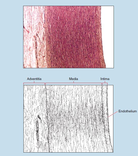 Figure 8-5 (50X): Aorta (elastic artery).
Muscular Arteries
Figure 8-6 is a photomicrograph of a muscular artery (right) and a vein (left), and Figure 8-7 is a higher magnification of the wall of a muscular artery.
Note the conspicuous intima in the muscular artery. The external limit of the intima is marked by an internal elastic membrane, which is convoluted because of the contraction of the elastic membrane during the fixation process.
The media of a muscular artery is composed of many circumferentially arranged smooth muscle cells. The media ends abruptly with a well-defined but thinner external elastic membrane.
Beyond the media is the adventitia. The adventitia contains circumferentially arranged fibrocytes and collagen fibers arranged both circumferentially and longitudinally along the length of the vessel. Although the thickness of the adventitia will vary from vessel to vessel, in some muscular arteries it is equal in thickness to the media.
Figure 8-8 is a photomicrograph of a muscular artery stained specifically for elastin. Note the prominent internal elastic membrane and external elastic membrane within the wall of this vessel.
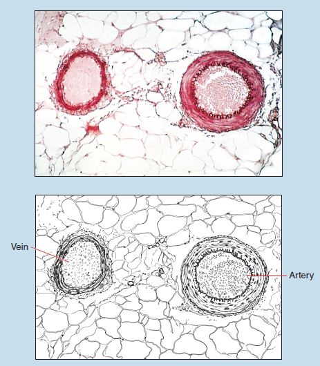
Figure 8-6 (50X): Cross section of artery and vein.
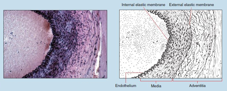 Figure 8-7 (100X): Cross section of a muscular artery.
Arterioles
Arterioles serve as the transition vessel between muscular arteries and capillaries. Three distinguishable coats (intima, media, and adventitia) are still evident in an arteriole.
Although a few, isolated elastic fibers may be seen in arterioles, an internal elastic membrane is not visible in small arterioles. In larger arterioles, however, a faint internal elastic membrane may be visible.
The media of an arteriole possesses only one or two layers of smooth muscle. The adventitia is thin and quite difficult to distinguish from any surrounding connective tissue.
Because of the difficulty and ambiguity in distinguishing between small and large arterioles, in this text an arteriole will be defined as a vessel that:
- Has the wall–thickness-to-lumen ratio that qualifies it as being a part of the arterial system
- Possesses one or two layers of smooth muscle
- Lacks an internal elastic membrane
Figure 8-9 demonstrates two arterioles within the adipose tissue that surrounds the pancreas. Close examination will show that these arterioles possess one to two layers of smooth muscle and lack an internal elastic membrane.
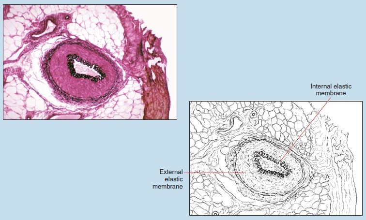 Figure 8-8 (100X): Cross section of a muscular artery, stained specifically for elastin.
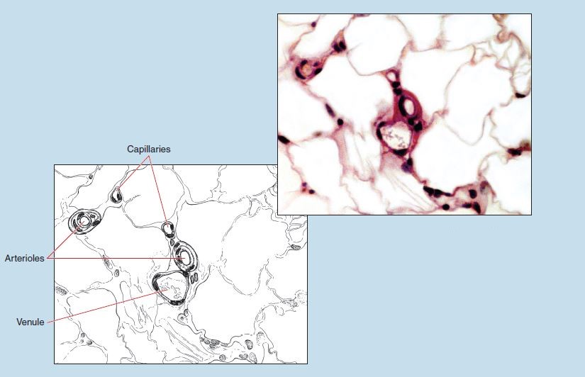
Figure 8-9 (200X): Arterioles, venules, and capillaries within adipose tissue.
Capillaries
Figures 8-9 and 8-10 are photomicrographs of capillaries of varying diameters within adipose tissue. Note that the lumen diameter ranges in size from slightly smaller than a single red blood cell (RBC) to one and one-half times the diameter of an RBC.
The wall of a capillary has only one layer—the intima. The intima of a capillary is composed of one to three circumferentially arranged endothelial cells.
Because a capillary lacks a media, no smooth muscle cells will be found within its wall.
Note the sectioning and staining artifacts in the lower left corner of Figure 8-10.
Table 8-1: Summary of Histological Characteristics of Capillaries and Arteries
| Blood Vessels | Intima | Media | Adventitia |
| Capillary | Single layer of endothelial cells and underlying subendothelial layer of loose (areolar) connective tissue. | None present | None present |
| Arterioles | Endothelial cells and underlying connective tissue. Small arterioles lack an internal elastic membrane; larger arterioles may have a very delicate internal elastic membrane. | Only 1 to 2 layers of smooth muscle | Dense, irregular connective tissue compose of longitudinally and circumferentially arranged collagen and elastin fibers |
| Muscular artery | Endothelial cells on underlying connective tissue; internal elastic membrane present | Smooth muscle and some elastin and collagen; external elastic membrane present | Composed of longitudinally and circumferentially arranged collagen and elastin |
| Elastic artery | Relatively thick layer with endothelium on underlying connective tissue; internal elastic membrane present but not distinct because of numerous layers of elastic tissue within media | Thick layer of smooth muscle with fenestrated layers of elastin; external elastic membrane present but not distinct because of numerous layers of elastic tissue within media | Thin layer composed of collagen longitudinally arranged; often interspersed with elastic fibers |
Venous System
Vessels of the venous system possess the same three-layered organization as the arterial system. However, there are several histological characteristics of the venous system that easily distinguish its vessels from those of the arterial side of the cardiovascular system:
- The ratio of wall thickness to lumen diameter is considerably smaller for vessels of the venous system.
- The media is relatively thin and poorly developed in vessels of the venous system.
- Internal and external elastic membranes are more difficult to distinguish in vessels of the venous system.
Veins
Return to Figure 8-6. The vessel on the left is a vein. Use this photomicrograph to distinguish the histological differences between an artery and a vein. Note that the size of the lumen in both vessels is approximately equal. However, the artery has a thicker wall than the vein. This wall–thickness-to-lumen ratio is a major feature to consider when trying to identify arteries and veins. An artery will always have a larger wall–thickness-tolumen ratio than a vein of corresponding size.
Keeping in mind the differences in the wall-to-lumen ratios between the two vessels in Figure 8-6, now examine the vein in the lower left corner of Figure 8-11. Only three to four layers of smooth muscle are seen within the media of this vein. You will also note that the adventitia and intima are more prominent in the vein as compared with that seen in an artery of comparable size (Figure 8-7).
Venules
Figures 8-9, 8-11, and 8-12 demonstrate the histology of an arteriole and venule. A venule is a transition vessel between a capillary and a vein. Because venules are part of the venous system, they have all of the histological characteristics of veins.
First, note the magnification of each photomicrograph. Figure 8-11 demonstrates the relative size of an arteriole and venule compared with a vein and muscular artery, and Figure 8-12 demonstrates the relative sizes of an arteriole, capillary, and venule.
Examination of Figures 8-9 and 8-12 clearly demonstrates the wall-to-lumen ratio in arterioles and venules, as well as the thickness of the media of a venule. Both of these characteristics are major histological features of a venule.
Table 8-2: Summary of the Histology of the Cardiovascular System: Veins
| Blood Vessel | Intima | Media | Adventitia |
| Venules | Endothelial cells on layer of areolar connective tissue | 1 to 2 layers of smooth muscle | Relatively thick layer compared with intima or media |
| Veins: medium diameter | Layer composed of endothelium resting on subendothelial layer of thin collagen bundles interspersed with elastin. Internal elastic membrane present | Smooth muscle interspersed with elastic fibers that are often arranged longitudinally; external elastic membrane present but not prominent |
Forms bulk of wall; composed of collagen and elastin and some longitudinally arranged smooth muscle fibers |
| Veins: large diameter | Similar to that of medium-diameter veins. | Poorly developed layer; collagen and some elastic fibers interspersed between smooth muscle | Relatively thick layer; bundles of irregularly arranged collagen and elastic fibers interspersed with longitudinally arranged smooth muscle |
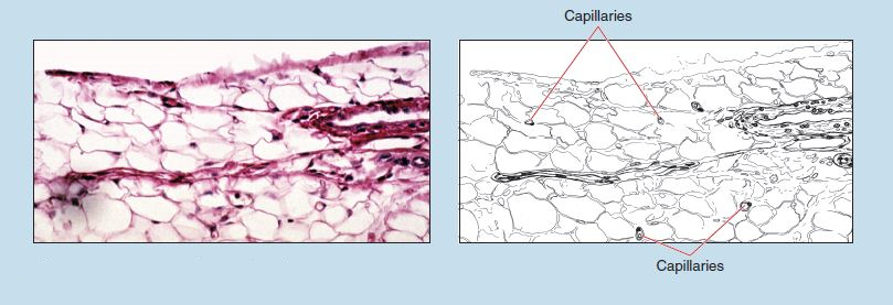 Figure 8-10 (200X): Capillaries within adipose tissue.
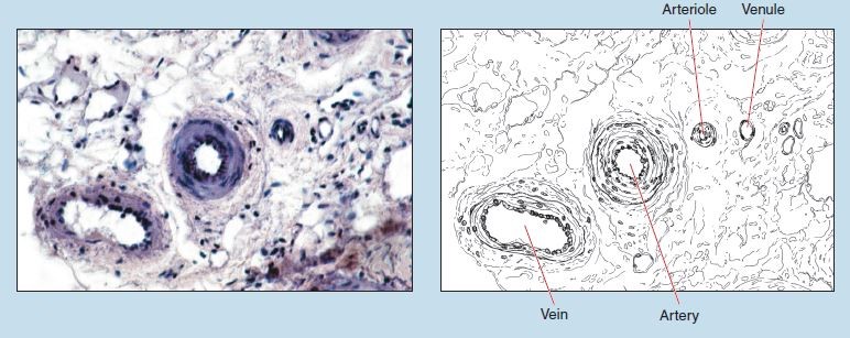 Figure 8-11 (200X): Cross section of a vein.
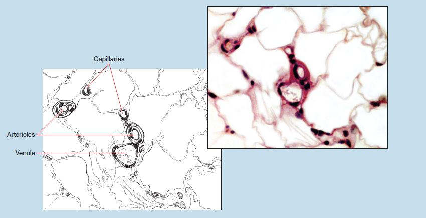
Figure 8-12 (200X): Cross section of a venule and arteriole.