Chapter Objectives
This chapter will enable you to:
-
- Recognize peripheral nerves (including neuronal somas and peripheral processes).
- Discuss the differences seen in nervous tissue stained with hematoxylin and eosin (H & E) and heavy metal (silver, gold) stains.
- Understand the organization of the Central Nervous System (CNS) and Peripheral Nervous System (PNS) as observed in the specimens presented in this chapter.
- Differentiate the various specimens of the CNS and PNS presented in this chapter, as well as their characteristics and cellular components.
The nervous system is composed of two cell types: neurons and glial supporting cells. Both of these cell types are found within the two subdivisions of the nervous system, the PNS and CNS. The CNS is composed of the brain and spinal cord, and the PNS consists of everything else.
The brain and spinal cord consist of white matter and gray matter. White matter is composed mostly of myelinated neuronal processes and supporting cells. Some unmyelinated processes may also be found within white matter. The gray matter is composed of neuronal somas, unmyelinated neuronal processes, and supporting cells.
While reading through this chapter you will notice that most of the specimens are represented twice: once stained with H & E and then with a heavy metal stain (silver). (See Appendix.) Study both types of specimens equally, because each of these stains will enable you to see different structures within each specimen. You should make constant mental notes as to which stain represents which structures most clearly. It is also important for you to keep in mind the gross anatomy of the central and peripheral nervous systems as you proceed through this chapter.
Central Nervous System
Spinal Cord
The spinal cord, when viewed in cross section, is composed of central gray matter and peripheral white matter. The gray matter resembles a butterfly in shape and may be subdivided into three pairs of horns: dorsal, lateral (found only between the first thoracic and second lumbar spinal levels), and ventral. Sensory neurons enter the spinal cord via the dorsal horn, whereas motor neurons originate within the lateral (autonomic nervous system, sympathetic branch) and anterior (somatic nervous system and autonomic nervous system, parasympathetic branch) horns.
Figure 7-1 is a line drawing of a cross section of the spinal cord. Use this line drawing to refresh your understanding of the gross anatomy of the spinal cord, paying particular attention to the following structures in this figure: ventral horn, dorsal horn, white matter, gray matter, meninges, central canal, dorsal root ganglion, dorsal root of the spinal nerve, and the ventral root of the spinal nerve.
Figure 7-2 is a photomicrograph of the anterior (ventral) horn of the spinal cord stained with H & E. The neuronal cell bodies of the anterior horn cells are quite large. They contain a single, large, centrally located nucleus. Several large neuronal processes exiting from the anterior horn cells are also evident in this section.
Many of the other nuclei found within the gray matter of the cord belong to neural glial cells. Their cytoplasm is not evident in these photomicrographs. The identification of the three types of glial cells within the CNS (microglia, astroglia, and oligodendroglia cells) requires the use of special stains and is not possible in these photomicrographs.
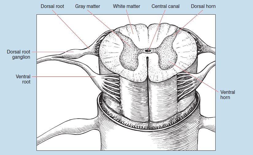
Figure 7-1: Gross anatomy of a cross section of the spinal cord.
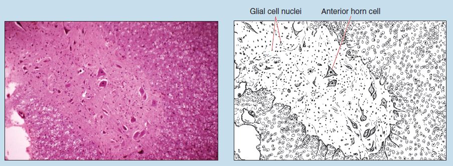
Figure 7-2 (50X): Ventral (anterior) horn of the spinal cord.
Figure 7-3 is a photomicrograph of the anterior (ventral) horn of the spinal cord, viewed in cross section and stained with silver. As you compare this with Figure 7-2, what structures are stained more readily with silver than H & E? For which is the reverse true?
A cross section of the spinal cord at the junction of the anterior horn and peripheral white matter (see Figure 7-1) and stained with H & E is seen in Figure 7-4. Several large neuronal cell bodies may be seen within the central gray matter. Many of the smaller nuclei present within the white matter represent various neuroglia. The axons in true cross section appear as a dot surrounded by a white halo because the peripherally located myelin has dissolved during the fixation process and therefore is not available for staining.
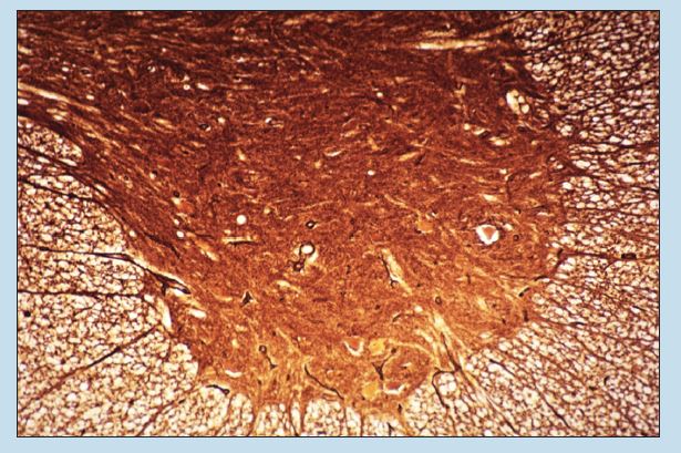
Figure 7-3 (50X): Ventral (anterior) horn of the spinal cord (silver stain).
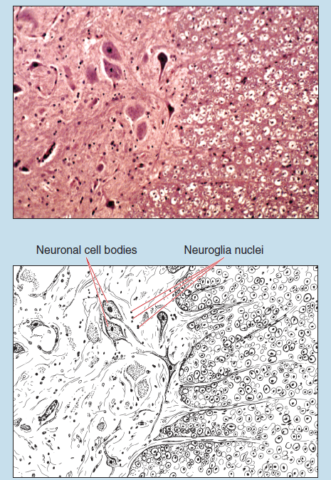
Figure 7-4 (100X): Cross section of the spinal cord at the junction of the anterior horn and peripheral white matter.
Figure 7-5 represents a silver stain of a cross section of the spinal cord at the junction of the anterior horn and peripheral white matter (see Figure 7-1). In the central gray matter you will note several large neuronal cell bodies. Central nuclei are visible in some of these cells. You will also see several neuronal processes exiting from the gray matter and entering the white matter.
Now compare the images seen in Figures 7-4 and 7-5. As you view the neurons and axons seen in Figure 7-4, what structures are clearly visible that are not seen in Figure 7-5? For which is the reverse true?
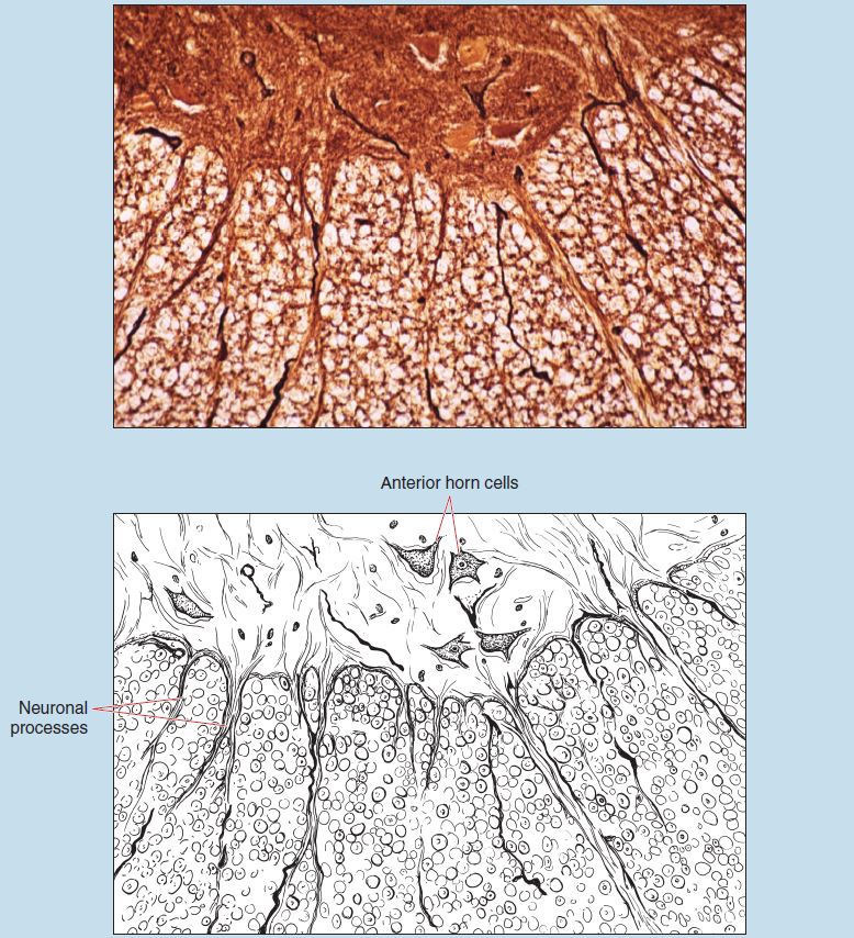
Figure 7-5 (100X): Cross section of the spinal cord at the junction of the anterior horn and peripheral white matter (silver stain).
Cerebellum
The surface of the cerebellum of the CNS is thrown into a large number of folds, termed folia cerebelli, which serve to increase the surface area of the cerebellum. The cerebellum is divided into a cortex of gray matter and a medullary center of white matter. The cerebellar cortex is divided into three layers: molecular, Purkinje, and granular. Each layer is named for the predominant cell type found within that layer.
Figures 7-6 and 7-7 are H & E photomicrographs of the cerebellar cortex. Note that the cerebellar cortex is composed of three layers: the outer molecular layer (sometimes termed the plexiform layer), which contains few cells and no myelinated fibers, an intermediate Purkinje layer, and the inner granular layer. Deep to the cortex is the histologically quite indistinct white matter.
In Figure 7-7 the border between the granular and molecular layers is the site of rather large neurons called Purkinje cells, which are characteristic of the cerebellum. Within the molecular layer you may be able to see basket cells. These neurons are quite small and demonstrate a small amount of cytoplasm around a centrally located nucleus.
In the granular layer, near its junction with the molecular layer, you may see occasional stellate neurons (also termed Golgi cells). These neurons are characterized by nuclei that are larger than those of the granule cells. Granule cells do not demonstrate the characteristic structure of neurons in that they possess dark nuclei, have no obvious nucleolus, and show no clearly stained cytoplasm. They are quite difficult to distinguish from glial cells.
Figure 7-8 is a silver stain of the junction between the granular and molecular layers of the cerebellum. Which neurons are visible within this photomicrograph?
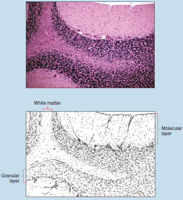
Figure 7-6 (50X): Cerebellum.
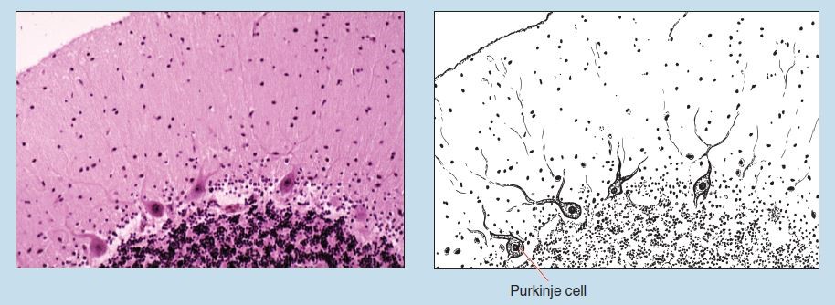
Figure 7-7 (200X): Cerebellum.
Cerebral Cortex
Ninety percent of the human cerebral cortex is neocortex (also termed isocortex), which is the phylogenetically newest and structurally most complex. The neocortex is composed of six layers that the beginning histology student will find difficult to distinguish. Each layer possesses several neuronal and glial cell types. These cells are not randomly arranged; one or more neurons dominate within each layer. Horizontal fibers associated with each layer also give a laminated appearance to the cortex.
The layers of the neocortex (from superficial to deep) and the neurons contained within them are as follows:
- Molecular layer: horizontal cells (also termed horizontal cells of Cajal)
- External granular layer: stellate (granule) cells
- External pyramidal layer: stellate cells and large pyramidal cells
- Internal granular layer: stellate cells
- Internal pyramidal layer: large and medium pyramidal cells
- Multiform layer: inverted pyramidal neurons (also termed Martinotti cells)
Figure 7-9 is a photomicrograph of the superficial layers of the cerebral cortex. You will note that meninges and associated blood vessels that normally cover the surface of the cerebral cortex are clearly visible. Within the molecular layer you will see the horizontal neurons (horizontal cells of Cajal). These relatively small cells are stellate or spindle shaped and their axons give rise to horizontally directed fibers. Deeper within the cerebral cortex are the large, triangular-shaped pyramidal cells.
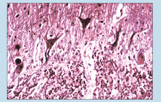
Figure 7-8 (200X): Cerebellum (silver stain).
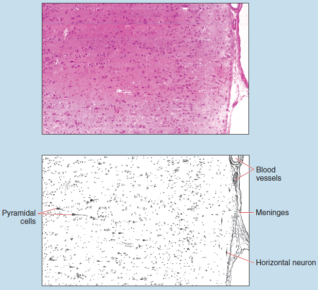
Figure 8-9 (50X): Cerebral cortex – superficial layers.
Peripheral Nervous System
Spiral Ganglion (Dorsal Root Ganglion)
Figures 7-10 and 7-11 demonstrate that the neuronal somas for sensory spinal nerves are located within the spinal (dorsal root) ganglia. Note the prominent, large, pale-staining nucleus and the smaller, darker-staining nucleolus within the soma. Also visible within these sections are horizontally aligned myelinated axons.
Surrounding the neuronal somas are satellite cells, a type of glial cell found with the peripheral nervous system.
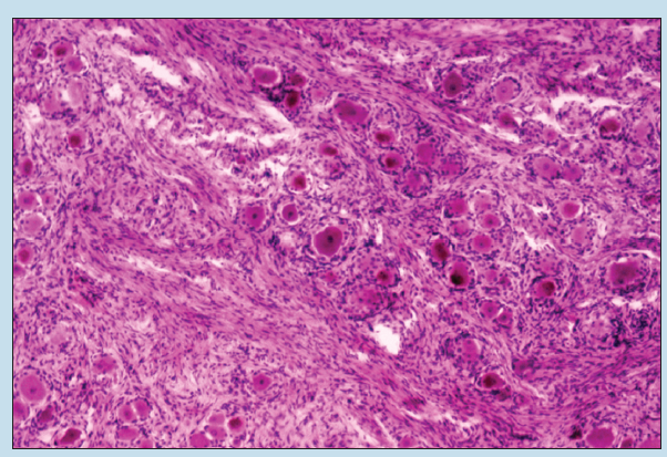
Figure 7-10 (50X): Dorsal root (spinal) ganglion.
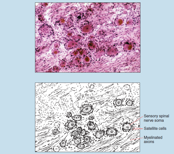
Figure 7-11 (100X): Dorsal root (spinal) ganglion.
Peripheral Nerve
Peripheral Nerve: Longitudinal Section
Figure 7-12 is a photomicrograph of a longitudinal section of peripheral nerve stained with H & E. At this magnification, several characteristics are apparent:
- Note the wavy appearance of the section. In addition, you will note several sections in which both longitudinal and cross sections are visible in this photomicrograph. Both of these are due to the recoil of the preparation during fixation and therefore are fixation artifacts.
- This longitudinal section is of a fasciculus of a peripheral nerve, thus the surrounding connective tissue is the perineurium.
- At this magnification, a longitudinal section of peripheral nerve may be easily confused with dense regular connective tissue (tendon) or smooth muscle.
Figure 7-13 is a photomicrograph of the same specimen taken at higher magnification. As you look at this specimen, note the following:
- The myelin sheath of these neurons has been dissolved away during the fixation process.
- A centrally located axon may be seen in some of the peripheral nerves.
- Several indentations may be seen in some of the myelin sheaths of the peripheral nerve. These indentations represent myelin sheath gaps (also termed Nodes of Ranvier).
- Numerous nuclei, which represent Schwann cell and fibrocyte nuclei, are evident in this figure.
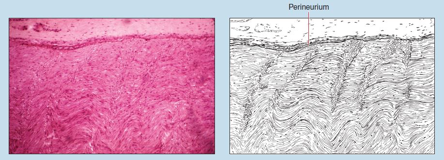
Figure 7-12 (100X): Peripheral nerve (longitudinal section).
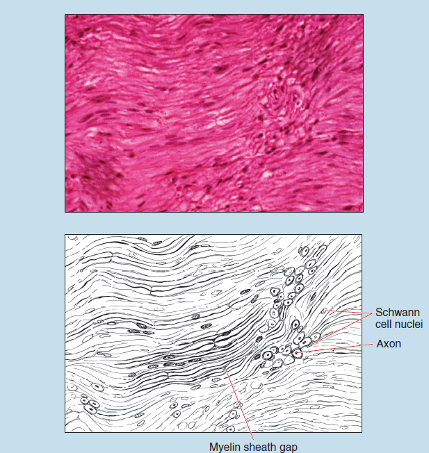
Figure 7-13 (200X): Peripheral nerve (longitudinal section).
Peripheral Nerve: Cross Section
Figures 7-14, 7-15, and 7-16 are photomicrographs of a large peripheral nerve bundle containing several smaller bundles within a connective tissue sheath. Each bundle, in turn, is composed of many large peripheral axons. Figures 7-14 and 7-15 were stained with H & E, and Figure 7-16 is a silver stain preparation.
As you progress from Figure 7-14 to 7-16, you will note the varying connective tissue investments: epineurium, perineurium, and endoneurium.
Figure 7-16 is a photomicrograph of peripheral nerve in cross section, stained with silver. Note the individual axons. The axons appear as a dot surrounded by a white halo because the myelin has been dissolved away during the fixation process.
As you compare Figures 7-14 through 7-16, what structures are more visible with silver stain than H & E? For which is the reverse true?
Several other questions are worth considering as you look at peripheral nerve in cross or longitudinal sections. In these photomicrographs, several nuclei are present. What cell types are represented by these nuclei? What cell type is definitely not represented by these nuclei here?
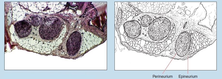 Figure 7-14 (50X): Peripheral nerve (cross section).
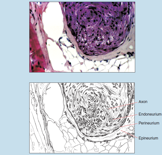
Figure 7-15 (200X): Peripheral nerve (cross section).
Peripheral Nerve: Autonomic Nervous System Ganglion
The gastrointestinal tract has rich autonomic nervous system innervation. Figures 7-17 and 7-18 demonstrate a myenteric neural plexus (also termed a Myenteric Plexus of Auerbach). This is a ganglion of the parasympathetic branch of the autonomic nervous system that is found between the circular and longitudinal layers of smooth muscle (see section on smooth muscle in Chapter 6 of the muscularis externa of the small intestine.) Individual neuronal somas with prominent nuclei are visible within this ganglion. Several blood vessels are also visible in these sections between the inner circular and outer longitudinal layers of smooth muscle.
Following are questions to consider for this structure:
- On the basis of what you know of anatomy, why is this a ganglion of the parasympathetic nervous system and not a ganglion of the sympathetic nervous system?
- The autonomic nervous system is a two-neuron system, composed of preganglionic and postganglionic neurons. Are the somas visible within this ganglion from preganglionic or postganglionic neurons? Why?
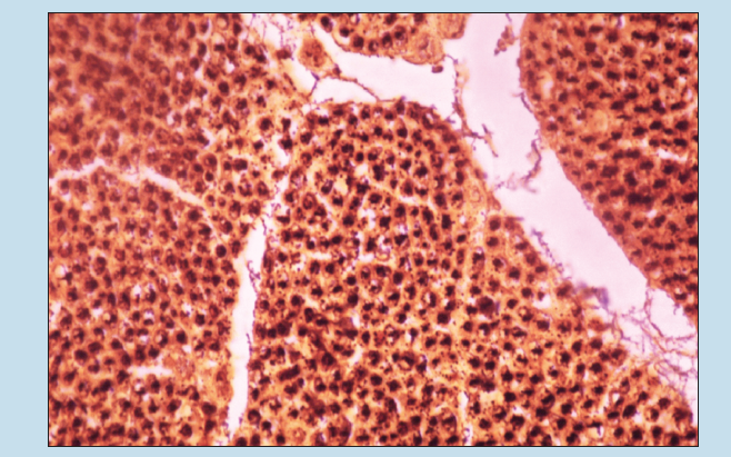
Figure 7-16 (200X): Peripheral nerve (cross section) (silver stain).
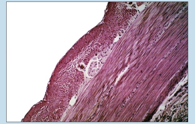
Figure 7-17 (100X): Myentric neural plexus of the small intestine.
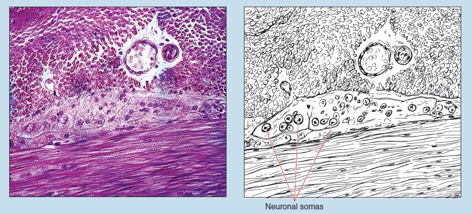
Figure 7-18 (200X): Myentric neural plexus of the small intestine.
Commonly Misidentified Tissues
Peripheral Nerve, Dense Regular Connective Tissue (Tendon and Elastic Ligament)
Nervous tissue typically presents some identification problems. Peripheral nerve, smooth muscle, and dense regular connective tissue are often mistaken for each other and as a result are incorrectly identified. Therefore it is important for you to keep the following differences in mind when trying to differentiate between these different tissues:
Peripheral Nerve (Longitudinal Section) (Review Figures 7-12 and 7-13 in section on Peripheral Nerve)
- Presence of myelin sheath gaps (Nodes of Ranvier) underhigh-dry or oil-immersion objective
- Generally “washed out” appearance of specimen
- Axons surrounded by myelin sheath, which has been dissolved away during fixation process
- Nuclei numerous and rounded (Schwann cells), or flat and dark (fibroblasts and fibrocytes
- Generally wavy appearance
- Epineurium or perineurium present
Dense Regular Connective Tissue (Tendon and Elastic
Ligament) (Longitudinal Section) (Review Figures 3-8 to 3-11 in section on Dense Regular Connective Tissue in Chapter 3)
- Absence of myelin sheath gaps (Nodes of Ranvier) under high-dry or oil-immersion objective
- Fibers stained darkly; less wavy than peripheral nerve; more irregular in appearance
- Nuclei more numerous and more evenly stained
- Acidophilic collagen fiber bundles
- Long, thin nuclei arranged in parallel
Peripheral Nerve and Smooth Muscle
Peripheral Nerve (Longitudinal Section) (Review Figures 7-12 and 7-13 in section on Peripheral Nerve)
- Presence of myelin sheath gaps (Nodes of Ranvier) under high-dry or oil-immersion objective
- Generally “washed out” appearance of specimen
- Nuclei numerous and rounded (Schwann cells), or flat and dark (fibroblasts and fibrocytes)
- Generally wavy appearance
- Blood vessels lacking (except in large nerves)
- Epineurium or perineurium present
Smooth Muscle (Longitudinal Section) (Review Figures 6-8 and 6-9 in section on Smooth Muscle in Chapter 6)
- Absence of myelin sheath gaps (Nodes of Ranvier)
- More uniform staining to general specimen
- More uniformity in size, shape, and staining qualities of nuclei within specimen
Peripheral Nerve (Cross Section) (Review Figures 7-14 and 7-15 in section on Cross Section of Peripheral Nerve)
- Regular arrangement to connective tissue investment
- Presence of “halos” around axons
- Variation in staining qualities of nuclei within tissue
- Large degree of uniformity in size of structures (axons) represented in cross section
- Epineurium or perineurium present
Smooth Muscle (Cross Section) (Review Figure 6-9 in section on Smooth Muscle in Chapter 6)
- Decreased amount of connective tissue within specimen
- Lack of myelin “halos”
- Uniformity in staining qualities of nuclei within tissue
- Considerable size variation of structures (myocytes)
represented in cross section
