Chapter Objectives
At the completion of this chapter you should be able to:
-
- Distinguish among the various formed elements found in human blood
Red Blood Cells and Platelets
Red blood cells (RBCs or erythrocytes) are anucleated, biconcave cells that perform their major functions within the blood vessels of the cardiovascular system. Figure 9-1 is an oil-immersion photomicrograph demonstrating red blood cells and platelets. Some of the red blood cells stain quite homogeneously, whereas a few may show a pale center. Other than this minor staining difference, red blood cells are unremarkable. Platelets are also visible as small purple dots between the RBCs in this photomicrograph.
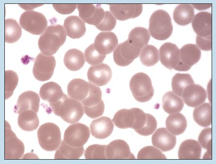
Figure 9-1 (350X): Red blood cells and platelets (Wright stain).
Leukocytes
Leukocytes (white blood cells) migrate out of the blood vessels to perform their functions (see Table 9-1). Unlike erythrocytes, leukocytes are nucleated blood cells that are subdivided into two groups on the basis of the presence (granulocytes) or absence (agranulocytes) of granules within their cytoplasms. Lymphocytes and monocytes are classified as agranulocytes, whereas neutrophils, basophils, and eosinophils are classified as granulocytes.
Table 9-1: Comparison of Blood Cells
| Cell | Cells per μl | Major Function |
| Erythrocyte | 4.5-6.5 × 10⁶ | Transport of oxygen and carbon dioxide |
| Leukocyte (WBC) | 4-11 × 103 | Defense mechanisms |
| Neutrophilic granulocytes | 50%-70% of total WBC count | First white blood cell to arrive at the site of an infection in any large numbers; phagocytose antigens |
| Eosinophilic granulocyte | 1%-4% of total WBC count | Release of histamines in inflammatory reactions |
| Basophilic granulocyte | Less than 1% of total WBC count | Involved in acute inflammation and allergic reactions by releasing histamine and interleukins |
| Monocyte | 2%-8% of total WBC count | Migrate to sites of infection in large numbers; able to leave the bloodstream and ingest bacteria and cellular debris. Are termed macrophages when they exit the cardiovascular system and enter the extracellular space. |
| Lymphocyte | 20%-40% of total WBC count | Appear at infection site at approximately the same time as monocytes. Some transform into plasmocytes. Only WBC able to recognize specific antigens and only WBC with an immunological memory. |
WBC, White blood cell.
Agranulocytes
Lymphocyte
Lymphocytes (Figure 9-2) are the second most common cell type among the leukocytes, with neutrophils being the most common. (See Table 9-1.) The most obvious feature of a lymphocyte is a rounded nucleus that contains a small dimple on one side. A rim of cytoplasm surrounds the nucleus, which is variable in both amount and staining characteristics, depending on the size of the lymphocyte.
Monocytes
Monocytes (Figure 9-3) are not as numerous as lymphocytes. The cell is quite large and possesses a rather blue-gray, irregularly staining cytoplasm. The nucleus will vary from kidney shaped to horseshoe shaped and is usually mottled in appearance.
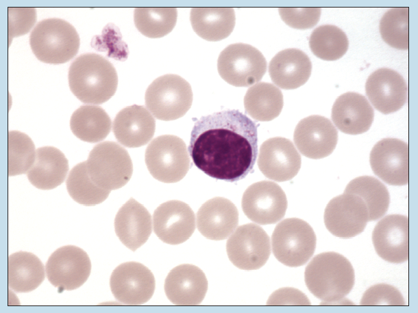
Figure 9-2 (350X): Lymphocyte (Wright stain).
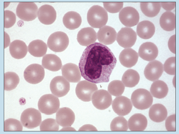
Figure 9-3 (350X): Monocyte (Wright stain).
Granulocytes
Granulocytes are subdivided into different categories according to (1) the presence or absence of cytoplasmic granules, (2) the staining properties of those granules, and (3) the shape of the nucleus of the cell. Because the cytoplasmic granules of eosinophils and basophils often obscure the nucleus from view, it is best to base your identification mostly on the staining properties of the cytoplasmic granules, if they are present.
Eosinophilic Granulocyte
Eosinophilic granulocytes (eosinophils) have a bilobed nucleus, with each lobe being quite smooth in outline and approximately the same size (Figure 9-4). The cytoplasm contains rather large, eosinophilic granules. In some eosinophils (such as the one in this photomicrograph), the granules are so prominent that they obscure some or all of the nucleus from view.
Basophilic Granulocyte
Basophilic granulocytes (basophils) (Figure 9-5) are quite rare in a normal blood smear. Such cells contain a bilobed nucleus and prominent basophilic cytoplasmic granules, which normally obscure the nucleus from view.
Neutrophilic Granulocytes
Neutrophilic granulocytes (neutrophils) are by far the most numerous leukocytes (Figure 9-6). Neutrophils are approximately the same size as basophilic and eosinophilic granulocytes. Neutrophils contain a segmented nucleus and have a relatively clear cytoplasm because the granules are just at or below the resolution of the light microscope. The nucleus of a neutrophil may possess as few as two segments or as many as six. These segments, which are joined together by thin strands of chromatin, are quite irregular in shape. The shape of the nucleus and the relatively clear cytoplasm both serve as excellent histological characteristics.
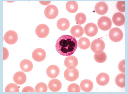
Figure 9-4 (350X): Eosinophilic granulocyte (Wright stain).
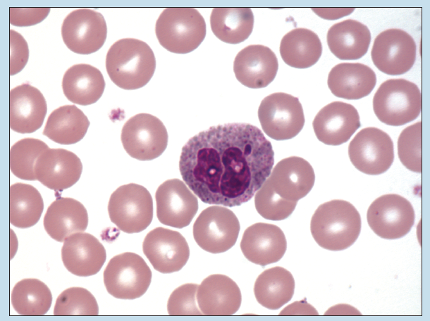
Figure 9-5 (350X): Basophilic granulocyte (Wright stain).
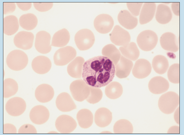
Figure 9-6 (350X): Neutrophilic granulocyte (Wright stain).
Commonly Misidentified Tissues
Monocytes and Large Lymphocytes
Assuming that the granules found in neutrophils, basophils, and eosinophils stain normally, thereby minimizing identification errors, the two blood cells that are most commonly misidentified are the monocyte and lymphocyte.
Monocyte (Review Figure 9-3 in section on Monocytes)
-
- Is the larger of the two cells in question (approximately 12 to 15 μM in diameter).
- Possesses more cytoplasm. Cytoplasm stains blue/gray in color.
- Nucleus usually possesses noticeable indentation.
Lymphocyte (Review Figure 9-2 in section on Lymphocytes)
-
- Is the smaller of the two cells in question (approximately 10 to 12 μM in diameter)..
- Has slightly less cytoplasm, often seen as a thin rim around the nucleus. Cytoplasm stains blue in color.
- Nucleus is more rounded. If indentation is present, it is considerably less prominent in the large lymphocyte.
