This chapter will enable you to:
1. Differentiate among the three types of cartilage
2. Differentiate among the different cell types normally found in cartilage
3. Identify the components of the perichondrium
Characteristics of Cartilage
Like other forms of connective tissue, cartilage is composed of cells and an extracellular matrix of fibers and ground substance. However, cartilage differs from previously studied forms of connective tissue in several ways:
- Mature cartilage is avascular.
- Cartilage is not penetrated by any element of the nervous system.
- The cells of cartilage, termed chondrocytes, are interspersed within the ground substance of cartilage and are to be found trapped within small, cavity-like spaces termed lacunae.
- The ground substance of cartilage has a gel-like consistency, which gives the tissue an elastic firmness capable of withstanding large amounts of pressure and shearing forces.
Subcategories of Cartilage
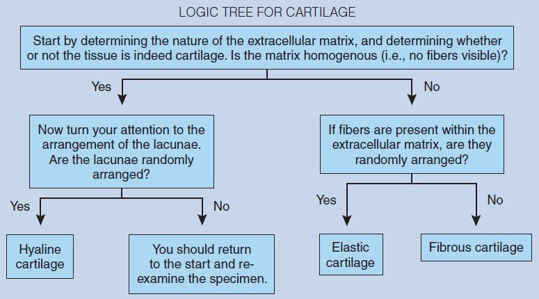
Cartilage is subdivided into three categories: hyaline, elastic, and fibrous cartilage (fibrocartilage). These different types of cartilage are distinguished by the appearance and components of the ground substance and fibers and by the presence or absence of an outer fibrous layer of connective tissue termed the perichondrium.
- Hyaline cartilage is the most common of the three forms of cartilage. It will be found in numerous locations, including the articulating surfaces of synovial joints, the cartilage model of developing long bones, the epiphyseal growth plates of long bones, and as supporting structures within the respiratory tree.
- Fibrous cartilage is typically found in areas of considerable stress. Because of the arrangement of fibers within the extracellular matrix, fibrous cartilage is better able to withstand compressive and shearing forces than the other forms of cartilage. Fibrous cartilage will be found within the pubic symphysis, annulus fibrosus of intervertebral discs, articular discs of the temporomandibular and sternoclavicular joints, and the menisci within the knee joint. It is also found in the areas of transition between cartilage and bone and forming the linkage between tendons and bones.
- Elastic cartilage is the least common of the three forms. It is found within the auricle of the external ear, the auditory tube of the middle ear, and the epiglottis.
Hyaline Cartilage
Hyaline Cartilage (Trachea)
As you examine these photomicrographs, it is important to keep in mind that hyaline cartilage should be the reference by which you judge all other cartilages. If you know the histological characteristics of hyaline cartilage, you can use them to compare and contrast all other unknown cartilage specimens and thereby arrive at a correct tissue identification.
As mentioned earlier, the best way to begin an initial examination of a new preparation or the identification of an unknown tissue is to start on scanning power (Figure 4-1) and then slowly progress to medium power (Figure 4-2) and high-dry (Figure 4-3) objectives.
The trachea is held open by C-shaped pieces of hyaline cartilage. As you begin your examination of Figure 4-1 you will see hyaline cartilage surrounded by adipose tissue and skeletal muscle. You will note that the hyaline cartilage is surrounded by dense regular connective tissue termed the perichondrium.
Continued examination shows that the interior of the cartilage is composed of cells, termed chondrocytes, which are trapped within spaces called lacunae. The prominence of these lacunae within the collagen is due to the shrinkage of the chondrocytes during fixation—a fixation artifact commonly seen in hyaline cartilage preparations. This fixation artifact creates an artificial space between the chondrocyte and the walls of its lacuna.
Hyaline cartilage stained with hematoxylin and eosin (H & E) demonstrates an unevenly stained matrix between the chondrocytes and their lacunae (Figure 4-2). The matrix between lacunae is typically homogeneous and lightly basophilic, although that may vary from preparation to preparation. The area immediately surrounding each lacuna or groups of lacunae typically stains more intensely and is termed the territorial matrix. The lighter-staining matrix between cell groups is termed the interterritorial matrix.
Because the fibers of hyaline cartilage are at or below the resolution of the light microscope and therefore are not visible with H & E, the matrix of hyaline cartilage appears “glassy,” a major histological feature of hyaline cartilage.
Now turn your attention to Figures 4-2 and 4-3, and begin your examination at the periphery of the cartilage. Hyaline cartilage is surrounded by a connective tissue structure termed the perichondrium. The perichondrium serves an important role during growth and repair of hyaline cartilage.
The perichondrium is composed of two layers. The outer layer, termed the fibrous perichondrium, is composed of dense, regular connective tissue. The spindle-shaped cells found within this layer of the perichondrium are fibrocytes. The inner layer of the perichondrium is termed the chondrogenic layer. The cells found within this layer are rounder and larger than the fibrocytes found within the outer, fibrous layer. These cells are termed chondroblasts and are responsible for the secretion of the ground matrix of the hyaline cartilage. Chondroblasts secrete large amounts of collagenous fibers and the hyaluronic acid and chondroitin of the matrix. Because the refractive indices for the chondroitin sulfate and collagen at the periphery of the cartilage matrix are quite similar, the collagenous fibers are poorly visible.
Now continue your examination of Figures 4-2 and 4-3 by moving deeper within the cartilage. As the chondroblasts continue to secrete the collagen, hyaluronic acid, and chondroitin sulfate, the cells become confined to restricted areas termed lacunae. When a chondroblast becomes trapped within a lacuna, it differentiates, becomes known as a chondrocyte, and is responsible for maintaining the cartilaginous matrix. Immediately surrounding the lacunae of hyaline cartilage you will note the capsule.
In mature cartilage, a chondrocyte will completely fill the lacuna. However, these photomicrographs present you with a very common fixation artifact—namely, the shrinkage of the chondrocyte such that it no longer fills the lacuna.
Note that some lacunae are occupied by two or more chondrocytes. Such a structure is termed a chondrocyte aggregate (isogenous group). Cartilage grows by two quite different processes—appositional growth and interstitial growth. These chondrocyte aggregates are the result of interstitial growth.
Toward the periphery of the cartilage there is a gradual transition from true cartilage to the surrounding perichondrium. This gives evidence of the second kind of cartilage growth, which is known as appositional growth. In young cartilage, interstitial growth predominates; in older cartilage, appositional growth predominates.
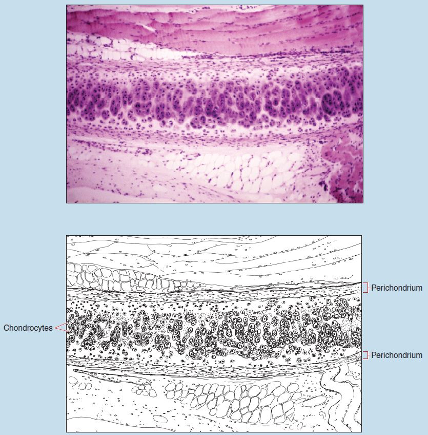
Figure 4-1 (50X): Hyaline cartilage (trachea).
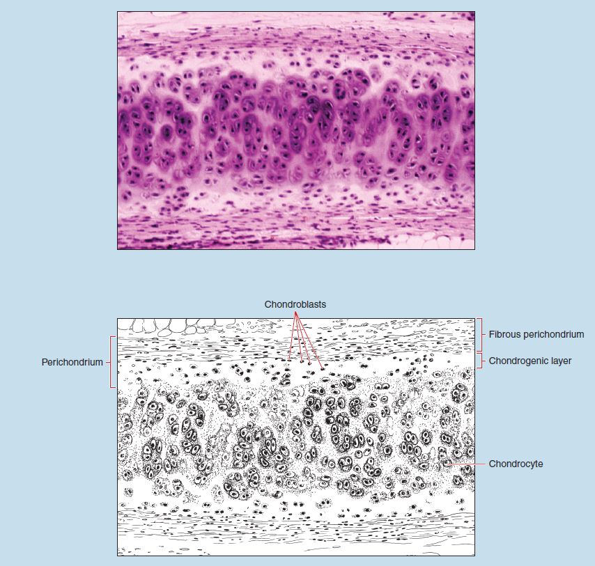
Figure 4-2 (100X): Hyaline cartilage (trachea)
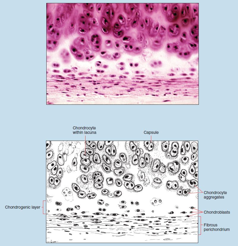
Figure 4-3 (200X): Hyaline cartilage (trachea).
Elastic Cartilage
Elastic Cartilage (Auricle of the Ear) (Elastin Stain)
As you examine Figures 4-4 and 4-5, you will note several similarities between hyaline cartilage (see Figures 4-1 to 4-3) and elastic cartilage:
- Elastic cartilage, like hyaline cartilage, possesses a perichondrium.
- As in hyaline cartilage, the perichondrium surrounding elastic cartilage is composed of an outer fibrous layer and an inner chondrogenic layer.
- The fibrous layer of the perichondrium in elastic cartilage is dense, regular connective tissue. The dominant cell type within this layer is the fibrocyte, just as in hyaline cartilage.
- The chondrogenic layer of the perichondrium in elastic cartilage possesses chondroblasts, with histological characteristics identical to those seen in the chondrogenic layer of hyaline cartilage.
Continued examination of elastic cartilage will show you that the chondrocytes of elastic cartilage are similar to the chondrocytes of hyaline cartilage (see Figures 4-1 to 4-3) in many ways:
- Chondrocytes of elastic cartilage are contained within lacunae.
- As in hyaline cartilage, a capsule surrounds the periphery of the lacunae.
- Chondrocytes of elastic cartilage may be found singly or in chondrocyte aggregates of two to four cells.
Closer examination of the chondrocytes within the lacunae of elastic cartilage will show several important differences when compared with those found within hyaline cartilage (see Figures 4-1 to 4-3):
- The chondrocytes of elastic cartilage are usually flatter.
- Chondrocytes of elastic cartilage have a more pointed shape.
Finally, the most obvious difference between hyaline and elastic cartilage is the appearance of the ground substance of elastic cartilage. In elastic cartilage, the ground substance is permeated with frequently branching elastic fibers. These elastic fibers form a network that often is so dense that it obscures the ground substance from view. This characteristic, when combined with the shape of chondrocytes within elastic cartilage, makes the identification of elastic cartilage quite easy.
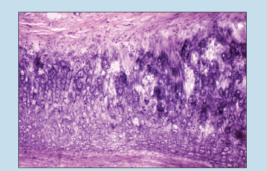
Figure 4-4 (50X): Elastic cartilage (auricle of the ear) (elastin stain).
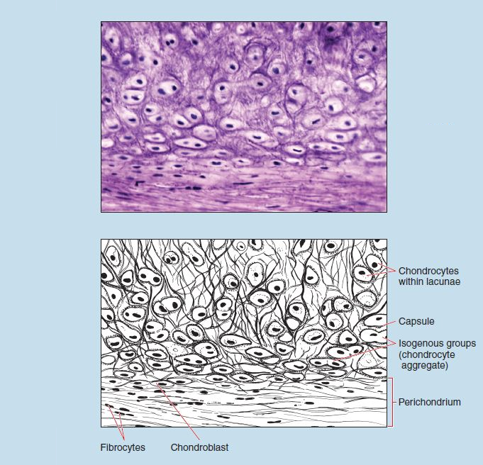
Figure 4-5 (200X): Elastic cartilage (auricle of the ear) (elastin stain).
Fibrous Cartilage
Fibrous Cartilage (Pubic Symphysis)
As you compare fibrous cartilage (fibrocartilage) in Figure 4-6 to hyaline cartilage (see Figures 4-1 to 4-3) and elastic cartilage (see Figures 4-4 and 4-5), you will note several striking similarities and differences.
As with hyaline and elastic cartilage, in fibrous cartilage the chondrocytes are contained within lacunae. However, there are several notable differences in the lacunae of fibrous cartilage:
- First, although capsules are present, they are not as prominent in fibrous cartilage as they are in hyaline or elastic cartilage.
- Note that the lacunae are more regularly arranged (often being arranged into rows) in fibrous cartilage, compared with the more random arrangement seen in hyaline and elastic cartilage.
Continued examination reveals that the interterritorial matrix of fibrous cartilage is not as homogenous as that of hyaline cartilage. Indeed, you will see collagen fibers within the cartilage matrix. You will also note that the collagenous fibers are more or less regularly arranged. These characteristics are important histological features of fibrous cartilage.
Fibrous cartilage is found in locations of high stress. The regular arrangement of the fibers within the matrix tends to follow the compression and shear forces to which the particular piece of fibrous cartilage is subjected.
Although elastic cartilage (see Figures 4-4 and 4-5) also presents a nonhomogenous matrix, fibrous cartilage differs from elastic cartilage in at least two ways:
- First, the fibers visible within the matrix of fibrous cartilage are more regularly arranged than those seen within the matrix of elastic cartilage.
- Second, the collagen fibers dominant within fibrous cartilage matrix stain acidophilic, whereas elastic fibers dominant within the matrix of elastic cartilage stain black in color.
Finally, fibrous cartilage often serves as a continuum between cartilage and bone or tendon and bone. In such instances, you will note that fibrous cartilage often lacks a peripheral perichondrium. This histological characteristic will vary, however, from location to location. The fibrous cartilage associated with many articulations, including the tibiofemoral menisci, glenoid labrum, and the discs of the acromioclavicular joint may possess a perichondrium, as evidenced by the capacity for limited cartilage repair at these joints.
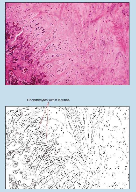
Figure 4-6 (100X): Fibrous cartilage (public symphesis).
Commonly Misidentified Tissues
Hyaline Cartilage and Fibrous Cartilage
If you do not closely examine sections of hyaline cartilage and fibrous cartilage, it is easy to confuse these two tissue types. Confusion may also arise if the section you are examining does not contain a perichondrium because of the plane of section. Therefore it is important to keep these characteristics in mind:
Hyaline Cartilage (Review Figures 4-1 to 4-3 in section on Hyaline Cartilage [Trachea])
- Matrix homogeneous in appearance
- Possesses a perichondrium
- Lacunae randomly arranged
- Capsule readily seen
- Chondrocyte aggregates commonly observed in tissue
Fibrous Cartilage (Review Figure 4-6 in section on Fibrous cartilage [Pubic Symphysis])
- Collagenous fibers are readily seen in matrix.
- Perichondrium is lacking.
- Lacunae are more widely spaced and more regularly arranged.
- Capsules are seldom seen.