Chapter Objectives
This chapter will enable you to identify the histological characteristics of the:
- Olfactory epithelium of the nose
- Cornea of the eye
- Retina of the eye
- Cochlea of the ear
Olfactory Epithelium
The cells responsible for the sense of smell are located within the posterodorsal regions of the nasal cavities. The olfactory epithelium is pigmented yellowish-brown, in contrast to the pinkish color of the respiratory epithelium. This yellowishbrown color is due to the presence of glands within the olfactory epithelium and pigment within the olfactory epithelial cells.
Olfactory epithelium (Figures 18-1 and 18-2) is similar to that of the respiratory tree (see section on Pseudostratified Epithelia in Chapter 2) in that it is a pseudostratifed columnar epithelium. However, the olfactory epithelium is lacking goblet cells and ciliated epithelial cells. The olfactory epithelium is composed of four cell types:
- Olfactory cells, which are bipolar neurons that span the entire thickness of the epithelium
- Supporting (sustentacular cells)
- Basal cells (stem cells)
- Brush cells, which are similar to those found within the respiratory epithelium
Deep to the olfactory epithelium you will see numerous capillaries, olfactory glands, unmyelinated and myelinated neurons. The lamina propria of the olfactory epithelium is continuous with the perichondrium or periosteum of the nasal cavity.
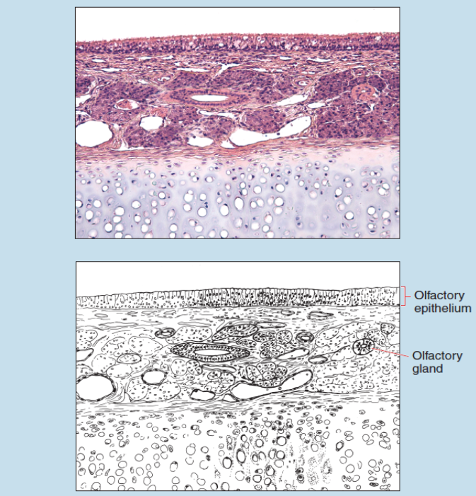
Figure 18-1 (35X): Olfactory epithelium.
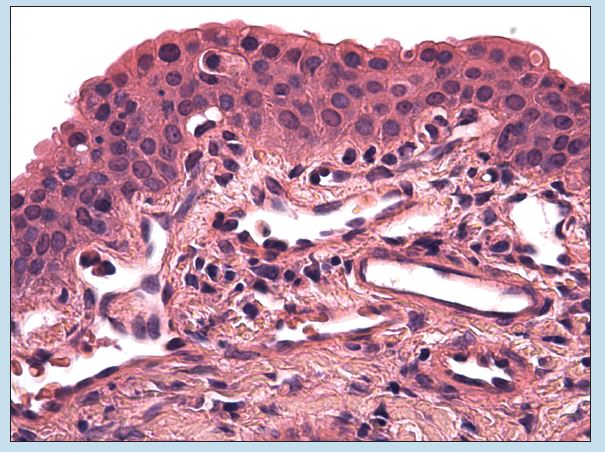
Figure 18-2 (70X): Olfactory epithelium.
Eye
Cornea
The cornea of the eye is a part of the fibrous tunic and is continuous with the sclera. Because the cornea is avascular, the superficial structures must obtain oxygen and nutrients from the tears that bathe its anterior surface.
The cornea is composed of five layers:
- The corneal epithelium (anterior epithelium) (Figures 18-3 and 18-4) is a stratified, nonkeratinized squamous epithelium.
- An anterior limiting membrane (Bowman’s membrane) (Figures 18-3 and 18-4) lies deep to the corneal epithelium.
This connective tissue layer is composed of randomly arranged collagenous fibers.
- The substantia propria (corneal stroma) (Figures 18-3 and 18-4) is a thick layer of parallel bundles of collagen fibers interspersed with fibrocytes. Each layer of connective tissue bundles within the substantial propria lies perpendicular to its adjacent layer.
- A posterior limiting membrane (Descemet’s membrane, or the posterior basement membrane) (Figure 18-3) is found deep to the substantia propria. This layer serves as the basal lamina for the endothelium of the anterior chamber.
- The endothelium of the anterior chamber (posterior epithelium) (Figure 18-3) is a simple squamous epithelium that faces the anterior chamber of the eye.
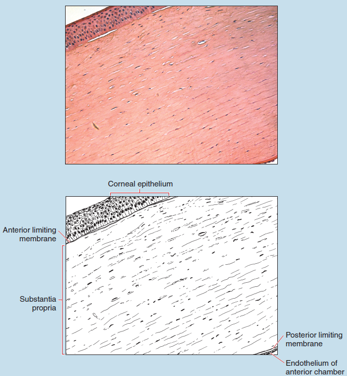
Figure 18-3 (35X): Cornea.
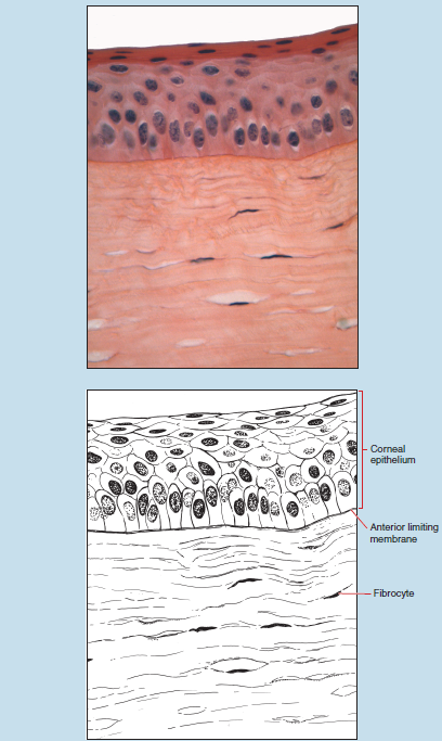
Figure 18-4 (40X): Cornea.
Retina
The retina of the eye consists of two layers: the neural retina (retina proper), which contains the photoreceptors, and the retinal pigment epithelium, an outer layer that is attached to the choroid.
The neural retina is also subdivided into two parts:
- The nonvisual retina, which is found anterior to the ora serrata
- The optic part of the retina, which contains the photoreceptors and lines the posterior surface of the eye
The optic portion of the retina is subdivided into 10 layers (Figures 18-5 and 18-6):
- The pigment epithelium, which rests on the lamina vitrea, is immediately superficial to the choroid and contains melanincontaining cells.
- Rods and cones
- Outer limiting membrane (not visible on this photomicrograph), which is a sieve-like sheet of connective tissue
- Outer nuclear layer, which contains the cell bodies of the rods and cones
- The outer plexiform layer (not visible on this photomicrograph), which contains the axonal processes of the rods and cones
- The inner nuclear layer, which contains the cell bodies of the bipolar cells (the first order neurons of the optic tract), supporting (Muller’s) cells, horizontal cells, and amacrine cells
- The inner plexiform layer, which contains the neural processes of bipolar, supporting, horizontal, and amacrine cells
- The ganglion cell layer, which contains the soma of the ganglion cells, the second order neurons of the optic tract
- The optic nerve layer, which contains the neuronal processes of the ganglion cells that will ultimately form the optic nerve
- Inner limiting membrane layer, which is formed by the basal lamina of the Muller’s cells
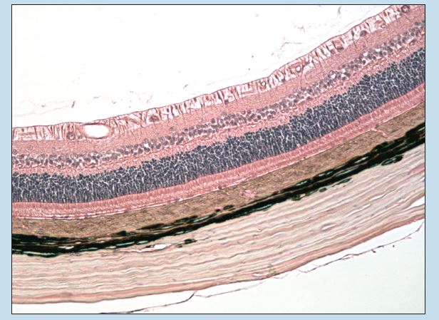
Figure 18-5 (35X): Retina.
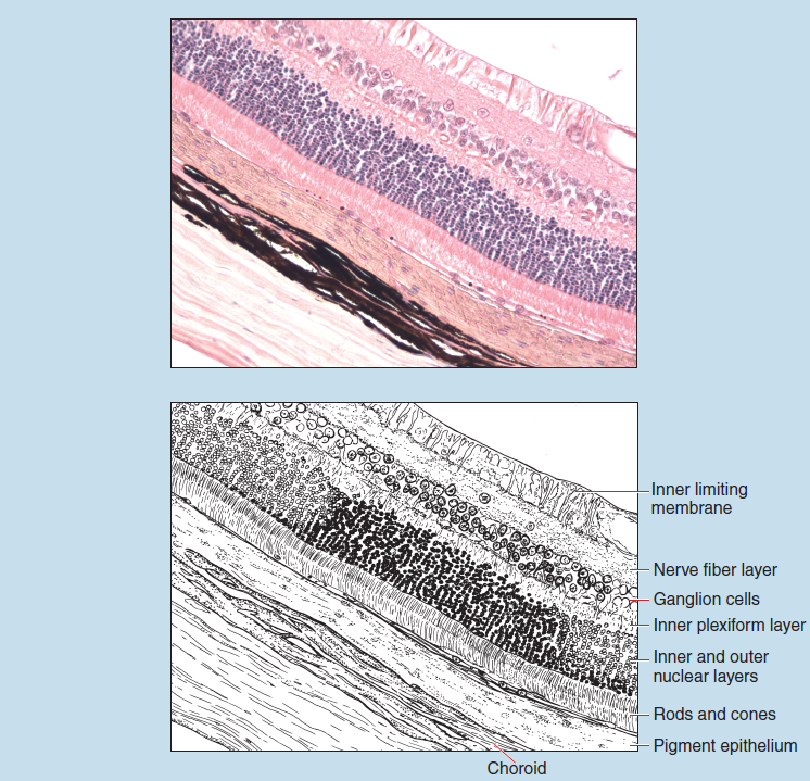
Figure 18-6 (70X): Retina.
Organ of Corti of the Ear (Guinea Pig)
The Organ of Corti is the sensory structure of the auditory portion of the ear. It is found within the cochlear duct (scala media), which is within the membranous labyrinth of the inner ear. The membranous labyrinth is divided into three segments, or ducts (that are not labeled on this photomicrograph), by two membranes of the inner ear. The basilar membrane separates the cochlear duct (scala media) above from the tympanic duct (scala tympani) below. The vestibular membrane separates the cochlear duct below from the vestibular duct (scala vestibuli) above.
The Organ of Corti (Figure 18-7) rests on the basilar membrane, which separates the cochlear duct above from the tympanic duct below. Hair cells rest on the basilar membrane and are in contact with the tectorial membrane. (Note that the tectorial membrane is not intact in this photomicrograph, because of a sectioning artifact.)
Also visible on Figure 18-7 are the spiral limbus and the stria vascularis of the inner ear. The stria vascularis forms the outer wall of the cochlear duct and is a highly vascular area. A unique stratified epithelium that contains an intraepithelial capillary network covers the stria vascularis. The floor of the cochlear duct is partly formed by the spiral limbus, which is a thickened form of periosteal tissue.
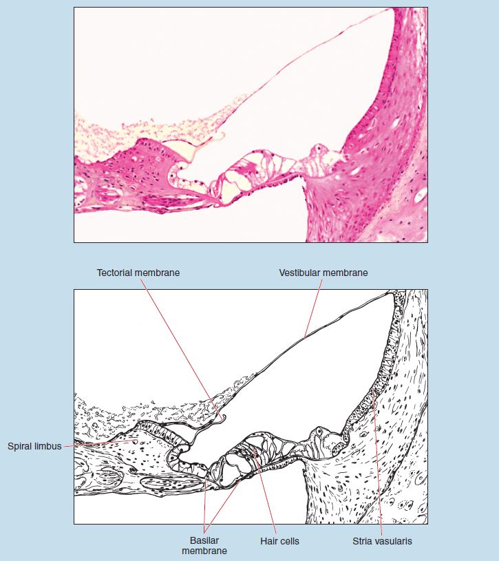
Figure 18-7 (70X): Organ of Corti.
