Chapter Objectives
This chapter will enable you to:
- Differentiate among the varying types of muscle tissue when viewed in cross and longitudinal sections.
- Differentiate among endomysium, perimysium, and epimysium and be able to explain the various components found within these connective tissue investments.
- Identify myosatellite cells and explain their function.
- Identify intercalated discs in cardiac muscle and explain their function.
Types of Muscle
Human muscle tissue is classified into the following categories: skeletal striated muscle and noncardiac visceral striated muscle,[1] smooth muscle, and cardiac striated muscle. These categories are based on the location, function, and structure of the tissue.
Skeletal striated muscle (skeletal muscle) is the most abundant of the muscle types in the human body. It is responsible for appendicular and axial skeletal movements. In addition to moving skeletal structures, skeletal muscles are also responsible for moving the eyes, tongue, deep fascia, and skin of the face. Skeletal muscle cells possess “stripes” or striations. These striations (A band and I band) are the result of differences in light passage and absorption in the tissue. Skeletal muscle is also termed voluntary muscle in that contractions can be initiated via the “voluntary” or somatic nervous system.
Cardiac muscle is limited to the heart and exhibits a banding pattern quite similar to that of skeletal muscle. Cardiac muscle is commonly called a functional syncytium in that excitation of one cardiac muscle cell will quickly cause the excitation of all adjacent cells via the intercalated discs found between the cells.
Smooth muscle is the only type that does not possess striations. Smooth muscle is typically found within the hollow visceral organs and blood vessels of the body and is functionally subdivided into “single unit” or “multi-unit” smooth muscle, depending on how easily action potentials may spread from one cell to another.
Skeletal Muscle
Skeletal muscle fibers demonstrate the following histological characteristics:
- Cells are long and cylindrically shaped.
- Cells are multinucleate.
- Nuclei are peripherally located.
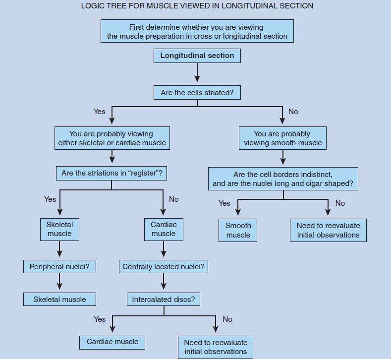
- When viewed in longitudinal section, skeletal muscle cells demonstrate a regularly arranged banding pattern.
As you examine the following photomicrographs, it is important to keep in mind that skeletal muscle should be the reference by which you judge all other muscular tissue. If you know the histological characteristics of skeletal muscle (in both cross and longitudinal sections), you can then use these characteristics to identify all other unknown muscle specimens.
Skeletal Muscle – Cross Section
Figure 6-1 is a cross-sectional view of skeletal muscle. Because skeletal muscle cells are cylindrical in shape, you will see very little size variation between the individual myofibers, a major histological feature of this tissue. Although this cross section does not demonstrate the multinucleate nature of skeletal muscle fibers, it clearly demonstrates that the nuclei are peripherally located, another major histological feature of skeletal muscle when viewed in cross section.
Skeletal muscle has a rather complex connective tissue investment. The external surface of the entire muscle is covered with connective tissue, termed epimysium, which is not visible in this section. The epimysium then extends into the interior of the muscle as perimysium, which divides the skeletal muscle into fascicles. The endomysium is found surrounding each individual skeletal muscle cell, external to the basal lamina.
Figure 6-2 is a higher magnification of Figure 6-1 and demonstrates the perimysium, endomysium, and accompanying slender fibrocytes.
As you center your attention on the individual myofibers, you will see the peripherally located nuclei. In addition, you can distinguish myosatellite (satellite) cells. Myosatellite cells are stem cells that may divide and form additional myofibers following muscle injury. Myosatellite cells are found beneath the basal lamina of skeletal muscle cells and external to the sarcolemma (muscle cell membrane). They may be distinguished from fibrocytes by their more slender nuclei.
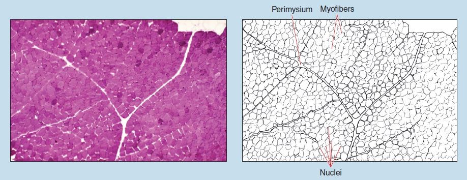
Figure 6-1 (25X): Skeletal muscle (cross section).
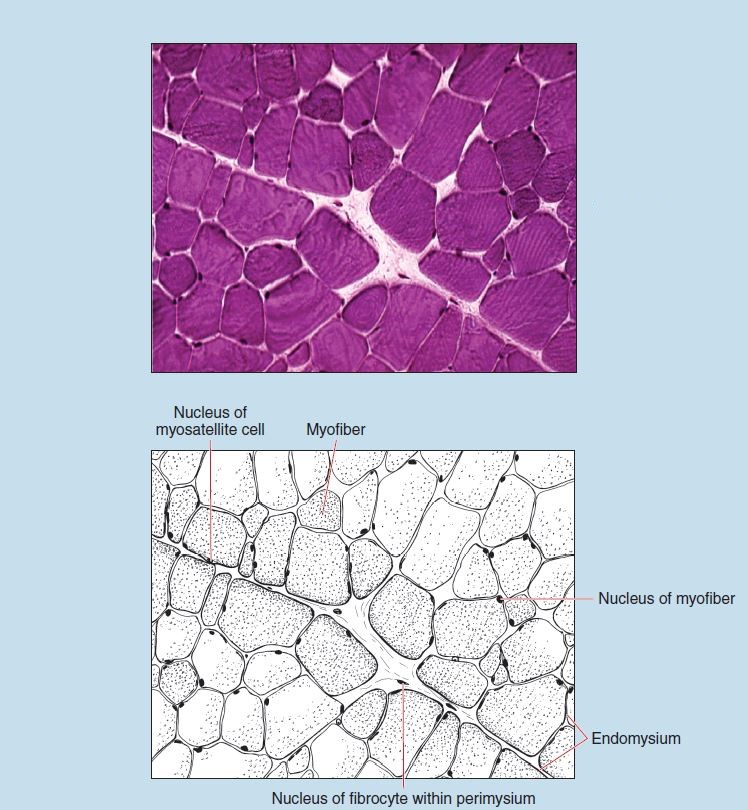
Figure 6-2 (100X): Skeletal muscle (cross section).
Skeletal Muscle – Longitudinal Section
Figures 6-3 and 6-4 demonstrate skeletal muscle in longitudinal section. The banding pattern is clearly evident, as are the peripheral nuclei. Note how the banding pattern in skeletal muscle is “in register” (lined up) from one cell to another. This is a major histological feature of skeletal muscle when viewed in longitudinal section.
Nuclei of fibrocytes located within the perimysium are also evident in Figure 6-3.
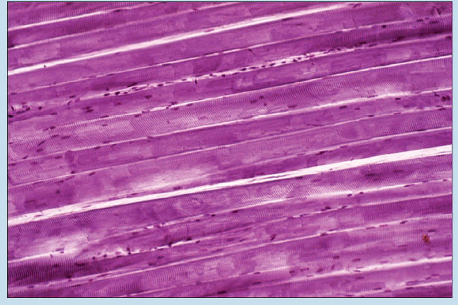
Figure 6-3 (50X): Skeletal muscle (longitudinal section).
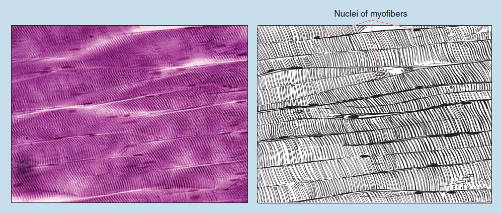
Figure 6-4 (100X): Skeletal muscle (longitudinal section).
Cardiac Muscle
Cardiac muscle, like skeletal muscle, is striated; however, that is where the histological similarities end. As you examine the following photomicrographs of cardiac muscle, keep in mind the following histological differences between skeletal and cardiac muscle:
- Cardiac muscle cells are considerably smaller than skeletal muscle fibers. The exception to this rule is Purkinje fibers, which will be studied in Chapter 8, the Cardiovascular System.
- Cardiac muscle cells branch frequently and therefore demonstrate a greater size variation when viewed in cross section.
- Cardiac muscle cells (except for Purkinje fibers) have a single, centrally located nucleus.
- Cardiac muscle cells possess specialized cell junctions called intercalated discs, and these are visible in longitudinal sections of cardiac muscle.
Figure 6-5 is a photomicrograph of cardiac muscle in longitudinal section. Note that cardiac muscle usually has only one centrally located nucleus per cell. In addition, note the sectioning artifacts at the periphery of this photomicrograph.
Figure 6-6 clearly demonstrates the centrally located nuclei and the intercalated discs found between adjacent cardiac muscle cells. Intercalated discs are specialized junctions that facilitate the spread of electrical impulses from one cardiac muscle cell to another. They also anchor cardiac muscle cells to one another, thereby allowing the spread of contraction forces.
Compare this figure to skeletal muscle in Figure 6-4. Note:
- How cardiac muscle cells branch
- That the banding pattern of cardiac muscle is not as
prominent as that of skeletal muscle - That the banding pattern between individual cardiac muscle
cells is not “in register,” as was seen in skeletal muscle
These three characteristics are all major histological features of cardiac muscle when viewed in longitudinal section.
You will also note, as you compare Figures 6-4 and 6-6, that cardiac muscle exhibits fibers running in several different directions. This results in the presence of cross, longitudinal, and oblique sections on the same slide because of the helical arrangement of cardiac muscle within the heart. This can be a useful identification tool.
Figure 6-7 is a photomicrograph of cardiac muscle in cross section. This figure clearly demonstrates two major histological features of cardiac muscle when viewed in cross section:
- Note that the nuclei in cardiac muscle cells are centrally located.
- In addition, this tissue demonstrates an increased variation in cell size compared to skeletal muscle in cross section (see Figure 6-1). This increased size variation is due to the branching of cardiac myocytes.
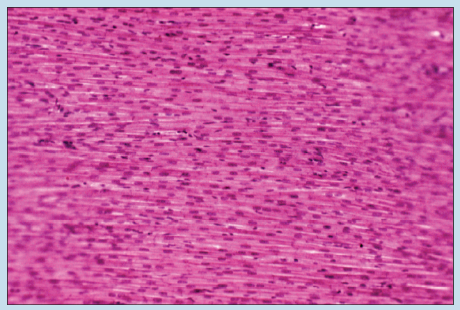
Figure 6-5 (50X): Cardiac muscle.
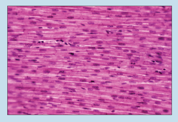
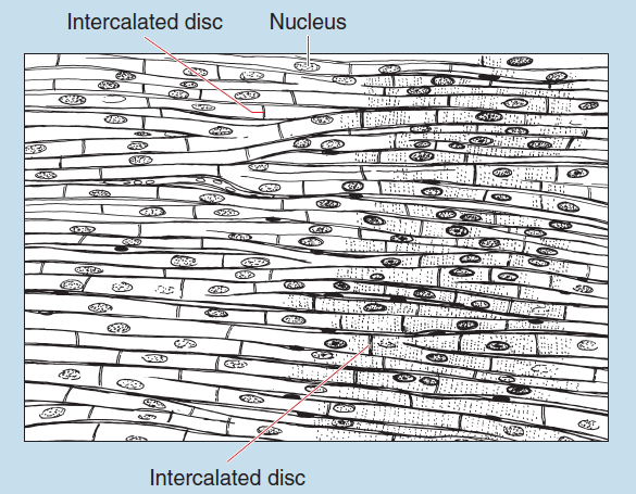
Figure 6-6 (100X): Cardiac muscle.
Smooth Muscle
As you examine the following photomicrographs of smooth muscle, you must compare smooth muscle to skeletal and cardiac muscle. As you make these comparisons, you will see the following similarities and differences:
- Smooth muscle, unlike cardiac and skeletal muscle, is not striated.
- Like cardiac muscle, smooth muscle has a single, centrally located nucleus.
- Smooth muscle cells, unlike skeletal and cardiac muscle, are fusiform in shape. Therefore they will demonstrate the greatest amount of size variation when viewed in cross section.
- Smooth muscle cells have long, football-shaped nuclei.
- The lateral borders of smooth muscle cells are quite difficult to see in hematoxylin and eosin preparations.
- Smooth muscle cells are much smaller than cardiac or skeletal muscle cells.
Figure 6-8 was taken from the smooth muscle found within the muscularis externa of the ileum of the small intestine. You can see smooth muscle in longitudinal section in this photomicrograph. (Note the sectioning artifacts at the periphery of this photomicrograph.)
When viewed in longitudinal section, smooth muscle cells are long and cigar shaped, appearing almost to interdigitate with each other. This reflects the overlapping of the individual smooth muscle cells.
Smooth muscle, when viewed in cross section, presents a considerable variation in cell and nucleus size. In addition, because of the shape of smooth muscle cells and the resulting plane of section, the number of cells possessing visible nuclei will also vary considerably throughout the field.
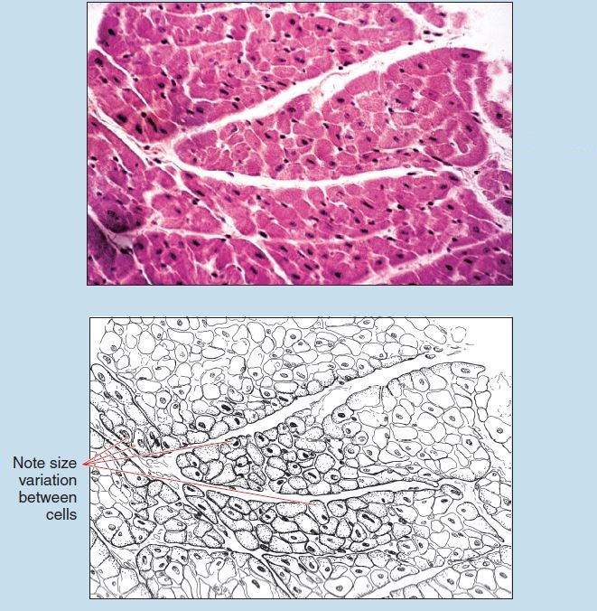
Figure 6-7 (100X): Cardiac muscle (cross section).
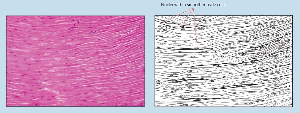
Figure 6-8 (50X): Smooth muscle from muscularis externa of the ileum of the small intestine.
Figure 6-9 is a photomicrograph of smooth muscle in longitudinal and cross section taken at a higher magnification. Note the artifacts between the two layers of smooth muscle in this photomicrograph.
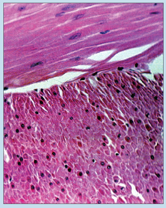
Figure 6-9 (100X): Smooth muscle from muscularis externa of the ileum of the small intestine (longitudinal and cross section).
Commonly Misidentified Tissues
Skeletal, Smooth, and Cardiac Muscle
When viewing skeletal, smooth, and cardiac muscle it is important to keep in mind multiple aspects for comparison. The characteristics listed below are not the only factors that may be used for comparison but are the most obvious and will yield the best results with the least amount of effort.
| Muscle and Section Type | Nucleus | Shape and Size Variation |
| Skeletal muscle (x.s.) | Multiple and peripheral | Rounded; 15-90 μM; minimal |
| Skeletal muscle (l.s.) | Multiple and peripheral | Rounded; 10-20 μM; minimal |
| Smooth muscle (x.s.) | Single and central | Circular; 7 μM; considerable |
| Smooth muscle (l.s.) | Single and central | Spindle shaped; considerable |
| Cardiac muscle (x.s.) | Single and central | Rounded; 10-20 μM; moderate |
| Cardiac muscle (l.s.) | Single and central | Branched; moderate |
x.s, Cross section; l.s., longitudinal section.
Note: The diameter of a red blood cell is 7 μM; this is a useful measurement tool for comparison when looking at these tissues.
Smooth Muscle, Tendon, and Ligament when Viewed in Longitudinal Section
Smooth Muscle/ Longitudinal section (Review Figures 6-8 and 6-9 in section on Smooth Muscle)
- Fusiform or spindle-shaped cells with a single, centrally located, football-shaped nucleus
- Nuclei aligned parallel with each other
- Noticeable size variation in cells within preparation
- Lack of “wavy” fibers in preparation
- Relative homogeneous staining of preparation
- Connective tissue investment surrounding smooth muscle cell bundles and individual cells
Dense Regular Connective Tissue: Tendon and Ligament/Longitudinal Section (Review Figures 3-8 to 3-11 in section on Dense Regular Connective Tissue)
- Long, thin, flattened nuclei within cells
- Cells present distinctively smaller in size
- Nuclei parallel with each other
- “Wavy” appearance in preparation
- Relative lack of homogeneous staining characteristics in
preparation
References
Wareham AC, Whitmore I: A comparison of the mechanical properties of oesophageal striated muscle with skeletal muscles of the guinea pig, Plflugers Arch 395:312-317, 1983.
Whitmore I: The ultrastructure of oesophageal striated muscle in the guinea pig and marmoset, Cell Tiss Res 234:365-376, 1983.
Whitmore I: 2006 Terminologia histologica. International terms for human cytology and histology, Philadelphia, Lippincott Williams & Wilkins.
Whitmore I, Gosling JA, Gilpin SA: A comparison between the physiological characteristics of urethral striated muscle in the guinea pig, Pflugers Arch 400:40-43, 1984.
Whitmore I, Notman JA: A quantitative investigation into some ultrastructural characteristics of guinea pig striated muscle, Ann Anat 153:233-240, 1987.
- As per the Terminologia Histologica (TH, 2008): “The following classification acknowledges the presence of nonskeletal, noncardiac striated muscle, such as esophageal striated muscle and external anal and urethral sphincters.” ↵
