PROCEDURAL STEPS
Cervical Preparation
- Review patient history, EGA, US, labs, & consents (procedure, sedation, contraception).
- Introduce yourself (and trainee or trainer), establish rapport, elicit and answer patient’s questions: “What questions do you have for me?” Reassurance and details as patient desires.
- Complete pelvic exam if chosen cervical prep requires: perform time-out, don gloves, then provide verbal guidance to position the patient.
- Initiate cervical preparation by either of the following:
- Place misoprostol vaginally (and/or invite patient to self-administer) or buccally. OR
- Place osmotic dilators (or foley) using ring forceps:
- Insert speculum
- Apply antiseptic to cervix
- Administer paracervical block
- Slowly place tenaculum
- May place 1-2 synthetic osmotic dilators (Dilapan-S®) without mechanical dilation, or dilate cervix to 10-13 mm before placing multiple osmotic dilators (or foley).
- Remove tenaculum and place 1-2 gauze sponges into the vagina abutting dilator ends to absorb secretions and maintain dilators in position
- Remove speculum (holding gauze sponges in place with a ring forceps)
- Replace patient’s feet on table, and re-cover their legs.
Procedural Positioning and Dilation
- Re-introduce yourself to the patient after appropriate wait time for cervical prep, answer any new questions, assess vitals, perform time-out, and administer IV medications.
- Appropriate (and relaxed) patient positioning (hips slightly beyond table end) will allow you to more safely visualize the cervix and maneuver instruments.
- Perform bimanual exam, although some providers determine uterine position by US or tactile feedback with dilators instead. Exam benefits of >14 week procedures include evaluation of adequacy of dilation, cervical consistency, and uterine mobility. If performed, remove gauze sponges, osmotic dilators if using, ensuring the same count as placed.
- Initiate intra-procedure ultrasound with abdominal probe (NAF CPGs 2022), ensuring staff are positioned in an ergonomically safe manner. An US machine with a dual monitor arm is ideal.
- Insert speculum, evaluate, and collect screening samples as needed (STI, pap). If no bimanual performed previously, remove gauze sponges, osmotic dilators (or foley) if using, ensuring the same count as placed.
- Apply antiseptic solution to cervix (if using).
- Administer paracervical block. Consider adding vasopressin for less EBL at >14 wk EGA (Schulz 1985), although cost may be prohibitive.
- Dilate cervix sequentially up to appropriate size (see chart for recommendations), while applying gentle traction to straighten cervical canal. Greater dilation allows easier instrumentation with forceps and decreases risk of uterine and cervical injury.
- If planning uterine evacuation with aspiration only, use a large cannula (14 – 17mm), dilate up to equivalent size = weeks +/- 1mm. Note: cannula sizes >15mm necessitate use of large-bore tubing, however passage of a smaller cannula (i.e. flexible 8mm) at end of procedure can be achieved with an MVA so single tubing can be used.
Table 3. Dilation, Cannula Size, and Recommended Forceps by EGA |
|||
| EGA | Dilate to | Cannula | Recommended Forceps |
| Up to 14w | 14 Denniston
43 Pratt |
14 curved
May follow w/ 8 flexible |
None
(If needed Ring, Hern Van Lyth) |
| 14w0-14w6 | 43-45 Pratt | Either 14 curved or
12 curved w/forceps May follow w/ 8 flexible |
None
(If needed Ring, Hern Van Lyth) |
| 15w0d – 15w6d | 45-47 Pratt | Either 15 curved or 12 curved w/ forceps
May follow with 8 flexible |
None or Small finks (or Ring, Hern Van Lyth) |
| 16w0d – 16w6d | 49 Pratt | Either 16 or 12 curved w/ forceps
May follow with 8 flexible |
Small finks (or Regular Finks or Sophers) |
| 17w0d – 17w6d | 49/51 Pratt | Either 16 curved w/ forceps or 12 curved and forceps
May follow by 8 flexible |
Small finks (or Regular Finks or Sophers) |
- Uterine evacuation under ultrasound guidance using a) aspiration alone (many find this adequate through 16 wks or b) D&E.
a) Aspiration Technique:
-
-
- Introduce cannula of appropriate size (12- 14-mm for ≥ 14 weeks, 14- 15-mm for 15 weeks, and 16-mm for 16 weeks) through cervix into lower uterine segment (LUS)
- Connect cannula to large bore tubing, and empty uterus under US guidance.
- With steady patience, suction to remove all fetal tissue, including decompressing and removal of the calvarium.
- If the calvarium is not moving into the cannula, slowly pull it into the lower uterine segment (LUS) using suction while gently withdrawing the cannula to avoid pushing it further into the fundal space while attempting to decompress.
- If tissue gets caught and is not moving into the cannula, gently place the cannula end (with tissue) against the inside of the LUS or internal os (avoiding 3 and 9 o’clock areas) and “push” tissue into cannula by compressing it against uterine wall using an outward (away from the fundus) motion. This action is like pulling a squeegee against the inside of a windshield while standing outside a car.
- Alternatively, pull the clogged items into the LUS and attempt to remove by pulling the cannula through and out the cervix. Consider pressing the cannula’s distal tip against sterile gauze to de-clog.
- If still unable to remove calvarium or all tissue through cannula, proceed to forceps extraction (below)
- Ensure empty uterus, good tone, and no bleeding with ultrasound prior to removal of instruments (or placement of IUD).
- Check POC for adequacy (4 extremities, spine, calvarium, placenta).
- Inform patient of complete procedure & initiate the recovery process as described in Chapter 7: Abortion Aftercare
-
b) D&E (Primary forceps technique)
-
-
- Drain amniotic fluid by either by a) placing a cannula through internal os, or b) inserting forceps just through cervix and opening, allowing fluid to drain into basin or fluid-collecting drape below
- Use US to determine location of fetal tissue, most frequently in a longitudinal view including cervix and entirety of uterus
- Applying tenaculum traction to straighten the endocervical canal, close forceps to enter into the LUS, remaining mindful of the uterine arteries at 9 and 3 o’clock.
- To begin removing tissue, using US guidance, as soon as forceps jaws extend beyond the internal os, open as widely as possible to surround and grasp tissue without pushing it into fundus. Best to aim to grasp torso/thorax, and apply steady outward traction while repeatedly supinating/pronating the forceps hand.
- At 14-16 weeks, fetal parts may be soft and non-calcified, making tactile feedback from forceps difficult to assess. At this gestation, forceps with smaller/finer teeth are appropriate to use.
- At 16-18 weeks, fetal parts may have more calcification and may be easier to sense in forceps’ jaws. Larger teeth allow easier grasping.
-
 |
| Figure 6. When inserting forceps through cervix into uterus, always insert with jaws closed, shanks oriented vertically with top jaw at 12 o’clock and bottom jaw at 6 o’clock. Both jaws will be visualized on ultrasound in this orientation. |
-
-
- Forceps handling to maximize range of motion and stability, and minimize trauma:
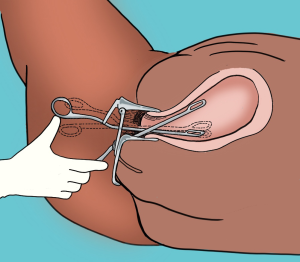
- Remove thumb from forceps loop, instead using thumb to push against medial (palmar) aspect of loop (see image)
- Minimize passes through cervix
- Keep hinge at level of the cervix
- Use forceps in LUS, and remain vigilant about distal tip orientation
- If deeper insertion needed to explore fundus, use traction & follow uterine axis
- Be cautious grasping tissue near the cervix, to avoid accidental traction with toothed forceps. Instead push tissue back into LUS and observe forceps grasp via US.
- Continue traction with rotation to LUS, through cervix, and out until tissue is free.
- Drop grasped fetal tissue into the basin sterilly.
- Removal of an extremity could bring the torso into the LUS. If tissue is still connected, advance the forceps up the torso and grasp for removal.
- Close forceps, reenter cervical canal; repeat until all fetal tissue is removed.
- To remove calvarium, place forceps through the cervix, open, and attempt to encircle it placing serrated jaws on opposite sides; then close around it to grasp and decompress.
- If it “floats” high in the uterus, grasp any part (with forceps or with 12-14mm cannula) and apply slow traction to move into LUS before (re)attempting decompression. Grasping it may require opening forceps wider than expected.
- Thick, bright white fluid (neural tissue) can sometimes be seen leaking from external os after collapsing the calvarium.
- To remove the placenta, grasp and remove with light traction +/- fundal massage while observing US to ensure no uterine wall movement concerning for myometrium entrapment in forceps. The placenta is best removed intact, and will feel thicker, softer, and bulkier than fetal tissue. Alternatively, remove the placenta with suction alone.
- A final suction curettage using a smaller cannula (8mm-12mm;) can be used to empty the uterus of residual blood and tissue until empty. Ensure no bleeding with US prior to removal of instruments (or placement of IUD).
- Check POC for adequacy (4 limbs, spine, calvarium, placenta) using visual inspection during evacuation, intraoperative ultrasound, and / or inspection in the lab.
- Inform patient of complete procedure & initiate the recovery process as described in Chapter 7: Abortion Aftercare
- Forceps handling to maximize range of motion and stability, and minimize trauma:
-
Additional Pearls
- When coaching staff on ultrasound guidance, be direct and deliberate with feedback to ensure they always see the instrument in the uterus.
- DON”T HESITATE TO PAUSE briefly if you are uncertain or concerned that something is not going well. Breathe, roll your shoulders, and reassess the following:
- What can you optimize?
- Can you improve sedation for patient comfort?
- Does the patient need to be repositioned?
- Do you need a different speculum for better visualization?
- Can you add more misoprostol or consult with a colleague?
- Micro-movements are usually sufficient to approach the tissue in a different way to achieve removal. Newer providers usually make larger movements than necessary.
- Sometimes rotating the forceps 180 degrees or changing to a different type of forceps can be sufficient to achieve grasp.