The Cell Through the Microscope
parts of a Typical Human Cell
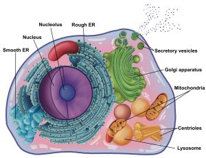
By this point in your biology education, you should be familiar with the parts of animal cells. As an example, we have included an illustration of a generalized cell below:
Figure 1: A generalized animal cell
the cell under the microscope
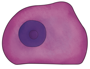
However, this elaborate illustrated view, with its complex organelle depictions, is not what cells look like through a compound light microscope. The resolving power of a compound light microscope is not high enough to be able to see individual organelles. We can easily visualize the cell’s plasma membrane, nucleus, and nucleolus (Figure 2). Often organelles and cytoplasmic inclusions, like secretory vesicles, appear as a granular texture of staining in the cytoplasm.
Figure 2: Illustration rendering of what the generalized animal cell in Figure 1 would look like under the microscope.
SLIDE PREPARATION – THE BASICS
To visualize most human tissues/cells using a compound light microscope, the tissue must be fixed/embedded, sectioned, and stained. Fixing and embedding is a process that preserves the tissue to prevent degrading and embeds it into a hard substance like resin or wax which provides stability. Alternatively, tissue can be frozen instead of embedded. The tissue must be sectioned, which produces very thin slices of the tissue around 4-10 µm in thickness. The goal of sectioning is to create slices of tissue that are the thickness of a single cell. Many types of stains are used to highlight different molecules in a cell.
A common technique is H&E (hematoxylin and eosin):
- Hematoxylin – purple/blue, stains nucleic acids (basophilic) – highlights nucleus and rough ER (ribosomes have RNA)
- Eosin – pink, stains proteins (acidophilic) – highlights connective tissue proteins (collagen), plasma membrane, cytoplasm
One thing you have to keep in mind is that the colors you will see on each slide is determined by the stain that was used.
IMPORTANT NOTE: Different stains can be used for the same tissue type, so you should always look at cell shape and arrangement to determine tissue type, and not simply the color pattern.
HUMAN CELL TYPES – DIFFERENT FORM, DIFFERENT FUNCTION
The human body contains many different cell types. The structure or form the cell has determines the function it can carry out. For example, skeletal muscle cells are very long and contain contractile proteins, allowing it to generate tension through contraction. However, an epithelial cell is cube-shaped and tightly connected to its neighbors allowing it to form boundaries. Figure 4 illustrates examples of four dramatically different cell types in the human body: epithelial cells, skeletal muscle cells, a neuron, and a sperm. Each of these cells carries out dramatically different functions: maintaining boundaries (epithelial cells), contraction (muscle cells), sending information (neurons), movement (sperm).
Figure 4: Various cell types found in the human body: epithelial cells (top left), skeletal muscle cells (top right), a neuron (bottom left), and a sperm (bottom right)
Tissue sectioning
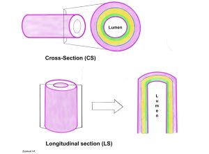
Organs are 3-dimensional structures and how they are sectioned can impact how the tissue looks on the microscope slide. For example, let us imagine a long tube-like organ like the intestines, stretching the tube out to resemble a drinking straw. If we cut along the length of the straw, this would be a longitudinal section and would produce a long piece of tissue (Figure 5). The entire length of the tissue section would be uniform and would have all the layers of tissue from the inside of the tube (top) to the outside of the tube (bottom). If we cut perpendicular or across the straw, this would be a cross section and would produce a circular ring of tissue (Figure 5). The inside of the circular ring represents the inside of the organ, which we call the lumen.
Figure 5: Illustration comparing the cross section (top panel) and longitudinal section (bottom panel) of a tube-shaped organ showing multiple tissue layers (pink, blue, yellow, green, purple) with the organ’s lumen in the center
Chapter Illustrations By:
Edna Martinez Sanchez
Georgios Kallifatidis, Ph.D.
Soma Mukhopadhyay, Ph.D.
hematoxylin and eosin staining protocol
Hematoxylin - purple/blue, stains nucleic acids (basophilic) - highlights nucleus and rough ER (ribosomes have RNA)
Eosin - pink, stains proteins (acidophilic) - highlights connective tissue proteins (collagen), plasma membrane, cytoplasm
cut along the long axis of a tissue
a transverse section, cutting perpendicular to the length of a structure. If cutting a tubular structure, a cross section would produce a ring of tissue
the interior of a tube-shaped organ, like a blood vessel, kidney tubule, or intestines
