Simple Epithelial Tissues
Simple epithelial tissues consist of a single layer or sheet of cells that is exposed to a lumen on the apical side and anchored to the basement membrane on the basal side. Because they are a single layer of cells in thickness, simple epithelial tissues are found in organs where substances are passed across the layer by processes such as diffusion, absorption, filtration, or secretion.
Simple Epithelial Tissues are Named According to Cell Shape
Squamous – scale-like, flattened cells that resemble the scales on a fish with a flat disc nucleus
Cuboidal – roughly cube-shaped cells (or as cube-shaped as a water balloon can be!) with a round nucleus
Columnar – tall, rectangle cells with an egg-shaped nucleus located near the basal side
As you learn how to recognize each type of epithelial tissue, remember to look at the shape of the nucleus and the number of cells layers!
Simple squamous epithelium
Simple squamous epithelium can be found in many locations in the body (e.g., lining blood vessels, lining the alveoli (air sacs) of our lungs, and in Bowman’s capsule of the kidney). To help you understand how to identify simple squamous epithelium, we have included two examples of this tissue. Table 1 and Figure 1 show simple squamous epithelium in the alveoli of the lung, while Table 2 and Figure 2 show simple squamous epithelium in Bowman’s capsule of the kidney. In both organs, you will see that this epithelium is made of a single layer of flattened cells.
Table 1: Simple squamous epithelium of the alveoli
| Tissue Type | Simple Squamous Epithelium |
|---|---|
| General Description | Single layer of flattened, disc-shape cells |
| Location | Alveoli of the lung |
| Function | Allows for gas exchange between lungs and blood vessels |
| Helpful Hints | This tissue is easily confused with adipose tissue – look for the flattened nuclei lined up in a row |
Figure 1: Simple squamous epithelium of the alveoli with and without illustration overlay
Table 2: Simple squamous epithelium of Bowman’s capsule of the kidney
| Tissue Type | Simple Squamous Epithelium |
|---|---|
| General Description | Single layer of flattened, disc shape cells |
| Locations | Bowman’s capsule in the nephron of the kidney |
| Function | Forms a thin boundary which gives structure to the cup-like structure |
| Helpful Hints | Bowman’s capsule looks like a round bowl – look for the single row of flattened nuclei that touch the white space |
Figure 2: Simple squamous epithelium of Bowman’s capsule of the kidney with and without illustration overlay
Check out our YouTube videos to help you understand simple squamous epithelium:
YouTube Video – Alveoli of the Lung
YouTube Video: Bowman’s Capsule of the Kidney
SPECIALIZED SIMPLE SQUAMOUS EPITHELIUM – ENDOTHELIUM
Simple squamous epithelium also forms the lining of all of our blood vessels, where it is called endothelium. The picture below shows the cross-section of a blood vessel. The small pink dots are red blood cells (RBC) inside the vessel. The single layer of nuclei surrounding that space (lumen) is the layer of simple squamous epithelium called the endothelium.
Figure 3: Simple squamous epithelium, endothelium, lining a blood vessel lumen
Simple cuboidal epithelium
Simple cuboidal epithelium can look a little different depending on the direction the tissue has been sectioned. To help you understand how to identify simple squamous epithelium, we have included two examples of this tissue. While both figures show simple cuboidal epithelium of the renal tubules in the kidney, Figure 4 shows a cross section and Figure 5 shows a longitudinal section. In both examples, you will see that this epithelium is made of a single layer of cube-shaped cells with round nuclei.
Table 3: Simple cuboidal epithelium
| Tissue Type | Simple Cuboidal Epithelium |
|---|---|
| General Description | Single layer of cube-shaped cells with round nuclei |
| Locations | Tubules of kidney, follicles of the thyroid gland, salivary gland, surface of ovaries |
| Function | Absorption and/or secretion |
| Helpful Hints | Cells surround the lumen (inside) of the kidney tubule, so they may not look perfectly cube-shaped – look for the round nuclei |
Figure 4: Simple cuboidal epithelium of the renal tubule of the kidney (cross section) with and without illustration overlay
Figure 5: Simple cuboidal epithelium of the renal tubule of the kidney (longitudinal section) with and without illustration overlay
Check out our YouTube videos to help you understand simple cuboidal epithelium:
YouTube Video: Simple Cuboidal Epithelium
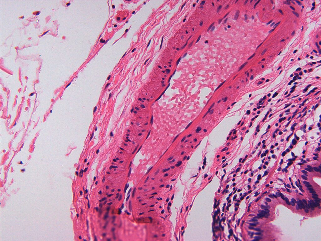
Simple columnar Epithelium
Table 4: Simple columnar epithelium
| Tissue Type | Simple Columnar Epithelium |
|---|---|
| General Description | Single layer of column (rectangular)-shaped cells with oval nuclei usually located near the basal side of the cell |
| Location | Lining of intestines, stomach, and gallbladder |
| Function | Absorption and/or secretion |
| Special Features | May contain goblet cells to secrete mucus, often cells have microvilli which appear as a fuzzy apical edge (called the brush border in the small intestines) |
| Helpful Hints | Depending on the direction of the tissue section, the appearance of the tissue may change. Longitudinal section will have a long row of cell with empty space at the apical side. Cross section will have a ring of cells with empty space in the center where the apical side touches the lumen. |
Figure 6: Simple columnar epithelium of the small intestine (ileum) with and without illustration overlay
Check out our YouTube videos to help you understand simple columnar epithelium:
YouTube Video – Simple Columnar Epithelium
Chapter Illustrations By:
Juan Manuel Ramiro-Diaz, Ph.D.
the interior of a tube-shaped organ, like a blood vessel, kidney tubule, or intestines
a thin layer of connective tissue that anchors the cells of epithelial tissues to the deeper structures of our body
the specialized simple squamous epithelium that lines the lumen of blood vessels
