How to use this book
The purpose of this book is to help students learn the complex field of tissue histology. Please take a moment to familiarize yourself with the features of our book.
How to navigate within the book
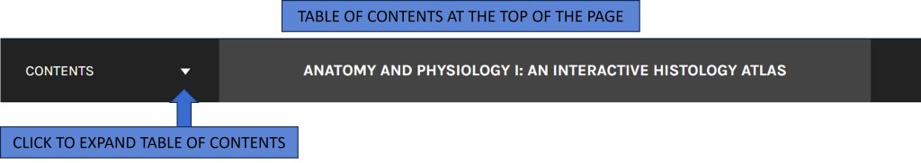
Navigation within the book is simple. The table of contents can be found at the top of the page. Click the upside-down triangle to expand the table of contents (Figure 1). The book is divided into three parts, which can each be expanded by clicking on the + sign with the Contents:
- Part I – Introduction to Microscopy and the Cell
- Part II – Types of Tissues
- Part III – Organ Systems and Their Unique Histology
Figure 1: The table of contents can be found at the top of the page. Click to expand the table of contents
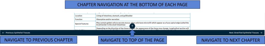
When you have reached the end of a chapter, you will find navigation tools at the bottom of the page (Figure 2). On the left and right sides of the page you will see tabs to navigate to the previous (left side) and next (right side) chapters. Clicking the up arrow in the center of the page will return you to the top of the page (Figure 2).
Figure 2: Chapter navigation can be found at the bottom of the page. Click the arrow in the center to return to the top of the page.
Using the text features
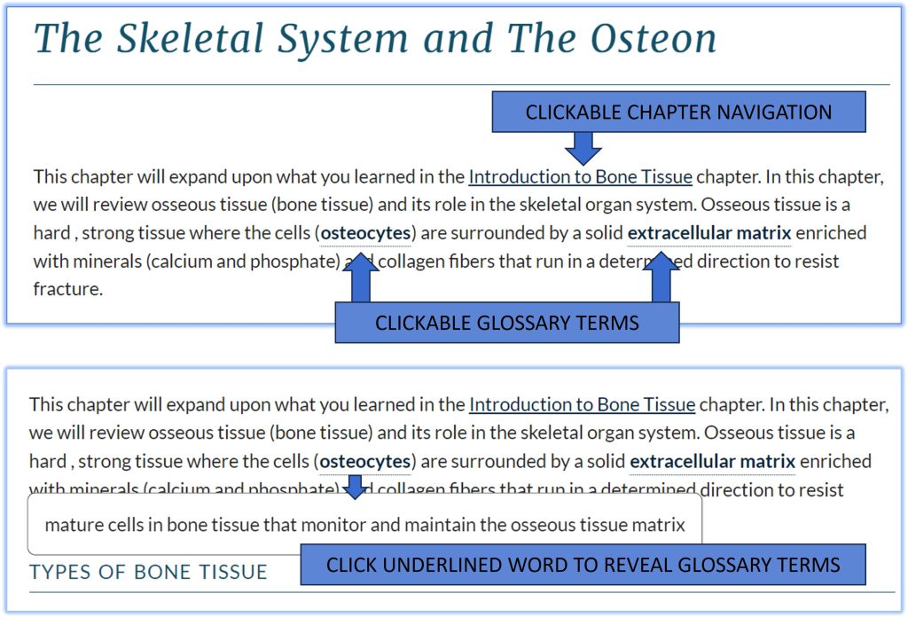
This book contains two types of clickable text. Clickable text to jump between chapters is underlined with a solid line. Clickable text to open glossary term definitions is underlined with a dotted line (Figure 3). The complete Glossary of Terms can be found at the end of the book: Glossary of Terms.
Figure 3: Clickable text features include glossary terms and chapter navigation
using the image features
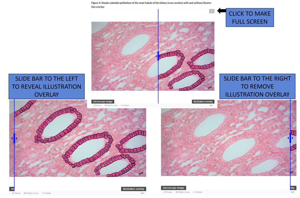
We encourage students to use our interactive images as study resources! All images can be expanded to full-screen by clicking on the arrows at the top right of the image (Figure 4). The vertical bar running through the image is a slider. Click and drag the slide bar to the right to reveal the microscope image without the illustration overlay. Click and drag the slide bar to the left to reveal a full illustration overlay of the microscope image. The illustration overlays are designed to help you better understand the cells and structures you see under the microscope.
Figure 4: Using the interactive slide bar on microscope images with and without illustration overlay
We hope you find this book helpful in your studies of the tissue histology in Anatomy and Physiology I.
Augusta University Anatomy and Physiology Youtube channel
Embedded within the chapters of this book are links to YouTube videos that walk students through each microscope slide. Please consider subscribing to the Augusta University Anatomy and Physiology YouTube Channel: http://www.youtube.com/@AugustaUniversityAandP
Contact the authors
We hope you find this book helpful in your studies of the tissue histology in Anatomy and Physiology I. If you find an error, or have a suggestion about how we could make this book more helpful to students, please contact us by email:
Karen Wiles: kawiles@augusta.edu
Christina Wilson: chrwilson@augusta.edu
