The Skeletal System and The Osteon
This chapter will expand upon what you learned in the Introduction to Bone Tissue chapter. In this chapter, we will review osseous tissue (bone tissue) and its role in the skeletal organ system.
Types of Bone Tissue
Osseous tissue is divided into two types: compact bone and spongy (also known as cancellous or trabecular) bone.
Compact bone is incredibly strong at resisting tension and compression, which makes it perfect for our skeleton. Compact bone consists of closely packed structures called osteons. Osteons consist of a central (Haversian) canal surrounded by concentric rings/layers of bone matrix called lamellae, where collagen fibers run in alternating directions. The central canal provides an opening for blood vessels and nerves. Figures 1 and 2 show illustrations of lateral and superior views of compact bone under low power magnification. For simplicity the vessels and nerves found within the central and perforating canals are shown as red lines in Figure 1.
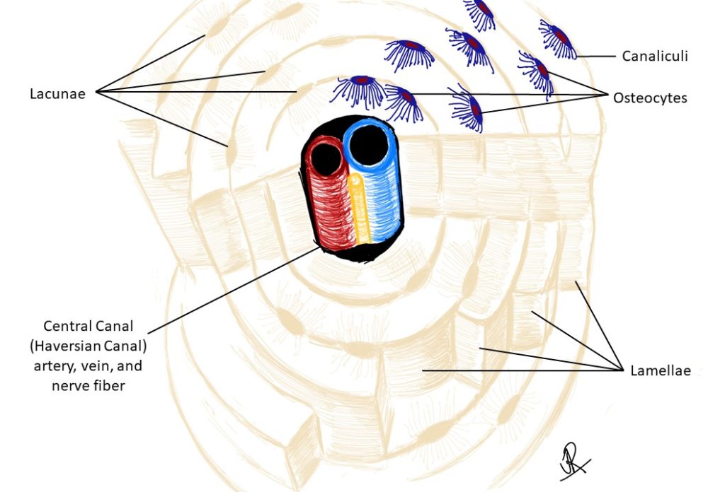
Sitting between the lamellae are pockets called lacunae in which the osteocytes are located (shown in blue in Figures 1, 2, and 4). Radiating out from the lacunae are tiny channels called canaliculi that allow communication between osteocytes and the central canal (allowing access to oxygen and nutrients) (Figure 3). The central canals are connecting to one another by perpendicular canals called perforating (Volkmann’s) canals.
Figure 1: Lateral view of compact bone at low power showing the central and perforating canals of several osteon
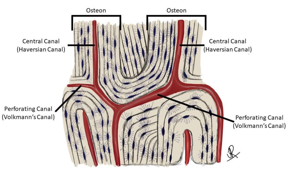
Figure 2: Superior view of compact bone at low power showing the central and perforating canals of several osteon
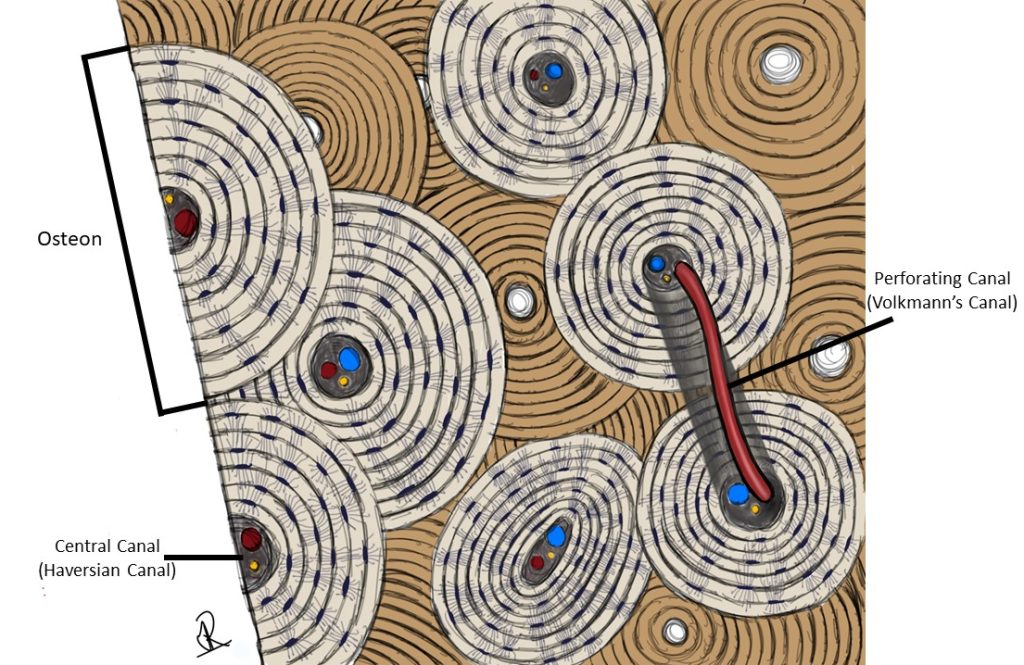
Figure 3: Osseous tissue with and without illustration overlay
Figure 4: Superior view of compact bone at high power showing the structural composition of a single osteon
Spongy bone is less dense/heavy than compact bone because it consists of very small plates/spikes of bone matrix called trabeculae. Between the trabeculae are spaces filled with red bone marrow for blood cell production.
Check out our YouTube video to help you understand Osseous Tissue:
YouTube Video – Osseous Tissue
Skeletal Organ System Organization
Our bones are part of our skeletal organ system, but they contain more than just osseous tissue. In addition to osseous tissue, our skeleton contains blood vessels, nerves, and several types of connective tissue. We will use long bones as an example of the general internal structure of bone. The ends of the long bone that articulate with other bones are covered by articular cartilage (hyaline).
Long bones have a long central shaft (diaphysis) containing the marrow-containing medullary cavity surrounded by compact bone. The osteons in this compact bone run parallel with the long-axis of the bone. For example, the osteons in the humerus or femur would run vertically up your arm or leg, respectively. The ends of long bone [epiphysis (singular) or epiphyses (pleural)] contain a core of spongy bone surrounded by an outer covering of compact bone.
Membranes of Bone
The internal surfaces are lined by connective tissue called the endosteum. The external, non-articular surfaces are lined by connective tissue called the periosteum. Both of these layers contain osteogenic cells and osteoblasts, which deposit bone matrix.
Chapter Illustrations by:
Jaylan Richardson
Soma Mukhopadhyay, Ph.D.
mature cells in bone tissue that monitor and maintain the osseous tissue matrix
structural proteins and surrounding ground substance
extracellular matrix containing collagen, calcium-binding proteins, calcium and phosphate depositions
sheets or layers
running the length of the osteon, containing blood vessels and nerves
A gap, cavity, or depression. Osteocytes (bone) and chondrocytes (cartilage) are found within lacunae.
meaning "tiny canal"
running at right angles to the central canal, containing blood vessels and nerves
plates/spikes of bone matrix found in spongy bone
two bones connecting to form a joint
cartilage that surrounds the articular surface of bones (surfaces where bone meets bone to form a joint). This cartilage decreases friction in movement and provides cushioning
a long central shaft of a long bone, containing the medullary cavity surrounded by compact bone
the end of a long bone, containing a core of spongy bone surrounded by an outer covering of compact bone
connective tissue membrane lining the internal surfaces of bone
connective tissue membrane lining the external surfaces (non-articular) of bone
stem cells in bone that can form osteoblasts and osteocytes
immature bone cells that deposit new bone matrix
