Tissue Type Look-Alikes
Some tissue types look similar to one another, which can be very confusing for students. As you learn how to identify each tissue type, pay attention to tissue type look-alikes. Be sure you know how to tell them apart, so you don’t get confused on your exams!
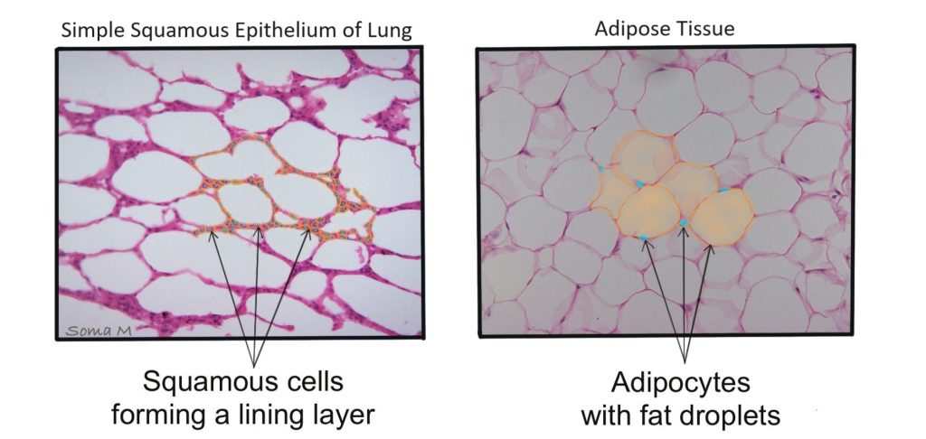
Adipose tissue vs Simple squamous epitehlium of the alveoli of the lung
Students often confuse the empty spaces in adipose tissue for the empty spaces of the air sacs (alveoli) of the lung. Because of this, students confuse adipose tissue and simple squamous epithelium of the alveoli of the lung. Figures 1 and 2 show these two tissues in side-by-side comparison.
Figure 1: Comparison of adipose tissue (top panel) with simple squamous epithelium of the lung (bottom panel). Both images show individual cells outlined by black dotted lines.
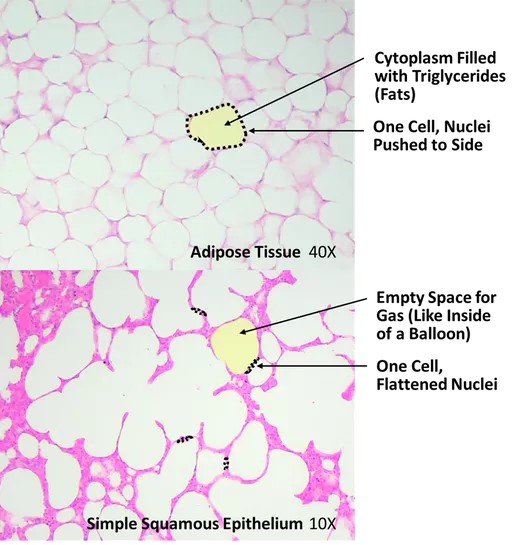
Figure 2: Comparison of simple squamous epithelium of the lung (left panel) with adipose tissue (right panel). Both images have individual cells in orange with the nuclei highlighted in turquoise.
Stratified squamous epithelium vs Transitional Epithelium
Because stratified squamous epithelium and transitional epithelium both contain multiple layers of cells, students often confuse the two tissue types. It is important to examine the cell shape at the apical surface. In stratified squamous epithelium (keratinized and non-keratinized), the cell shape becomes squamous in the cell layers close to the apical surface (Figure 3 left panel). In transitional epithelium the apical cell shape is cuboidal when relaxed (bulbous oversized cells or squamous when stretched (flattened but very long) as seen in the right panel of Figure 3.
Figure 3: Comparison of stratified squamous epithelium (left panel) with transitional epithelium (right panel)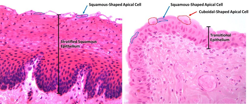
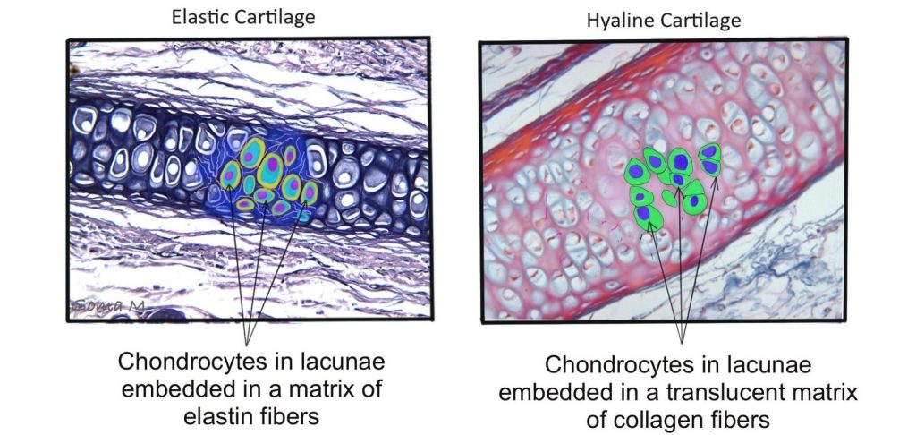
Elastic Cartilage vs Hyaline Cartilage
The overall pattern of elastic cartilage and hyaline cartilage is very similar. Both tissues have chondrocytes in lacunae surrounded by extracellular matrix. However, the extracellular matrix of elastic cartilage contains darkly-staining elastic fibers that are not seen in the smooth, glassy extracellular matrix of hyaline cartilage (Figure 4).
Figure 4: Comparison of elastic cartilage (left panel) with hyaline cartilage (right panel)
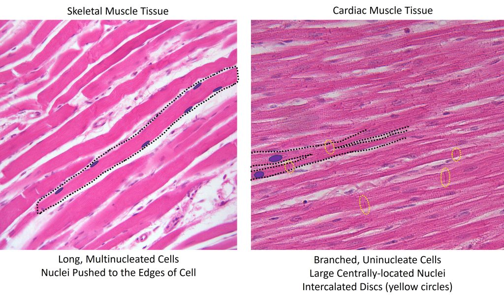
skeletal muscle Tissue vs cardiac muscle Tissue
Skeletal muscle tissue and cardiac muscle tissue share many similarities (elongated striated cells) so it is important to pay close attention to the differences between these tissue types to accurately identify them. Skeletal muscle tissue contains very long multinucleated cells, with flattened peripherally located nuclei (Figure 5 left panel). Cardiac muscle tissue contains branched cells, with typically one large centrally located nuclei, that are connected by darkly staining intercalated discs (Figure 5 right panel). Both images show individual cells outlined by black dotted lines.
Figure 5: Comparison of skeletal muscle tissue (left panel) with cardiac muscle tissue (right panel)
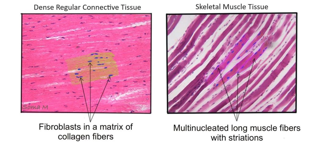
skeletal muscle Tissue vs dense regular connective tissue
The elongated muscle cells in skeletal muscle tissue are often confused with the elongated collagen fibers arranged in parallel rows in dense regular connective tissue. Figure 6 shows these two tissues in side-by-side comparison.
Figure 6: Comparison of dense regular connective tissue (left panel) with skeletal muscle tissue (right panel)
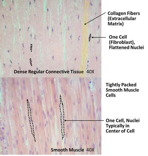
smooth muscle Tissue vs dense regular Connective Tissue
The spindle-shaped muscle cells in smooth muscle tissue are often confused with the elongated collagen fibers arranged in parallel rows in dense regular connective tissue. Figure 7 shows these two tissues in side-by-side comparison, with individual cells outlined by black dotted lines.
Figure 7: Comparison of dense regular connective tissue (top panel) with smooth muscle tissue (bottom panel)