Central and Peripheral Nervous Systems
Neurons can be classified structurally or functionally. These important distinctions are discussed in the following sections.
Structural classifications
Neurons can be structurally classified as multipolar, bipolar, or unipolar (pseudo-unipolar). Structural classification is based on the number of neuronal process (axons and dendrites) that extend from the soma (cell body) of the neuron.
Functional classifications
Neurons can also be classified by their function as sensory neurons, motor neurons, or interneurons. Sensory neurons (usually unipolar) carry information from sensory receptors in the body through the peripheral nerves to the central nervous system. Motor neurons (usually multipolar) carry information from the central nervous system through the peripheral nerves to stimulate muscles or glands. Interneurons (usually multipolar) relay and integrate the information between the sensory neurons and motor neurons, and are primarily located in the central nervous system.
Examples of Multipolar Interneurons in the Central Nervous System
The nervous tissue of the central nervous system can be divided into gray matter and white matter. Gray matter consists primarily of cells bodies while white matter consists of many myelinated axons. The convoluted outer layer of the cerebrum (cerebral cortex) is made up of gray matter. Scattered in this gray matter are large neurons called pyramidal cells. Pyramidal cells are multipolar neurons with a pyramid shaped soma (cell body).
Figure 1: Cerebrum highlighting pyramidal cells with and without illustration overlay (version A)
Figure 2: Cerebrum highlighting pyramidal cells with and without illustration overlay (version B)
Similar to the cerebrum, the cerebellum also has an outer layer of gray matter (cerebellar cortex). Purkinje cells, large multipolar neurons with pear-shaped soma, are lined up between two distinct layers of the cerebellar cortex (the molecular and granular layers).
Figure 3: Cerebellum highlighting Purkinje cells with and without illustration overlay (low power)
Figure 4: Cerebellum highlighting Purkinje cells with and without illustration overlay (high power)
Helpful hints: To determine whether you are looking at the cerebrum or the cerebellum, pay attention to the pattern of the cells. Notice that for the cerebrum, the triangular cell bodies of the pyramidal cells are located in what appears to be a random organization within the gray matter of the cortex (Figures 1 and 2). For the cerebellum, the cell bodies of the Purkinje cells are lined up in a single row within the gray matter (Figures 3 and 4).
Example of Unipolar (Pseudo-Unipolar) Neurons in the Peripheral Nervous System
The dorsal root ganglion refers to the collection of unipolar sensory neuron (pseudo-unipolar) cell bodies that are part of the peripheral nervous system, observed as a swelling in the dorsal root (lateral to the spinal cord). These neurons bring the sensory information to the central nervous system. Satellite cells are a type of glial cells found in surrounding the bodies of the sensory neurons (Figures 5 and 6).
Figure 5: Dorsal root ganglion with and without illustration overlay (version A)
Figure 6: Dorsal root ganglion with and without illustration overlay (version B)
In the peripheral nervous system axons, both myelinated and non-myelinated, are bundled together into peripheral nerves. It is important to remember that neurons and nerves are not the same – nerves contain the axons of many neurons. Bundles of axons, each surrounded by an endoneurium, are called a fascicle. Each fascicle is surrounded by a layer of connective tissue called the perineurium (Figure 7 and 8).
The entire nerve, bundling all the fascicles and blood vessels, is surrounded by a thicker layer of connective tissue called the epineurium (Figure 7).
Figure 7: Peripheral nerve in cross section (highlighting the epineurium and perineurium) with and without illustration overlay (low power)
Figure 8: Peripheral nerve in cross section (highlighting the perineurium and endoneurium) with and without illustration overlay (high power)
The axons of neurons are often myelinated. That is they are insulated by layers of myelin (protective layer of lipids and proteins). In the peripheral nervous system, myelination occurs when a Schwann cell wraps itself around the axon multiple times resulting in several layers of myelin. The cytoplasm and nucleus of the Schwann cells gets pushed to the outer layer called the outer collar of perinuclear cytoplasm or neurilemma. Along the axon, visible in longitudinal section, there are gaps in the myelin sheath called myelin sheath gaps or nodes of Ranvier (Figures 9 and 10). These act to facilitate the electrical impulses along the axon.
Figure 9: Peripheral nerve in longitudinal section (highlighting the nodes of Ranvier) with and without illustration overlay (version A)
Figure 10: Peripheral nerve in longitudinal section (highlighting the nodes of Ranvier) with and without illustration overlay (version B)
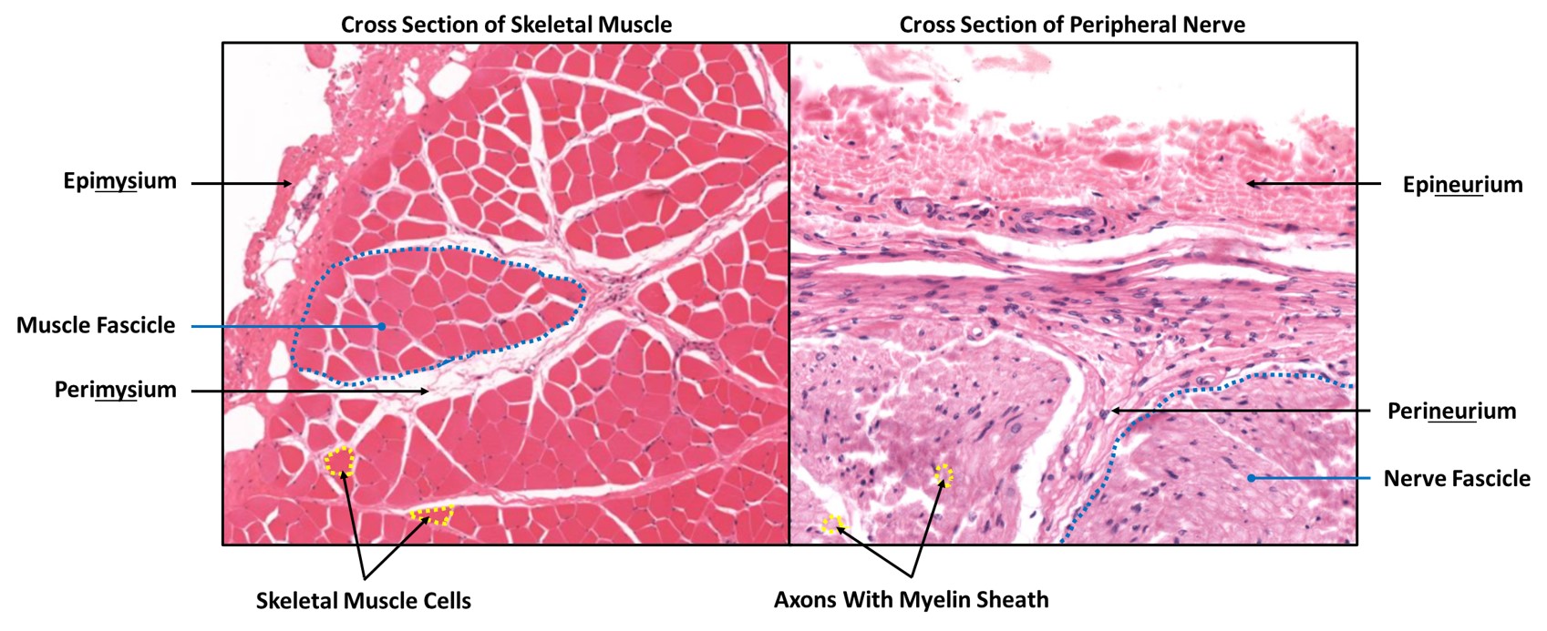
Comparison of nerve and skeletal muscle connective tissue sheaths
Students often confuse the epimysium and perimysium of the skeletal muscle with the epineurium and perineurium of the peripheral nerve. Be sure that you can identify the features of skeletal muscle histology and peripheral nerve histology that will allow you to tell the difference between the two slides.
Figure 11: Comparison of cross section of skeletal muscle and peripheral nerve