Unique Epithelial Tissues
In this chapter, we will discuss two unique types of epithelial tissues: pseudostratified columnar epithelium and transitional epithelium. We refer to these as “unique” because they do not match the cell layer patterns seen in simple and stratified epithelial tissues.
Pseudostratified Columnar Epithelium
The prefix pseudo- (from Greek ψευδής, pseudes, “false”) is used to mean that this tissue appears to be stratified, but it is not. In a stratified epithelium, the cells would be stacked on each other in layers. In pseudostratified columnar epithelium, cell size is not uniform so the nuclei appear at different heights, which gives this tissue the appearance of being stratified. However, if we look closely, the cells all touch the basement membrane basal side, but not all reach the apical surface. This causes some of the cells to appear wider at the basal side (nucleus at the bottom) and some appear wider at the apical side (nucleus at the top). You can imagine making a wall of triangles, where the base layer is made of triangles with the flat side at the bottom and inverted triangles are placed to fill the spaces in-between. This tissue often has numerous mucous-producing goblet cells, shown in turquoise in the illustration overlays for Figures 1 and 2. As seen in both of these examples, the cells of pseudostratified columnar epithelium often contains cilia on their apical side, which help to sweep/move the mucous across the surface of the tissue.
Table 1: Pseudostratified columnar epithelium
| Tissue Type | Pseudostratified Columnar Epithelium |
|---|---|
| General Description | Single layer of column-shaped cells that has the appearance of multiple layers as not all cells reach the surface, this results in nuclei appearing at multiple levels of the tissue |
| Location | Trachea, bronchi, nasal cavity, and sperm-carrying ducts |
| Function | Absorption and/or secretion |
| Special Features | Often contain goblet cells to secrete mucus, often cells have cilia that helps move substances across the cell surface |
| Helpful Hints | Look for the distinctive cilia on the apical surface of the cells |
Figure 1: Ciliated pseudostratified columnar epithelium, of the trachea (version A) with and without illustration overlay
Figure 2: Ciliated pseudostratified columnar epithelium, of the trachea (version B) with and without illustration overlay
Check out our YouTube video to help you understand ciliated pseudostratified columnar epithelium:
YouTube Video – ciliated pseudostratified columnar epithelium
Transitional Epithelium
IMPORTANT NOTE: Students often confuse transitional epithelium with stratified epithelium. In stratified epithelium, the shape of the cell looks different due to cellular changes that occur as the cell migrates from the basal side to the apical side. This “transition” is NOT what the name transitional epithelium refers to! Transitional epithelium is made of cells that can transition or change their shape depending on whether the tissue is relaxed (big round balloon cells or umbrella-shaped) or stretched (cells are pulled flat).
Transitional epithelium is found in organs that must stretch a lot (urinary bladder and ureters). Think about how small your urinary bladder is when it is empty (holds about 20 mL) and when it is full (typically holds 500-700 mL but can hold more!). Connective tissue and muscle tissue are quite stretchy/elastic. Epithelial tissue typically resembles a brick wall and is not very stretchy/elastic. The special property of the transitional epithelial tissue lining the inside of the bladder allows it to stretch so the urinary bladder to hold a large volume.
This phenomenon is most visible in the cells of the apical layer, which look enormously oversized with a tiny nucleus when the cell is relaxed. In some slides, these cells may have an umbrella shaped nucleus (flat on bottom, curved on top). We can see this phenomenon in Figure 3. On the left side of Figure 3 the tissue is stretched and the cells looked flattened. However, on the right side of Figure 3 the tissue is relaxed and the apical cells look bulged and oversized.
Table 2: Transitional epithelium
| Tissue Type | Transitional Epithelium |
|---|---|
| General Description | Multiple layers of cells, apical cells may appear either cuboidal or squamous depending on stretch |
| Location | Urinary bladder and ureters |
| Function | Allows stretch |
| Helpful Hints | Look for cube-shaped cells on the apical surface |
Figure 3: Transitional epithelium of the urinary bladder – stretched (left) and relaxed (right) with and without illustration overlay
Check out our YouTube videos to help you understand transitional epithelium:
YouTube Video – Transitional Epithelium
Summary of the major types of epithelial tissue
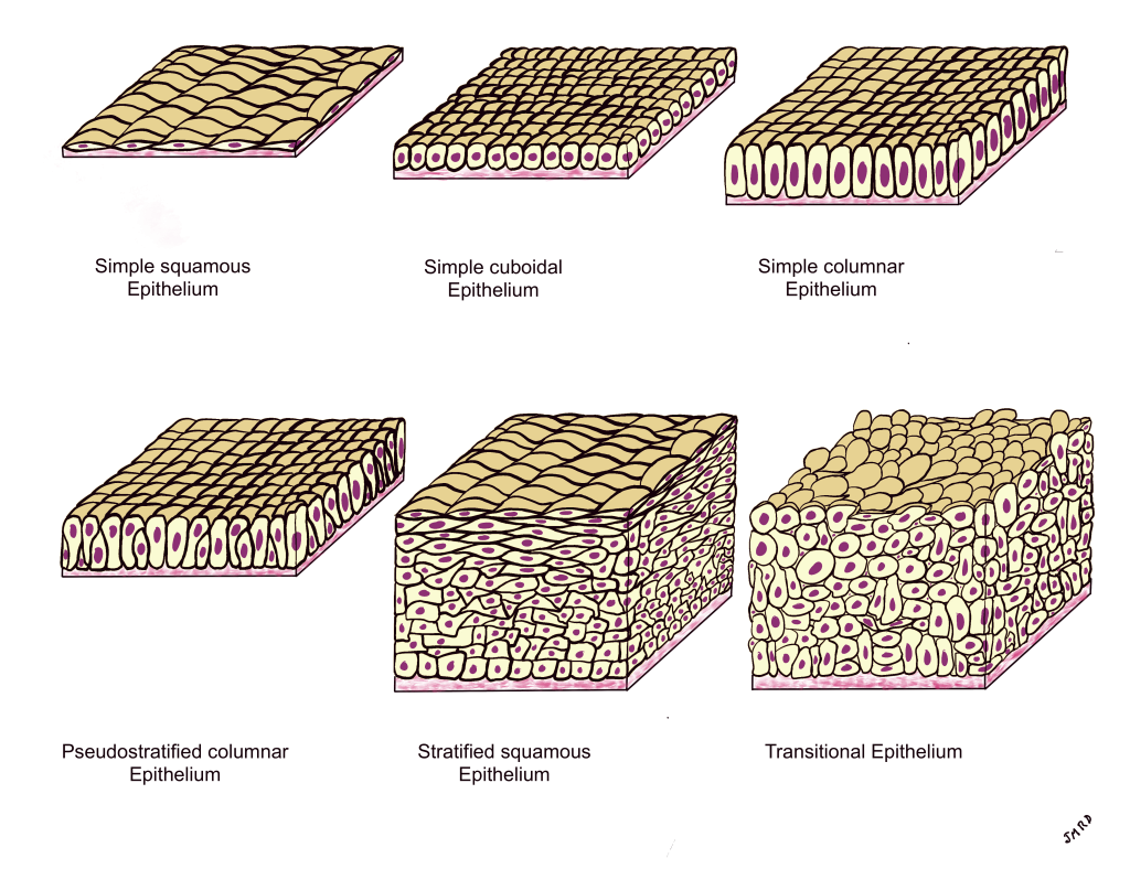
When you are learning how to identify types of epithelium, it is helpful to compare them to each other. You should take note of the similarities and differences between the different types. What distinguishing characteristics can help you identify the tissue type you are looking at? The image below (Figure 4) shows the types of epithelium presented in this textbook side-by-side to help you learn how to identify each type.
Figure 4: The major types of epithelium
Chapter Illustrations By:
Juan Manuel Ramiro-Diaz, Ph.D.
uni-cellular mucous-producing gland found in epithelium
surface appendages found in some epithelial cells that move in a rhythmic manner to move mucous across the apical suface of the tissue
