The Spinal Cord
Located in the central nervous system, the spinal cord is a cylindrical structure that runs down the center of your back. It carries information between the peripheral nervous system and the brain. The spinal cord runs vertically through the vertebral foramen of the vertebrae. This delicate organ is surrounded and protected by three-layer connective tissue meninges and the bony tissue of the vertebra. Spinal nerves containing axons of both sensory and motor neurons extend laterally, perpendicular to the spinal cord. The dorsal root contains sensory neurons (incoming, afferent signals) and the ventral root contains motor neurons (outgoing, efferent signals). The dorsal root and ventral root merge to form the spinal nerve. More information on the dorsal root ganglion and peripheral nerves can be found in the previous chapter, Central and Peripheral Nervous Systems.
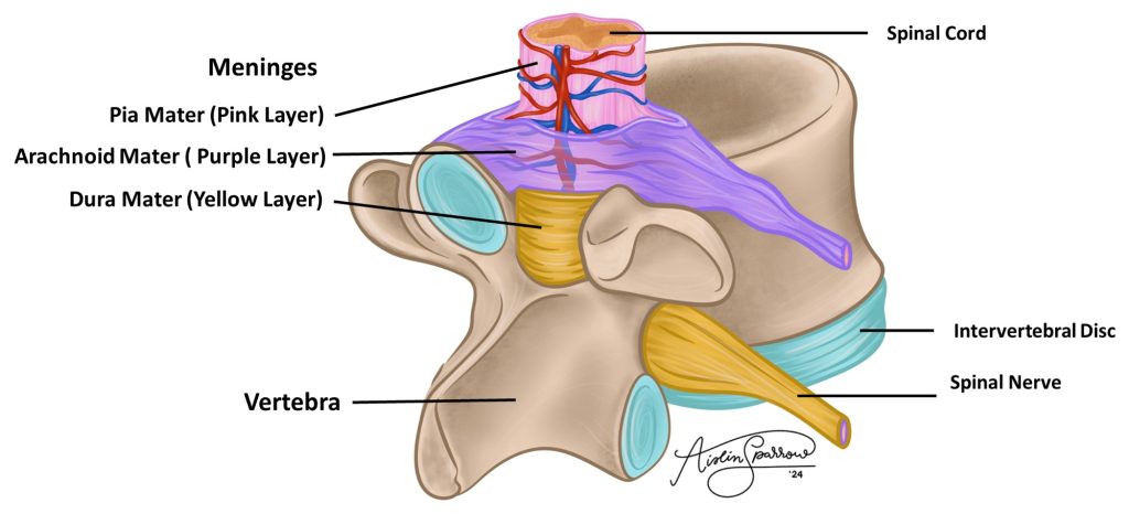
Figure 1: The illustrated rendering of a spinal cord protected by the meninges and bony tissue of the vertebra
Structure of the spinal cord
In the center of the spinal cord, containing cerebral spinal fluid (CSF), is the central canal. The central canal is lined by ependymal cells, glial cells that contain cilia which circulate the cerebral spinal fluid. The spinal cord contains an inner area of gray matter surrounded by an outer area of white matter. It is important to note that this pattern is reversed from the cerebrum and cerebellum, which both contain inner white matter and outer gray matter. The gray matter of the spinal cord consists of non-myelinated axons and neuron cell bodies. Gray matter primarily contains many local or short-distance interneuron connections. The white matter of the spinal cord consists primarily of myelinated axons in columns that run vertically to relay impulses to/from the brain.
Similar to the brain, the gray matter and white matter of the spinal cord are symmetrically distributed bilaterally. This means the left and right side of the spinal cord are mirror images of one another. The white matter areas of the spinal cord are given the name funiculus and the gray matter areas are referred to as horns. The funiculi and horns are named according to their position relative to the central canal – dorsal, lateral, and ventral. It is important to note that the lateral horn is only found in the thoracic and lumbar regions of the spinal cord.
Figure 2: The spinal cord in cross section highlighting general structure (scanning power), with and without illustration overlay
Figure 3: The spinal cord in cross section highlighting the central canal (high power), with and without illustration overlay
The meninges
The brain and spinal cord are both protected by meninges and bony skeleton (cranium and vertebrae, respectively). The inner-most layer of the meninges, which touches the tissue of the brain/spinal cord and consists of a thin layer of connective tissue, is the pia mater. The outer-most layer of the meninges, which sits closest to the bone and consists of a layer of dense connective tissue, is the dura mater. Sandwiched between the pia mater and dura mater is a delicate network of connective tissue fibers called the arachnoid mater, named for its spiderweb appearance. Cerebral spinal fluid and blood vessels fill the space between the pia mater and arachnoid mater, providing cushioning for the brain and spinal cord.
Figure 4: The spinal cord in cross section highlighting the meninges (high power), with and without illustration overlay
Chapter Illustrations By:
Aislin Sparrow
Georgios Kallifatidis, Ph.D.
