Introduction to Epithelial Tissues
Epithelial tissues are a special tissue type used to form boundaries in the human body. This means that epithelial tissues separate one compartment from another. Epithelial tissues line all of our open body cavities (inside your stomach, your nose, your blood vessels) and the external surfaces of your body (your skin).
General Characteristics of Epithelial tissues
All types of epithelial tissue have the same general characteristics:
- Polarity – epithelial cells have apical and basal sides with different structure/function
- Supported by connective tissue – anchored to connective tissue layer or basement membrane
- Avascular, but innervated – contains nervous supply, but no blood supply
- Specialized contacts – cells are joined by desmosomes and tight junctions
- High capacity for regeneration – cells are highly mitotic to repair and renew
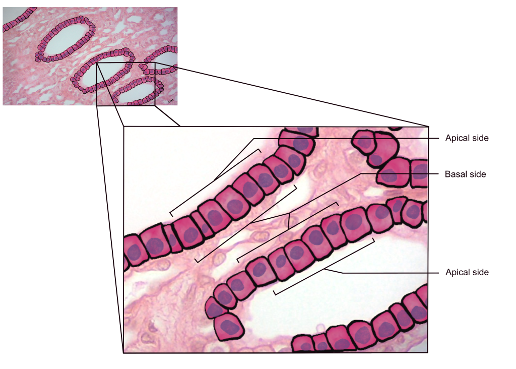
One of the characteristics that makes epithelial tissue special is its polarity. Polarity means that the cell has sides with different structure/function, which makes the cell polar. In epithelial tissue, the sides are the apical and basal sides.
The apical side of the tissue will be open and exposed to the environment/organ cavity. On a microscope slide, the apical surface is adjacent to empty space (white space), like the lumen of the structure.
The basal side of the layer/sheet of cells will be anchored to a connective tissue layer we called the basal lamina or basement membrane. Blood supply for the epithelial tissue is found beyond the basal lamina in the deeper connective tissues.
Figure 1: Simple cuboidal epithelium of the kidney (cross-section of renal tubule), highlighting the apical and basal sides of the cell
Naming epithelial tissues
Epithelial tissues are named by two criteria:
- Number of cells in the layer
- Shape of the cell at the apical side
Number of Cells in the Layer
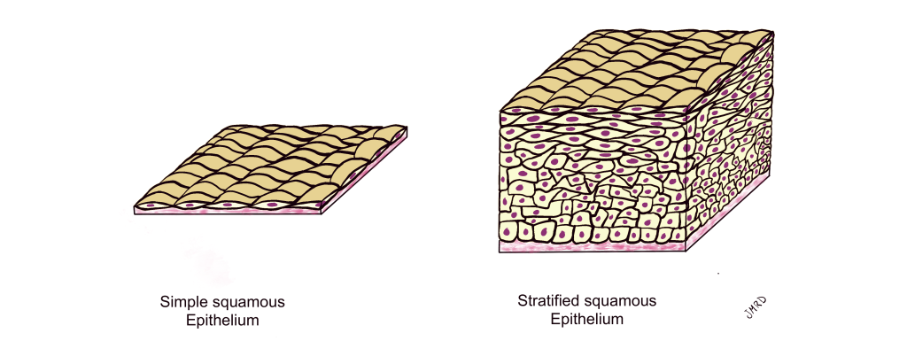
Simple Epithelial Tissues – If the tissue is a single layer of cells in thickness, it is a simple epithelium
Stratified Epithelial Tissues – If the tissue has more than one layer of cells in thickness, it is a stratified epithelium
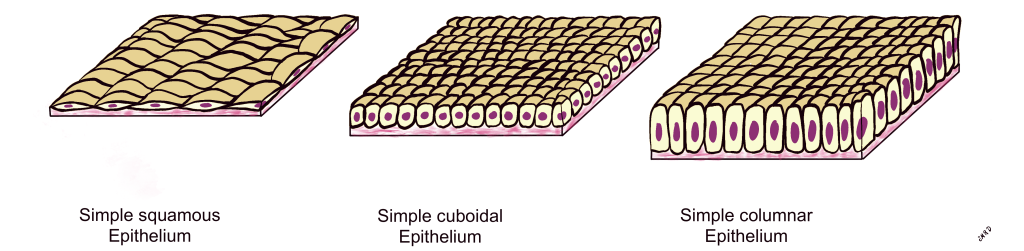
Shape of the Cell at the Apical Side
Figure 2: Simple epithelium illustrating three cell shapes: squamous (left), cuboidal (center), and columnar (right)
Locations of epithelial tissues
Epithelial tissues form the boundaries in our body and separate one space from another. Epithelial tissues line the surfaces of our body that are:
- Exposed to the exterior – our skin
- Exposed to the contents of an open cavity – our mouth and nasal cavities (upper respiratory passages)
- Exposed to the contents of a hollow organ – inside of a blood vessel, a kidney tubule, the gastrointestinal tract (stomach, small intestine, etc.)
Functions of epithelial tissues
The functions of epithelial tissues depend on the structural type of epithelium.
- Simple epithelial tissues line surfaces where diffusion/filtration/absorption/secretion occur. A single layer of epithelial cells make movement of molecules easier!
- Stratified epithelial tissues line surfaces where protection is needed. Our skin is constantly being rubbed and scratched and many layers of cells provide protection!
Figure 3: Simple epithelium (left) and stratified epithelium (right)
The following chapters will discuss the many different types of epithelial tissues.
IMPORTANT NOTE: Epithelial tissues are divided into two functional groups, lining epithelium and glandular epithelium. The following chapters will focus primarily on lining epithelium and will not focus on examples of glandular epithelium.
Chapter Illustrations By:
Juan Manuel Ramiro-Diaz, Ph.D.
having two sides with different structural or functional characteristics.
without blood vessels
having nerve endings for sensory or motor function
the ability of a tissue to repair itself or to replace old cells with new cells
the exposed side of epithelial tissue
the anchored side of epithelial tissue
in epithelial tissues, a single layer of cells anchored to a basement membrane
in epithelial tissues, a layer of two or more cells with the basal layer anchored to a basement membrane
a scale-like, flattened cell with a disc-shaped nucleus
a cube-shaped cell with a round nucleus
a tall, rectangular cell with an egg-shaped nucleus
